

At a Glance
- Represents
- The first positive QRS deflection: early-to-mid ventricular activation. Its height in each precordial lead reflects the forces facing that lead.
- Normal (Adult)
- Small r in V1 (R/S <1); R grows from V1 toward V5–V6; transition (R = S) at V3–V4; R >3 mm in V3; aVR mostly negative.
- Measure It
- R height in mm (1 mm = 0.1 mV) from baseline to peak, V1–V6. Find the lead where R = S. Compare with a prior ECG.
- Abnormal → Think
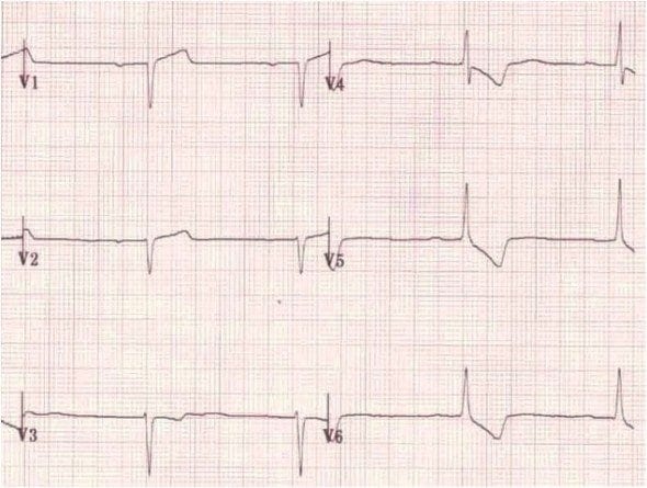
- Poor progression (R ≤3 mm in V3): old anterior MI, LVH, lead placement
- Tall R in V1: RVH, RBBB, posterior MI, WPW
- Dominant R in aVR: lead reversal, sodium-channel block, VT
- Red Flags
- New poor progression with chest pain: treat as possible ACS
- Tall R V1–V2 with ST depression V1–V3: check V7–V9
- Dominant R in aVR with a wide QRS: suspect sodium-channel block
- Bedside Action
- Fix lead errors first. Add V7–V9 for tall R in V1–V2, serial 12-leads for pain, and notify per local protocol. Sodium-channel block: poison control and medical direction.
Physiology & What It Represents
The R wave is the first positive deflection of the QRS complex. It records the early and middle part of ventricular depolarization. The His–Purkinje system has just delivered the impulse, and it now spreads through the ventricular muscle.
Follow the path. Sinoatrial node, atria, atrioventricular (AV) node, His bundle, both bundle branches, Purkinje fibers, then ventricular muscle. The septum lights up first, left to right. Next is the left ventricular free wall, which carries most of the muscle mass. The posterobasal left ventricle and the right ventricular outflow tract go last. The R wave is the net vector of that sequence, seen from each lead's angle.
Polarity depends on where the lead sits. A precordial lead over the left ventricle sees the wavefront coming at it, and that is the tall R. V1 sits over the right ventricular outflow and septum. So it usually shows a small r and then a deeper S wave. Any change in R height or progression reflects one of four things: ventricular mass, timing of activation, where the heart sits, or where the electrodes are.
- Why it matters at the bedside: missing R waves across the anterior leads can mark an old infarct. A tall R in V1 can mark RVH, conduction delay or posterior forces. A misplaced electrode imitates both.

Normal Values & How to Measure
| Parameter | Adult Normal | Pediatric Note | Where / How to Measure |
|---|---|---|---|
| R wave in V1 | Small r; R/S <1 (S deeper than R) | Dominant R in V1 can be normal in neonates and young children | V1; baseline to peak of the first positive deflection |
| R-wave progression | R rises from V2 to V4 and reaches V5–V6; R >3 mm in V3 | Progression pattern changes with age | V1–V6; compare each lead with the one before |
| Transition zone | R = S at V3–V4 | Earlier or later transition needs an age-based reference | The lead where R and S depths are equal |
| R wave in aVR | QRS mostly negative; R not dominant | — | aVR; if R is dominant, check lead I before further steps |
| LVH voltage limits | Sokolow-Lyon S V1 + R V5 or V6 <35 mm (3.5 mV); Cornell R aVL + S V3 ≤28 mm men, ≤20 mm women | Use age-based references; see Pediatrics | Measured on a 12-lead; not valid with QRS ≥120 ms |
- Confirm electrode placement first: V1 and V2 belong in the fourth intercostal space at the sternal edges, not higher on the chest. Check the limb leads too.
- Choose the beat: measure the same beat in every lead. In an irregular rhythm, average several beats.
- Measure the R wave: from the isoelectric baseline (PR or TP segment) to the peak of the R wave, in V1 through V6. Report in mm (1 mm = 0.1 mV).
- Find the transition zone: the lead where R and S depths are equal. Normal is V3–V4.
- Check progression: R should increase from V2 to V4. R ≤3 mm in V3 is poor R-wave progression.
- Check aVR: the QRS should be mostly negative. A dominant R in aVR needs lead I checked first.
- Compare with a prior ECG and review Q waves, ST segments and T waves in the same leads.
Poor R-Wave Progression (PRWP)
Poor R-wave progression (PRWP) means the R wave stalls. At 3 mm or less in V3, it has not grown the way it should from V1 to V4. It is a finding. It is not a diagnosis.
ECG Criteria
- Amplitude: R ≤3 mm in V3
- Pattern: R does not rise from V2 to V4, or stays small across V1–V4
- Context: check a prior ECG, Q waves and ST-T changes in V1–V4 before attributing the pattern to infarction
Causes
| Category | Examples |
|---|---|
| Ischemic | Prior anteroseptal MI, with Q waves and ST-T changes in the same leads |
| Structural | LVH; dilated or infiltrative cardiomyopathy affecting the anterior wall |
| Conduction | Left bundle branch block, which changes septal activation |
| Technical | V1–V2 placed too high on the chest wall |
| Position or lung | COPD with hyperinflation; clockwise rotation of the heart (late transition) |
| Normal variant | Common in young women and thin patients |
Clinical Significance
- Low sensitivity for scar: MESA was a community cohort of 2,950 adults who had cardiac MRI, not patients with chest pain. PRWP showed up in 5.5% and missed anterior scar. Here PRWP meant R in V3 of 3 mm or less, with R in V3 greater than R in V2. Sensitivity was 0.0% for strict scar criteria and 5.3% for liberal criteria. Specificity was 94.4%.
- Escalation: PRWP with new chest pain or ischemic symptoms gets worked up as possible acute coronary syndrome (2025 ACC/AHA ACS guideline, see References). New ST elevation or new Q waves go down the STEMI pathway per local protocol.
- Chronic pattern: same pattern on a prior ECG and the patient is stable? Document it. Compare every future tracing against it.
Look-Alikes
- Lead misplacement, lead reversal and normal variants: see Differential Diagnosis & Mimics.
- Rotation and COPD: see Causes above.
Dominant R Wave in V1
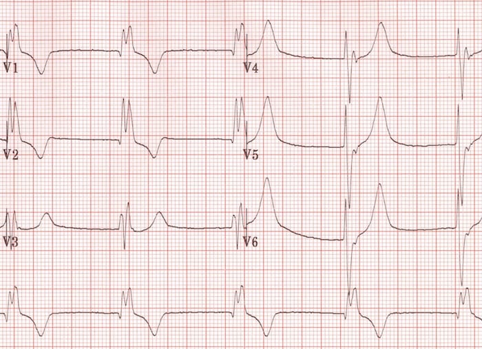
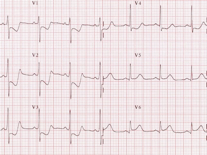
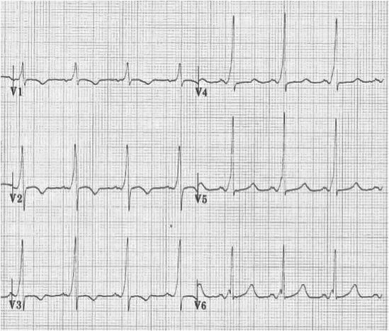
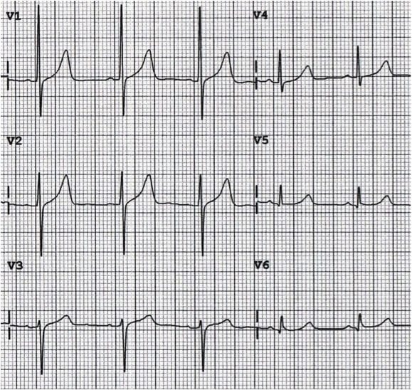
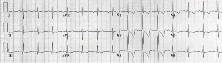
A dominant R wave in V1 means the R is taller than the S (R/S ≥1). In an adult that is abnormal, and it needs an explanation. In neonates and young children it is normal.
ECG Criteria
- R/S ≥1 in V1: R taller than S
- R ≥7 mm in V1: a voltage threshold used in RVH criteria (see the LVH and RVH section)
- rSR′ pattern with QRS ≥120 ms: right bundle branch block
- Tall R with ST depression in V1–V3: posterior MI pattern, when posterior leads confirm it
- Short PR with a delta wave: WPW type A
Causes
| Cause | Key Features |
|---|---|
| Right ventricular hypertrophy (RVH) | Tall R in V1, right axis deviation, right ventricular strain (ST depression and T inversion V1–V3) |
| Right bundle branch block (RBBB) | rSR′ ("rabbit ears") in V1 with QRS ≥120 ms; wide S in I and V6 |
| Posterior MI | Tall R in V1–V2 with ST depression V1–V3; ST elevation in V7–V9 confirms |
| WPW type A | Short PR, delta wave and tall R in V1 from a left-sided accessory pathway |
| Dextrocardia | Reversed precordial progression, inverted lead I and P wave |
| Lead misplacement | V1 and V3 swapped (biphasic P wave in V3), or V1–V2 placed too high |
| Normal variant | Neonates and young children |


Dominant R Wave in aVR
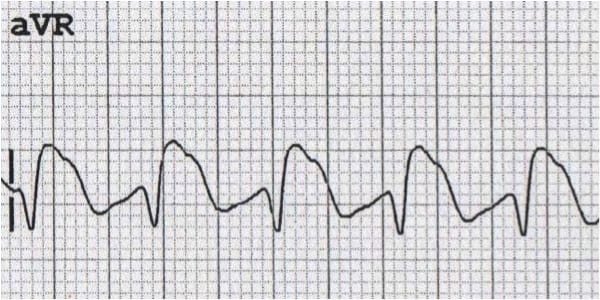
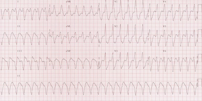
Expect aVR to run mostly negative. When a positive R dominates there, something is off. Four causes deserve your attention: arm-lead reversal, dextrocardia, sodium-channel blockade and ventricular tachycardia.
Causes
- Left and right arm lead reversal: a technical cause, and a common one. Lead I flips negative while aVR and aVL trade places.
- Dextrocardia: the heart sits on the right side. Precordial R-wave progression runs backwards.
- Sodium-channel blocker toxicity: tricyclic antidepressants and Class Ia or Ic antiarrhythmics, and other sodium-channel blockers such as diphenhydramine, cocaine, carbamazepine and bupropion.
- Ventricular tachycardia: a wide-complex rhythm. A dominant R in aVR is one VT feature among several.

Left and Right Ventricular Hypertrophy (LVH and RVH) Voltage Criteria
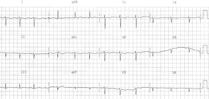
R and S amplitude climb with ventricular muscle mass. Voltage criteria are screening tools with limited sensitivity. They stop being reliable when conduction is abnormal.
Left Ventricular Hypertrophy (LVH) Criteria
| Criterion | Threshold | Notes |
|---|---|---|
| Sokolow-Lyon | S in V1 + R in V5 or V6 ≥35 mm (3.5 mV) | Common screening rule with limited sensitivity |
| Cornell voltage | R in aVL + S in V3 >28 mm (2.8 mV) men; >20 mm (2.0 mV) women | Sex-specific thresholds |
- Associated findings: left axis deviation, lateral ST depression and T inversion (strain pattern), and a late transition zone
- Under-detection: a 2026 general-population study checked guideline voltage criteria against cardiac CT. The criteria missed LVH most often in women and in people with overweight
Right Ventricular Hypertrophy (RVH) Criteria
No single RVH criterion is sensitive. Stack up more criteria and the finding gets more specific. Echocardiography confirms RVH.
- R in V1 ≥7 mm
- R/S ratio in V1 ≥1
- S in V5 or V6 ≥7 mm
- R/S ratio in V5 or V6 ≤1
- Right axis deviation ≥+110°
Causes of RVH
- Chronic lung disease and pulmonary hypertension: COPD, pulmonary fibrosis, primary or secondary pulmonary hypertension
- Pulmonary embolism: acute RV strain can show up without classic hypertrophy. Look for strain and the clinical picture
- Congenital heart disease: tetralogy of Fallot, pulmonary stenosis, left-to-right shunts
- Valve disease: tricuspid or pulmonary valve disease
Differential Diagnosis & Mimics
Tall R in V1
| Feature | Posterior MI | RVH | RBBB | WPW type A |
|---|---|---|---|---|
| QRS duration | Usually normal | Usually normal | ≥120 ms with rSR′ in V1 | Often widened by the delta wave |
| PR interval | Normal | Normal | Normal | Short |
| ST and T in V1–V3 | ST depression with upright T | Strain: ST depression, T inversion | Secondary T inversion V1–V2 | Secondary changes possible |
| Axis | Usually normal | Right axis, ≥+110° | Usually normal | Variable |
| Key clue | ST elevation V7–V9; prior Q waves | Right axis plus other RVH criteria | Wide S in I and V6 | Short PR and delta wave |
Dominant R in aVR
| Feature | Arm-lead reversal | Dextrocardia | Sodium-channel block | VT |
|---|---|---|---|---|
| Lead I | Inverted | Inverted | Normal | Variable |
| Precordial progression | Normal | Reversed | Normal | Abnormal in many cases |
| QRS width | Normal | Normal | Wide | Wide |
| Key clue | Lead I inverted; aVR and aVL swapped | Reversed progression; inverted lead I and P | Wide QRS in a toxic context | AV dissociation or capture beats |
Artifact & Lead Misplacement
- V1–V2 too high: false poor progression or a false small R wave
- V1 and V3 swapped: biphasic P wave in V3
- Left and right arm reversal: inverted lead I with a dominant R in aVR and normal precordial progression
- Tremor or motion: it can mimic VT or shift R height. Get the tracing again once the patient holds still.
- Transport vibration: it adds artifact to both the monitor and the 12-lead. Repeat the tracing once the vehicle is stopped.
Normal Variants
- Young women and thin patients: poor R-wave progression is common. Compare with prior tracings
- Neonates and young children: a dominant R in V1 is what you expect
- Heart rotation: counterclockwise rotation moves the transition earlier. Clockwise rotation moves it later
Bedside Management & Escalation
An R-wave finding changes management when it flags ischemia, toxic ingestion or a recording error. Check the recording first. Then act on the pattern.
- New poor progression with chest pain or new symptoms: treat as possible acute coronary syndrome. Get serial 12-leads and notify the receiving team per local STEMI protocol (ALS/RN).
- Tall R in V1–V2 with ST depression in V1–V3: add V7–V9. Posterior MI is supported by ST elevation of 0.5 mm or more there. Escalate per local protocol (ALS/RN).
- Dominant R in aVR with a wide QRS, altered mental status or seizure: suspect sodium-channel blockade. Call medical direction and poison control per protocol. Under toxicology guidance, sodium bicarbonate is used for QRS widening. See the Sodium Bicarbonate page (Paramedic/RN/CCT).
- Dominant R in V1 with dyspnea, hypoxia or right axis deviation: consider acute RV strain, with pulmonary embolism among the causes. Notify the receiving team and get echocardiography or CT per protocol (ALS/RN).
- Suspected lead error: fix the leads and repeat the ECG before you make any activation decision.
Follow local protocols and online medical direction.
Pediatric & Special Populations
Pediatrics
- Neonates and infants: a dominant R in V1 is normal. It reflects relatively large right ventricular forces
- Older children: the dominant R in V1 normally shrinks with age. If it persists in an older child or adolescent, evaluate for RVH or congenital heart disease.
- Age-based references: progression and transition shift with age. Use a pediatric ECG reference. For rate context, see Pediatric Vital Signs.
Other Populations
- Athletes: high-voltage LVH patterns are common, and often benign. Confirm with echocardiography before you label disease.
- Chronic lung disease: hyperinflation lowers voltage and can produce poor progression. Don't call it infarction without other supporting findings.
- Pacemaker or ICD: paced beats change R-wave shape and progression. Compare with a prior ECG, and see the QRS Complex page.
- Women and thin patients: poor progression turns up more often, and it is often benign. Confirm it against a prior tracing.
- Pregnancy: the enlarging uterus pushes the diaphragm up and shifts the heart. That can change the axis and R-wave progression. Compare with a prior ECG before you call a new pattern abnormal.
- Breast tissue and obesity: in women, place V4–V6 in the standard position under the breast at the inframammary fold, and record where each lead was placed. Tissue, body habitus and misplaced leads all pull voltage down, and they can shift R height and progression. Voltage criteria miss LVH most often in women and in people with overweight.
Monitoring & Transport Considerations
| Issue | What to Do |
|---|---|
| Rhythm monitoring | Monitor lead II for rhythm and P waves. Use a 12-lead to judge R-wave progression. |
| Electrode placement | Confirm V1 and V2 in the fourth intercostal space at the sternal edges on every 12-lead. |
| Serial 12-leads | Repeat every 15–30 min with ongoing chest pain or changing symptoms, and compare with the first tracing. |
| Posterior and right-sided leads | Add V7–V9 when there are tall R waves in V1–V2 with ST depression in V1–V3. Add V4R when right ventricular involvement is suspected. |
| Transmission | Send the 12-lead with lead labels and the time. Include a prior ECG if available. |
| Artifact | Vibration, shivering or movement changes R height and polarity. Pause, stabilize the electrodes and repeat. |
Step-by-Step Approach & Pitfalls
- Confirm lead labels and placement: check V1 and V2 position and the limb leads before reading any R wave.
- Measure the R wave height: in V1 through V6, from baseline to peak, in mm.
- Find the transition zone: where R equals S. Normal is V3–V4.
- Check progression: R should rise from V2 to V4. R ≤3 mm in V3 is poor progression.
- Check R/S in V1: R/S ≥1 is a dominant R. Name the cause using QRS width, axis and PR interval.
- Check aVR: it should be mostly negative. If R is dominant, check lead I first, then QRS width.
- Compare with a prior ECG and review Q waves and ST-T changes.
- Act on stability and red flags, then repeat the ECG after any lead correction.
Common Pitfalls
- Calling PRWP an infarct without Q waves, ST-T changes or a prior comparison.
- Missing posterior MI when tall R waves in V1–V2 come with ST depression in V1–V3.
- Applying LVH voltage criteria with bundle branch block, in athletes or in thin young adults.
- Diagnosing toxicity before checking lead I for arm-lead reversal.
- Reading one beat in an irregular rhythm or with ectopy.
- Ignoring QRS width when a dominant R in aVR is present.
Clinical Pearls
References
- Hancock EW, Deal BJ, Mirvis DM, Okin P, Kligfield P, Gettes LS. AHA/ACCF/HRS recommendations for the standardization and interpretation of the electrocardiogram: part V: electrocardiogram changes associated with cardiac chamber hypertrophy (landmark standardization statement). Circulation. 2009;119:e251–e261. https://doi.org/10.1161/CIRCULATIONAHA.108.191097
- Boehnert MT, Lovejoy FH Jr. Value of the QRS duration versus the serum drug level in predicting seizures and ventricular arrhythmias after an acute overdose of tricyclic antidepressants (landmark study). N Engl J Med. 1985;313:474–479. https://doi.org/10.1056/NEJM198508223130804
- Thygesen K, Alpert JS, Jaffe AS, et al. Fourth Universal Definition of Myocardial Infarction (2018). Circulation. 2018;138:e618–e651. https://doi.org/10.1161/CIR.0000000000000617
- Rao SV, et al. 2025 ACC/AHA/ACEP/NAEMSP/SCAI Guideline for the Management of Patients With Acute Coronary Syndromes. Circulation. 2025;151(13). https://doi.org/10.1161/cir.0000000000001309
- de Alencar JN, Morales PAS. Poor R-wave progression fails to detect anterior myocardial fibrosis in MESA. Int J Cardiol. 2026;456:134498. https://doi.org/10.1016/j.ijcard.2026.134498
- Johansen AO, Kühl JT, Aspe AW, Lowes M, Loft JJ, Pham MHC, Sigvardsen PE, Nielsen JB, Larsen AF, Graff C, Paulsen RR, Kofoed KF. Addressing systematic underdetection of left ventricular hypertrophy by guideline-recommended electrocardiographic criteria in women and individuals with overweight. Int J Cardiol. 2026;464:134761. https://doi.org/10.1016/j.ijcard.2026.134761
- Liebelt EL, Francis PD, Woolf AD. ECG lead aVR versus QRS interval in predicting seizures and arrhythmias in acute tricyclic antidepressant toxicity. Ann Emerg Med. 1995;26(2):195–201. https://doi.org/10.1016/s0196-0644(95)70151-6
Medical Disclaimer
- For Educational Purposes Only: This content is intended for educational reference and should not be used for clinical decision-making.
- Not a Substitute for Professional Judgment: Always consult your local protocols, institutional guidelines, and supervising physicians.
- Verify Before Acting: Users are responsible for verifying information through authoritative sources before any clinical application.