

At a Glance
- Represents
- Ventricular depolarization, from the His–Purkinje system through the myocardium. Width separates a supraventricular origin (narrow) from a ventricular, conduction or toxic cause (wide).
- Normal (Adult)
- ≤110 ms (≤0.11 s; under 3 small boxes)
110–119 ms: incomplete block
≥120 ms: wide - Measure It
- Onset to J point in the widest lead (often II, V1 or V5–V6). In an irregular rhythm, average several beats.
- Abnormal → Think
- Bundle branch or fascicular block, ventricular rhythm, paced beat, hyperkalemia, sodium-channel blocker, pre-excitation
- Red Flags
- Adult wide QRS with tachycardia: VT until proven otherwise.
QRS ≥100 ms with a tricyclic or other sodium-channel blocker: seizure and arrhythmia risk. - Bedside Action
- Unstable: synchronized cardioversion. Stable: 12-lead, exclude sodium-channel toxicity and hyperkalemia first, then amiodarone 150 mg over 10 min or procainamide 20–50 mg/min.
Physiology & What It Represents
The QRS complex is ventricular depolarization. Follow the impulse. It leaves the SA node, crosses the atria, pauses at the AV node, then drops down the His bundle, both bundle branches and the Purkinje network into the ventricular muscle. The septum lights up first. The free walls follow. Width tells you how fast the impulse spreads through the ventricles.
A narrow complex means the normal conduction system is carrying the impulse. Both ventricles activate within about 110 ms. A bundle branch block delays one ventricle, and that side activates late, by slow cell-to-cell conduction. Ventricular origin is the other route to a wide complex. Pacing or ventricular ectopy skips the system altogether.
- Q wave: first negative (downward) deflection. R wave: first positive deflection. S wave: negative deflection after an R wave.
- Capital vs lowercase: Capital letters mean big deflections. Lowercase letters mean small ones. R′: a second positive deflection (rSR′). QS: entirely negative, with no R wave.

Normal Values & How to Measure
| Parameter | Adult Normal | Pediatric Note | Where / How to Measure |
|---|---|---|---|
| QRS duration | ≤110 ms (≤0.11 s; under 3 small boxes). 110–119 ms: incomplete block. ≥120 ms: wide | Shorter in infants and children; use age-specific percentile charts | Onset to J point in the widest lead; 1 small box = 40 ms |
| Morphology, V1 | rS or QS; a small r can be present (septal activation left to right) | Use age-specific norms | Compare V1 with V6 on the same beat |
| Morphology, V6 | Upright qR or R, narrow, not notched | Use age-specific norms | Same beat as V1; a notched, broad R points to LBBB |
- Pick the widest lead: scan the 12 leads and measure the widest QRS (often lead II, V1 or V5–V6).
- Mark the onset: where the QRS leaves the baseline after the PR segment.
- Mark the J point: where the QRS meets the ST segment.
- Count: 1 small box = 40 ms (0.04 s); 5 small boxes = 200 ms (0.20 s). Measure 3 or more beats; in an irregular rhythm, average several beats.
- Classify: ≤110 ms normal; 110–119 ms incomplete block; ≥120 ms wide.
- Check V1 and V6: name the bundle branch pattern only after the width is confirmed.
R-wave progression: normally the R wave grows from V1 toward V4, and the transition (R = S) sits at V3–V4. Poor progression and the voltage criteria for hypertrophy are on the R Wave page. Pathologic Q waves get their own page, Q Wave. Axis and fascicular blocks sit on the Cardiac Axis page.
Wide QRS: Causes
A QRS of 120 ms or more means ventricular activation took longer than it should. Start with the mechanism. The table groups the causes that way, and the rhythm types live on the Ventricular Rhythms page.
ECG Criteria
- Duration: ≥120 ms (≥0.12 s; 3 or more small boxes)
- Morphology: bundle branch-like, notched, or bizarre. Compare the complex with the sinus beats, and with an old ECG if one exists.
Causes
| Category | Examples |
|---|---|
| Ventricular origin | Ventricular tachycardia, ventricular ectopy, escape and accelerated idioventricular rhythm |
| Conduction block | Right or left bundle branch block, fascicular block, nonspecific intraventricular conduction delay |
| Pacing | Right ventricular or biventricular pacing |
| Pre-excitation | Wolff-Parkinson-White (short PR, delta wave); see PR Interval |
| Electrolyte | Hyperkalemia (a late sign; see below) |
| Toxic | Tricyclic antidepressants, class Ia and Ic antiarrhythmics, diphenhydramine, cocaine, local anesthetic systemic toxicity |
Clinical Significance
- Wide with tachycardia in an adult? That is ventricular tachycardia until proven otherwise.
- A new wide QRS with hypotension, syncope, shock or chest pain needs treatment now. See Bedside Management.
- A change from the patient's baseline matters more than the width alone. A new bundle branch block or a new wide QRS is that change.
Bundle Branch Blocks (RBBB and LBBB)
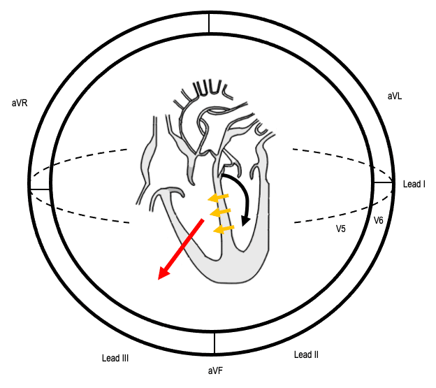
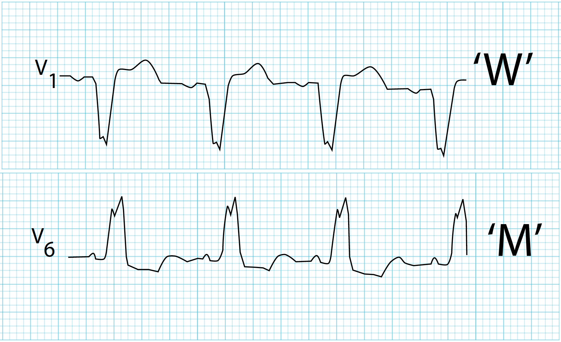
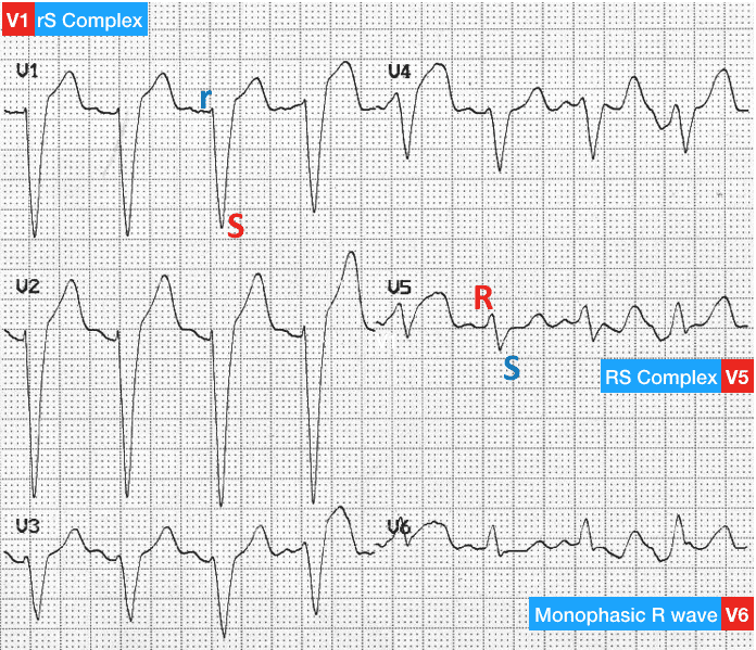
A bundle branch block delays one ventricle, and that ventricle activates late through slow cell-to-cell conduction. The QRS widens to 120 ms or more. Expect the ST segments and T waves to run opposite (discordant) to the terminal QRS. Figures 2 to 5 show the patterns.
ECG Criteria
| Feature | Right Bundle Branch Block (RBBB) | Left Bundle Branch Block (LBBB) |
|---|---|---|
| QRS duration | ≥120 ms | ≥120 ms |
| V1–V2 | rSR′ ("M-shaped"); the terminal R′ is late right ventricular activation | Broad QS or rS; no septal r |
| I, aVL, V5–V6 | Wide, slurred terminal S | Broad, monophasic or notched R; no septal q |
| ST-T | Discordant to the terminal QRS | Discordant to the QRS (ST elevation where the S is deep) |
Incomplete block, at 110–119 ms, looks the same with a narrower QRS.
Causes
- RBBB: normal variant in healthy and young people; right ventricular strain (pulmonary embolism, pulmonary hypertension); structural or ischemic heart disease.
- LBBB: ischemic heart disease; hypertensive heart disease with LVH; cardiomyopathy; aortic valve disease; degenerative conduction disease.
Clinical Significance
- Isolated RBBB in a patient without heart disease? Often benign.
- LBBB is usually structural. It hides the usual ST-segment clues. That is why you need a prior tracing to compare against.
Ventricular Paced QRS
Expect a wide QRS when one ventricular site does the pacing. The ventricles activate in a fixed order from that site. Its shape depends on where the lead sits. Seeing a paced complex is no sign of infarction or block. It is what pacing looks like.
ECG Criteria
- Pacing spike before each paced complex (look in leads II and V1)
- Wide QRS with a morphology set by the pacing site
- Discordant ST-T change is expected. Compare with the previous 12-lead
Clinical Significance
- Standard ST-segment criteria do not work on a paced beat. Compare with a prior paced 12-lead, or use the Smith-modified Sgarbossa approach.
- When pacing is needed, a spike alone does not prove capture. Confirm it with the pulse and the SpO₂ waveform.
- Missing spikes, or an escape rhythm in a paced patient, mean a device check is needed.
Sodium-Channel Blockade and Hyperkalemic QRS Widening
Sodium-channel blockers slow phase 0 of the cardiac action potential. The QRS widens. Hyperkalemia does it another way. It partly depolarizes the resting membrane and slows conduction. Both are reversible. Both can progress to ventricular arrhythmia or asystole if you do not treat them.
Sodium-Channel Blockade
- QRS ≥100 ms (maximal limb-lead width): in the landmark tricyclic overdose series, seizures showed up at this width. Ventricular arrhythmias showed up only at 160 ms or more.
- Terminal R wave in aVR ≥3 mm: it catches seizures and arrhythmia well, but its positive predictive value was about 43%. Treat it as a supporting sign, not a verdict.
- Serum drug level: it did not predict seizures or arrhythmia in that same series. Treat the ECG and the patient.
- Treatment: sodium bicarbonate is COR 1 (LOE B-NR) for adult tricyclic or tetracyclic cardiotoxicity, COR 2a (C-LD) for children with the same, and COR 2a (C-EO) for other sodium-channel blockers (2025 AHA Part 10, recommendations 1, 2 and 5). Whether the endpoint is pH about 7.50 or resolved hypotension and QRS widening is debated (AHA 2025 Part 10, supportive text). The ceilings (serum pH 7.55, serum sodium 155 mEq/L) are E3 house position, which cites AHA 2025 Part 10 Table 4; that table could not be checked, so they are not verified against AHA text.
Hyperkalemic QRS Widening
Expect this order: peaked T waves, then flattened P waves, then PR prolongation, then QRS widening, and finally a sine-wave pattern. QRS widening is a late sign. The T Wave page covers that progression. Serum potassium guides the treatment. Calcium stabilizes the myocardium while potassium-shifting therapy starts. In cardiac arrest, though, the 2025 AHA rates IV calcium, sodium bicarbonate and insulin with glucose COR 2b. That means effectiveness is not well established (Part 10, Hyperkalemia recommendations 1 to 3).
Low Voltage and Electrical Alternans
Low QRS voltage means small QRS deflections across the limb or precordial leads. It is nonspecific, so it will not name the cause for you. With tachycardia, hypotension or shock, think of pericardial effusion and tamponade.
ECG Criteria
- Low voltage: small QRS deflections across the limb or precordial leads. Compare with the prior ECG. Amplitude changes with body habitus and the lead set.
- Electrical alternans: beat-to-beat change in QRS amplitude (and sometimes axis) in an otherwise regular rhythm.
Causes
- Pericardial effusion, especially with tamponade physiology.
- Other causes exist. The pattern alone cannot name them.
Clinical Significance
- Electrical alternans with tachycardia and hypotension: treat as possible tamponade and call for physician evaluation and point-of-care ultrasound.
- Repeat the 12-lead ECG as the patient changes, and compare each tracing with the last.
Differential Diagnosis & Mimics
A wide QRS can have several causes. Separate the common mimics with the table before you act on a label.
| Feature | Bundle Branch Block | Ventricular Pacing | Hyperkalemia | Sodium-Channel Toxicity |
|---|---|---|---|---|
| Pacing spike | None | Present before each paced QRS | None | None |
| Setting | Chronic heart disease, or a normal variant | Pacemaker or ICD in place | Kidney failure, potassium-raising drugs | Tricyclic antidepressant, antiarrhythmic, cocaine, diphenhydramine |
| Key ECG clue | rSR′ (RBBB) or broad notched R (LBBB); discordant ST-T | Wide QRS with a morphology set by the pacing site | Peaked T, PR prolongation, QRS widening, sine wave | R in aVR ≥3 mm; QRS ≥100 ms |
| First action | Compare with an old ECG; apply Sgarbossa criteria if LBBB with chest pain | Confirm capture; compare with a prior paced 12-lead | Calcium for membrane stabilization; insulin 10 units IV with dextrose 25 g (see the Insulin page) | Sodium bicarbonate; see Bedside Management |
Artifact and Lead Misplacement
- Precordial placement: leads placed high or low shift the R and S waves. They can mimic a bundle branch or a hypertrophy pattern.
- Motion and shivering: tremor in transport can look like wide, bizarre complexes. Confirm on a second lead or a repeat 12-lead.
- Limb lead reversal: swapped limb electrodes flip the polarity of leads I and aVR, and that can mimic an abnormal pattern. Compare with the baseline.
Bedside Management & Escalation
In adults, a wide-complex tachycardia of uncertain origin gets managed as ventricular tachycardia. That is the E3 working rule. AHA 2025 Part 9 makes no recommendation for an uncertain rhythm. The table sorts the approach by stability. The medication table has the doses.
Stability and Actions
| Situation | BLS (EMT) | ALS (AEMT / Paramedic / RN) | Critical Care / Transport |
|---|---|---|---|
| Unstable (wide QRS with pulse) | Oxygen if SpO₂ <94%; 12-lead if your protocol allows; call ALS early | Synchronized cardioversion (100 J for regular monomorphic VT, 2025 AHA ACLS cardioversion algorithm); sedation per protocol | Pads on before moving; physician at the bedside; sodium bicarbonate for sodium-channel toxicity (see the bicarbonate row), or calcium for suspected hyperkalemia with ECG change: calcium gluconate 10% 30 mL (3 g) IV over 10 min if peripheral access, or calcium chloride 10% 10 mL (1 g) IV over 5 min if peri-arrest or central/deep access (UKKA 2026; differs from FDA label, see the calcium chloride row) |
| Stable (wide QRS with pulse) | 12-lead ECG if allowed; oxygen if SpO₂ <94%; notify ALS | In adults, manage a wide-complex tachycardia of regular or uncertain rhythm as VT (E3 working rule, not an AHA 2025 recommendation for an uncertain rhythm), but first exclude sodium-channel toxicity (TCA, class I drugs, diphenhydramine, cocaine, local anesthetic) and hyperkalemia (history, R in aVR ≥3 mm, peaked T, potassium). Treat suspected toxicity with sodium bicarbonate (COR 1 for TCA cardiotoxicity, Part 10) and suspected hyperkalemia with calcium: calcium gluconate 10% 30 mL (3 g) IV over 10 min if peripheral access, or calcium chloride 10 mL (1 g) over 5 min if peri-arrest or central/deep access (UKKA 2026; differs from FDA label). Then amiodarone 150 mg IV/IO over 10 min, or procainamide 20–50 mg/min until the arrhythmia is suppressed, hypotension develops, QRS widens more than 50%, or 17 mg/kg is given (AHA 2025 Part 9 WCT rec 3, COR 2b, LOE B-R). Adenosine may be considered only when the rhythm is stable, regular and monomorphic (AHA 2025 Part 9 WCT rec 4, COR 2b, B-NR). Avoid procainamide while the QRS is widening from a toxin or hyperkalemia. AHA 2025 Part 10 makes no recommendation for class Ia drugs in sodium-channel poisoning; it says lidocaine (class Ib) may be reasonable for cardiotoxicity from class Ia or Ic poisoning (COR 2b, C-EO). | Serial 12-leads; cardiology input; expert input before a second antiarrhythmic |
| Pulseless | CPR; AED shock | Defibrillate per device (manufacturer setting; 120–200 J biphasic per the 2025 ACLS cardiac arrest algorithm); for shock-refractory VF or pulseless VT, amiodarone 300 mg then 150 mg, or lidocaine 1–1.5 mg/kg, then 0.5–0.75 mg/kg (COR 2b, LOE B-R, Part 9) | Hyperkalemic arrest: IV calcium has uncertain benefit (COR 2b, Part 10); routine calcium in other arrests is not recommended (COR 3: No Benefit, Part 9). See the calcium chloride row. |
Medications and Energy
| Therapy | Adult | Pediatric | Key Notes |
|---|---|---|---|
| Sodium bicarbonate (sodium-channel toxicity) | 50–150 mEq IV/IO bolus (8.4%); repeat to narrow the QRS. Titrate to QRS narrowing; do not exceed serum pH 7.55 |
1–3 mEq/kg IV/IO bolus, never above the adult dose. Titrate to QRS narrowing |
Dilution: adults 1 mEq/mL (8.4%); children 0.5 mEq/mL (dilution not in the cached AHA text). 8.4% is hypertonic; push slowly in children. Neonates and infants: dilute per protocol and pharmacy. Check pH, potassium and ionized calcium. AHA 2025 Part 10: COR 1 for adult TCA cardiotoxicity; COR 2a for children with TCA cardiotoxicity and for other sodium-channel blockers. Ceilings (pH 7.55, sodium 155 mEq/L) and bolus doses: E3 house position, which cites AHA 2025 Part 10 Table 4 (not independently checked). Sodium Bicarbonate |
| Amiodarone (stable wide-complex, presumed VT) | 150 mg IV/IO over 10 min, then 1 mg/min for the first 6 hr (2025 AHA ACLS tachyarrhythmia algorithm). Repeat 150 mg only if VT recurs |
Stable wide-complex: 5 mg/kg IV/IO over 20–60 min, only after expert consultation (PALS 2020 tachycardia algorithm; the cached 2025 PALS text gives no stable dose; expert consultation is Part 8, COR 1, C-LD). Shock-refractory VF or pVT: 5 mg/kg IV/IO bolus. Max 300 mg; later doses max 150 mg |
High-alert Not for torsades or pre-excited AF. Exclude sodium-channel toxicity and hyperkalemia first. Watch for hypotension and bradycardia. AHA 2025 Part 9: amiodarone, procainamide or sotalol may be considered for adult wide-complex tachycardia (COR 2b, LOE B-R). Amiodarone |
| Procainamide (stable regular monomorphic wide-complex) | 20–50 mg/min IV infusion until the arrhythmia is suppressed, hypotension develops, or QRS widens >50%. Max 17 mg/kg |
15 mg/kg IV/IO over 30–60 min (PALS 2020 algorithm dose; the 2025 PALS algorithms list no procainamide dose). Specialist use only; monitor BP and QRS; stop for hypotension or QRS widening >50% |
High-alert Avoid with prolonged QT, heart failure, complete heart block, lupus, or recent tricyclic or sodium-channel blocker ingestion. Boxed warning (DailyMed label): positive ANA and lupus-like syndrome with prolonged use; CAST-based mortality concern (encainide and flecainide data), so reserve for life-threatening arrhythmias; blood dyscrasias, including agranulocytosis, with weekly blood counts for the first 3 months. Class Ia: adds to sodium-channel block, can widen the QRS further, and is proarrhythmic; exclude toxicity and hyperkalemia first. Infuse no faster than 50 mg/min (ACLS tachyarrhythmia algorithm sidebar); AHA 2025 Part 9 WCT rec 3, COR 2b, LOE B-R. Procainamide |
| Calcium chloride (hyperkalemia with QRS widening) | 10 mL (1 g) of 10% IV over 5 min (peri-arrest or central/deep access); IV/IO push in arrest. Repeat if ECG changes persist |
20 mg/kg IV/IO slowly. Max 1 g per dose |
Stabilizes the myocardium; does not lower potassium. Insulin 10 units IV with dextrose 25 g (UKKA 2026; AHA 2025 Part 10 Table 3 uses D50 50 g). Differs from FDA label: the label limits the rate to 1 mL/min (central or deep vein, no bolus); the rate here is from UKKA 2026 and is off-label. Extravasation causes tissue necrosis. Avoid in patients receiving digoxin (hypercalcemia raises digoxin-toxicity risk). Labeled for hypocalcemia; the label contraindicates use in VF, asystole and PEA, so arrest use is off-label. AHA 2025 Part 10 gives IV calcium in hyperkalemic arrest COR 2b (C-LD); Part 8 (COR 3: No Benefit, B-NR) reserves routine pediatric arrest calcium for hypocalcemia, calcium channel blocker overdose or hyperkalemia. Calcium Chloride |
| Synchronized cardioversion (regular monomorphic VT with pulse) | 100 J synchronized (2025 AHA ACLS cardioversion algorithm, monomorphic VT). Escalate per device |
0.5–1 J/kg, then 2 J/kg (2025 AHA Part 8, COR 2a, C-EO). Escalate per device |
Sedate per protocol; synchronize on every R wave. Synchronized Cardioversion |
Doses vary by protocol; follow local protocols and medical direction. Adult amiodarone, procainamide and cardioversion doses come from the 2025 AHA ACLS and Part 9 recommendations. Pediatric procainamide and stable pediatric amiodarone come from the PALS 2020 algorithm. Pediatric calcium comes from NASEMSO v3.0. Calcium chloride and insulin-dextrose rates come from UKKA 2026, and they differ from the FDA label. The sodium bicarbonate doses are the E3 house position. They cite AHA 2025 Part 10 Table 4, which was not independently checked.
Pediatric & Special Populations
Pediatrics
- Age-dependent width: the QRS runs shorter in infants and children, and it changes with age. Judge width against age-specific percentile charts, not the adult ≥120 ms cutoff alone.
- Wide complex with tachycardia: in children, SVT with aberrant conduction is the most common cause, but the rhythm can still be ventricular. With cardiopulmonary compromise, cardiovert urgently: 0.5–1 J/kg, then 2 J/kg if that fails (2025 AHA Part 8, COR 2a, C-EO). Without compromise, get expert consultation before any antiarrhythmic (2025 AHA Part 8, COR 1, C-LD). For a regular monomorphic WCT without compromise, adenosine can be useful, alongside expert consultation (2025 AHA Part 8, COR 2a, C-EO).
- Weight-based drugs: use Weight-Based Dosing and the Pediatric Vital Signs ranges.
Other Populations
- Pacemaker or ICD: a paced QRS is the finding to expect. Confirm capture, and compare with the previous paced 12-lead.
- Athletes and young adults: incomplete or complete RBBB is often a normal variant. Compare with an old ECG.
- Pregnancy: an unstable wide-complex tachycardia gets synchronized cardioversion, the same as in non-pregnant patients. For a stable one, the same agents apply: amiodarone or procainamide (AHA 2025 Part 9 WCT rec 3, COR 2b), after obstetric input.
- Kidney failure or potassium-raising drugs: check potassium early when the QRS widens.
Monitoring & Transport Considerations
| Issue | What to Do |
|---|---|
| Monitoring lead | Lead II for rhythm; V1 or MCL1 to judge QRS width and bundle branch morphology |
| Repeat 12-leads | Every 15–30 min during ongoing chest pain or a changing QRS; transmit when your protocol allows |
| Artifact | Shivering, vibration and movement can mimic wide complexes; confirm on a second lead |
| Pacing and transport | Anterior–posterior pads before moving an unstable bradycardic or tachycardic patient; check capture by pulse and SpO₂ waveform |
| Low voltage or alternans | Repeat the 12-lead; treat as possible tamponade and call for physician evaluation |
| Potassium | Obtain a potassium level with any new wide QRS; see the T Wave page for the hyperkalemia sequence |
Step-by-Step Approach & Pitfalls
- Measure the QRS in the widest lead, from onset to J point, and classify it as ≤110, 110–119, or ≥120 ms.
- Establish the rhythm: a narrow complex, a wide and regular complex, or a wide and irregular complex.
- Assess the patient. If unstable, go straight to the stability table.
- Look for a cause before any antiarrhythmic: a prior ECG, a pacing spike, pre-excitation (short PR, delta wave), drugs or toxins (sodium-channel toxicity), and potassium (hyperkalemia). Treat those causes first; see Bedside Management.
- Check V1 and V6 to classify a bundle branch block, only after the width is confirmed.
- For a new LBBB with chest pain, apply the Smith-modified Sgarbossa criteria and follow the local STEMI pathway.
Common Pitfalls
- Calling a wide complex a bundle branch block before you confirm the width and the rhythm.
- In adults, the habit of calling a wide, regular tachycardia SVT with aberrancy.
- Calling a new LBBB a STEMI, or waving it off as chronic with no prior ECG to check.
- Reaching for an AV-nodal blocker on a wide-complex tachycardia whose rhythm you do not know.
- Judging voltage or morphology from one lead or one beat in an irregular rhythm.
ECG Examples

Clinical Pearls
References
- Surawicz B, Childers R, Deal BJ, Gettes LS, et al. AHA/ACCF/HRS recommendations for the standardization and interpretation of the electrocardiogram: part III: intraventricular conduction disturbances. Circulation. 2009;119:e235–e240. https://doi.org/10.1161/CIRCULATIONAHA.108.191095
- Sgarbossa EB, Pinski SL, Barbagelata A, et al. Electrocardiographic diagnosis of evolving acute myocardial infarction in the presence of left bundle-branch block. N Engl J Med. 1996;334(8):481–487. https://doi.org/10.1056/NEJM199602223340801
- Smith SW, Dodd KW, Henry TD, Dvorak DM, Pearce LA. Diagnosis of ST-elevation myocardial infarction in the presence of left bundle branch block with the ST-elevation to S-wave ratio in a modified Sgarbossa rule. Ann Emerg Med. 2012;60(6):766–776. https://doi.org/10.1016/j.annemergmed.2012.07.119
- Boehnert MT, Lovejoy FH Jr. Value of the QRS duration versus the serum drug level in predicting seizures and ventricular arrhythmias after an acute overdose of tricyclic antidepressants. N Engl J Med. 1985;313(8):474–479 (landmark study). https://doi.org/10.1056/NEJM198508223130804
- Liebelt EL, Francis PD, Woolf AD. ECG lead aVR versus QRS interval in predicting seizures and arrhythmias in acute tricyclic antidepressant toxicity. Ann Emerg Med. 1995;26(2):195–201. https://doi.org/10.1016/s0196-0644(95)70151-6
- Cao D, Arens AM, Chow SL, Easter SR, Hoffman RS, Lagina AT III, Lavonas EJ, Patil KD, Sutherland LD, Tijssen JA, Wang GS, Zelop CM, Rodriguez AJ, Drennan IR, McBride ME. Part 10: adult and pediatric special circumstances of resuscitation: 2025 American Heart Association Guidelines for Cardiopulmonary Resuscitation and Emergency Cardiovascular Care. Circulation. 2025;152(suppl 2):S578–S672. https://doi.org/10.1161/CIR.0000000000001380 Available at cpr.heart.org.
- Wigginton JG, Agarwal S, Bartos JA, et al. Part 9: advanced life support: 2025 American Heart Association guidelines for cardiopulmonary resuscitation and emergency cardiovascular care. Circulation. 2025;152(suppl 2):S538–S577. https://doi.org/10.1161/CIR.0000000000001376
- Adler Y, Charron P, Imazio M, et al. 2015 ESC Guidelines for the diagnosis and management of pericardial diseases. Eur Heart J. 2015;36:2921–2964. https://doi.org/10.1093/eurheartj/ehv318
- Lue HC, Wu MH, Wang JK, et al. Normal ECG standards and percentile charts for infants, children and adolescents. Pediatr Neonatol. 2023;64:256–273. https://doi.org/10.1016/j.pedneo.2022.07.013
- Topjian AA, Raymond TT, Atkins D, et al. Part 4: Pediatric basic and advanced life support: 2020 American Heart Association guidelines for cardiopulmonary resuscitation and emergency cardiovascular care. Circulation. 2020;142(16 suppl 2):S469–S523. https://doi.org/10.1161/CIR.0000000000000901
- Lasa JJ, Dhillon GS, Duff JP, Hayes J, Kamath-Rayne BD, Levy A, Mahgoub M, Morgan RW, McCormick T, Roberts JS, Ross CE, Schexnayder SM, Sweberg T, Valdes SO, Topjian AA. Part 8: pediatric advanced life support: 2025 American Heart Association Guidelines for Cardiopulmonary Resuscitation and Emergency Cardiovascular Care. Circulation. 2025;152(suppl 2):S479–S537. https://doi.org/10.1161/CIR.0000000000001368 Available at cpr.heart.org.
Medical Disclaimer
- For Educational Purposes Only: This content is intended for educational reference and should not be used for clinical decision-making.
- Not a Substitute for Professional Judgment: Always consult your local protocols, institutional guidelines, and supervising physicians.
- Verify Before Acting: Users are responsible for verifying information through authoritative sources before any clinical application.