

Bedside Snapshot
- Normal (Adult)
- Men under 50: 0–15 mm/hr; women under 50: 0–20 mm/hr (SI: same, mm/h). Age 50 and older: up to 20 (men) and 30 (women). Lab-specific
- Critical Values
- No universal critical value: interpret by trend and clinical context. ESR ≥100 mm/hr is not a panic value but almost always has a cause
- High Suggests
- Infection: epidural abscess, osteomyelitis, endocarditis
- Giant cell arteritis (GCA) or polymyalgia rheumatica
- Multiple myeloma or other cancer
- Anemia, CKD, pregnancy, older age
- Low Suggests
- Rarely clinically significant on its own. Polycythemia, sickle cell disease, low fibrinogen (DIC) or IL-6 blockade can hide real inflammation
- Act Now If
- Age ≥50 with new headache, jaw claudication or visual symptoms and raised ESR or C-reactive protein (CRP): same-day emergency evaluation; steroids before biopsy. Fever, back pain and raised ESR: neuro checks, MRI
- Watch Out For
- A normal ESR does not exclude GCA, epidural abscess or early infection; the ESR lags CRP by days
Reference & Critical Ranges
Reference ranges
| Population | Conventional | SI | Notes |
|---|---|---|---|
| Men under 50 | 0–15 mm/hr | 0–15 mm/h | Westergren method |
| Women under 50 | 0–20 mm/hr | 0–20 mm/h | Higher than in men at every age |
| Men 50 and older | 0–20 mm/hr | 0–20 mm/h | The upper limit keeps rising with age (age-adjusted rule below) |
| Women 50 and older | 0–30 mm/hr | 0–30 mm/h | Some automated methods list higher limits (up to 40 mm/hr) |
| Pregnancy | 18–48 mm/hr (first half); 30–70 mm/hr (second half) | Same values in mm/h | 95% range in healthy women without anemia; with anemia up to 40–95 mm/hr late in pregnancy |
| Newborn | 0–2 mm/hr | 0–2 mm/h | Very low in the first days of life |
| Child (1 month to puberty) | 0–13 mm/hr | 0–13 mm/h | Upper limit varies by lab (about 13–14 mm/hr); adult ranges after puberty |
Take a 70-year-old man: his upper limit is 35 mm/hr. Take a 70-year-old woman: hers is 40 mm/hr. Both come from healthy adults. They are a rule of thumb, not a diagnostic cutoff.
Severity & critical values
| Level | Conventional | SI | What it means / typical findings |
|---|---|---|---|
| Normal | At or below the age- and sex-adjusted limit | Same | Does not exclude giant cell arteritis (GCA), early infection or spinal epidural abscess |
| Raised | Above the adjusted limit | Same | Nonspecific: anemia, chronic kidney disease (CKD), pregnancy, obesity or any inflammation |
| Kawasaki threshold | ≥40 mm/hr | ≥40 mm/h | Prolonged fever with incomplete Kawasaki criteria: ESR ≥40 mm/hr or CRP ≥3 mg/dL triggers the supplemental workup |
| GCA: ESR not above 40 | ≤40 mm/hr | ≤40 mm/h | In suspected GCA, an ESR of 40 mm/hr or less makes GCA less likely (negative likelihood ratio about 0.2 for absence of ESR above 40 mm/hr; van der Geest 2020). Not a diagnostic criterion |
| GCA criterion | ≥50 mm/hr | ≥50 mm/h | In patients 50 or older, ESR ≥50 mm/hr or CRP ≥10 mg/L (1 mg/dL) scores 3 of the 6 points needed in the 2022 ACR/EULAR GCA classification criteria |
| Osteomyelitis support | ≥70 mm/hr | ≥70 mm/h | Infected diabetic foot ulcer: supports osteomyelitis (sensitivity 81%, specificity 80%); combine with probe-to-bone and X-ray |
| Extreme | ≥100 mm/hr | ≥100 mm/h | A cause is usually found: infection, autoimmune or inflammatory disease, malignancy and renal disease are each common; categories overlap |
| Critical | No universal critical value | — | Interpret by trend and clinical context |
Conversion: There is none. mm/hr (conventional) and mm/h (SI) are the same number.
Causes of Abnormal Results
High (raised ESR: above the age- and sex-adjusted limit)
| Mechanism | Common causes | Bedside clue |
|---|---|---|
| Infection (fibrinogen rise) | Spinal epidural abscess, vertebral or diabetic foot osteomyelitis, endocarditis, septic arthritis, deep abscess, tuberculosis | Fever, focal back or bone pain, injection drug use, diabetes, new murmur |
| Large-vessel vasculitis | Giant cell arteritis, polymyalgia rheumatica (PMR) | Age 50 or older; new headache, jaw claudication, scalp tenderness, visual loss; shoulder and hip girdle stiffness |
| Other autoimmune disease | Rheumatoid arthritis, systemic lupus erythematosus (SLE), other vasculitis, inflammatory bowel disease | Joint swelling, rash, bloody diarrhea; in SLE flares CRP often stays low |
| Paraprotein (excess immunoglobulin) | Multiple myeloma, Waldenström macroglobulinemia | Bone pain, anemia, high calcium, renal failure; ESR far higher than CRP |
| Other malignancy | Lymphoma, metastatic cancer | Weight loss, night sweats, lymph nodes |
| Kidney disease | CKD, nephrotic syndrome | High creatinine, proteinuria, edema; ESR raised even without infection |
| Physiologic and red-cell factors | Anemia, macrocytosis, pregnancy, older age, female sex, obesity | Low hemoglobin or high MCV; a raised ESR without any inflammation |
| Pediatric vasculitis | Kawasaki disease | Fever for 5 days or more, conjunctival injection, rash, lip and hand or foot changes; ESR often ≥40 mm/hr |
Low (below the expected range)
A low ESR means very little by itself. It matters when it hides real inflammation.
| Mechanism | Common causes | Bedside clue |
|---|---|---|
| More red cells | Polycythemia, high hematocrit | Hemoglobin and hematocrit above range; crowded cells settle slowly |
| Abnormal red-cell shape | Sickle cell disease, spherocytosis | Misshapen cells cannot stack, so the ESR can stay low during infection |
| Low fibrinogen | Disseminated intravascular coagulation (DIC), macrophage activation syndrome, severe liver failure | An ESR that falls while a febrile patient worsens; check fibrinogen, platelets and ferritin |
| Suppressed acute phase | Glucocorticoids, interleukin-6 (IL-6) receptor inhibitors such as tocilizumab | Normal ESR and CRP despite infection or relapse |
Interpretation & Bedside Approach
Step-by-step approach
- Is it real? Make sure the tube was full and unclotted, and run it promptly. Then check hemoglobin and MCV. Anemia and macrocytosis raise the ESR with no inflammation at all.
- How sick is the patient? Urgency comes from the patient, never from the ESR alone. Fever with back pain, a new neurologic deficit, sepsis or visual symptoms set it.
- Acute or chronic, and the trend: The ESR lags by days. It can take weeks to come down, so one value may be a picture of last week. Trend beats a single value. Compare results from the same lab and method.
- Adjust for age, sex and context: Use age ÷ 2 for men or (age + 10) ÷ 2 for women. Pregnancy, CKD and anemia push the baseline up.
- Classify with named criteria: 2022 American College of Rheumatology (ACR)/European Alliance of Associations for Rheumatology (EULAR) GCA classification criteria; American Heart Association (AHA) 2017 incomplete Kawasaki algorithm; International Working Group on the Diabetic Foot (IWGDF)/Infectious Diseases Society of America (IDSA) 2023 foot osteomyelitis guidance.
- Escalate or dispose: Suspected GCA gets same-day specialist assessment and glucocorticoids. Fever, back pain and raised markers get an emergent MRI when neurologic signs are present. An unexplained extreme ESR needs a workup and follow-up. Reassurance is not a plan.
Pair the ESR with CRP
| Feature | ESR | CRP |
|---|---|---|
| Rises | Over days | Within about 6 hours; peaks near 48 hours |
| Falls | Slowly; weeks to normalize | Quickly; half-life about 19 hours |
| Skewed by | Anemia, red-cell shape, age, sex, pregnancy, paraproteins, CKD | Little affected by red cells, age or sex |
| Best use | GCA and PMR (with CRP), diabetic foot osteomyelitis, paraprotein clue, slow disease monitoring | Acute infection, early response to treatment, Kawasaki disease after IVIG |
Trend in PMR and osteomyelitis: On corticosteroids, the ESR in polymyalgia rheumatica usually settles back to normal over weeks. Still high? Revisit the diagnosis or the treatment. Osteomyelitis runs differently at first: the ESR usually climbs in the first week of antibiotics before it falls. A value that stays high or keeps rising suggests treatment failure or an undrained abscess.
Discordant patterns
| Pattern | Suggests |
|---|---|
| High ESR, normal CRP | Paraprotein (myeloma), anemia, CKD, pregnancy, SLE flare without serositis, or inflammation that is resolving (CRP has already fallen) |
| High CRP, normal ESR | Very early infection or inflammation (first day or two), low fibrinogen, polycythemia or sickle cell disease |
| Both normal | Active GCA, osteomyelitis and epidural abscess less likely, but not excluded when the story is strong |
| ESR rising after IVIG | Expected: intravenous immune globulin (IVIG) itself raises the ESR; judge Kawasaki response by fever and CRP |
Management Priorities
Emergency: Suspected giant cell arteritis (age 50 or older with new headache, jaw claudication, scalp tenderness or visual symptoms)
- Recognize: Send ESR and CRP with a CBC. When the story is strong, don't wait for results before you escalate.
- Protect vision: Any visual loss is a same-day problem, transient visual loss (amaurosis fugax) included. It goes to the emergency department for same-day ophthalmology review.
- Treat now (prescriber order; RN administers): Don't wait for biopsy or imaging before starting high-dose glucocorticoids. For acute or intermittent visual loss, give IV methylprednisolone pulses.
- Refer and confirm: Get a specialist assessment within 24 hours. Temporal artery ultrasound or biopsy follows soon after treatment starts, and a biopsy stays positive for weeks.
Medications
| Drug | Adult dose | Pediatric dose (max) | Onset / key notes |
|---|---|---|---|
| Prednisone (GCA without visual loss) | GCA: 40–60 mg PO once daily (max 60 mg/day at the start); taper per specialist | Not established in children: GCA occurs only from age 50 | Most patients improve within 1–7 days; no response should prompt a rethink of the diagnosis. Prednisolone is dosed mg for mg |
| Methylprednisolone (GCA with visual loss) | GCA with acute or intermittent visual loss: 500 mg–1 g IV once daily for up to 3 days, then oral prednisone (max 1,000 mg/dose) | Not established in children: GCA occurs only from age 50 | Infuse over at least 30 min; more than 500 mg in under 10 min has caused arrhythmias and cardiac arrest. Check glucose. If IV is not possible, oral high-dose glucocorticoid is a specialist decision (dose per local protocol) |
| IVIG (Kawasaki disease) | Not used for this indication in adults | Kawasaki disease: 2 g/kg IV once over 10–12 hr. Aspirin per pediatric cardiology and AHA 2017 (moderate 30–50 mg/kg/day or low 3–5 mg/kg/day; protocol-dependent) | Pediatric specialist decision. Boxed warning: thrombosis, renal dysfunction and acute renal failure; monitor vital signs during infusion and hydrate. Also hemolysis and aseptic meningitis; defer live-virus vaccines about 11 months after 2 g/kg. IVIG raises the ESR, so draw it before the infusion and follow fever and CRP afterward |
Prolonged glucocorticoid courses: Monitor glucose and blood pressure. Add gastric protection and bone protection. For prolonged courses, add PJP prophylaxis. The steroid-sparing use of tocilizumab, the taper and the prophylaxis are specialist decisions.
Definitive care & disposition
- Suspected GCA: rheumatology within 24 hours; same-day ophthalmology for visual symptoms; admit for IV pulses or threatened vision.
- Fever, back pain and raised ESR or CRP: emergent MRI when any neurologic deficit is present; blood cultures; spine surgery consult for a confirmed epidural abscess.
- Diabetic foot ulcer with ESR ≥70 mm/hr: probe-to-bone and plain X-rays, then MRI if osteomyelitis is still in doubt.
- Extreme ESR (≥100 mm/hr) without an obvious cause: CBC with smear, creatinine, urinalysis, blood cultures if febrile and serum protein electrophoresis with free light chains; discharge only with follow-up.
- Child with prolonged fever and ESR ≥40 mm/hr: evaluate for Kawasaki disease (supplemental labs, echocardiogram) with pediatric cardiology.
Doses are typical references. Follow local protocols, medical direction and institutional policy.
Special Populations
| Population | What changes | Clinical implication |
|---|---|---|
| Neonates & infants | ESR is very low at birth (0–2 mm/hr) | Rarely useful; cultures and CRP drive the neonatal sepsis workup |
| Children & adolescents | Upper limit about 13 mm/hr until puberty | Key in Kawasaki disease (ESR ≥40 mm/hr or CRP ≥3 mg/dL) and bone or joint infection. CRP falls faster and tracks early response better. IVIG raises the ESR |
| Pregnancy | Rises through gestation to 30–70 mm/hr in the second half; higher with anemia | A raised ESR alone means little in pregnancy; rely on the clinical picture and CRP |
| Older adults | Upper limit rises with age: age ÷ 2 (men), (age + 10) ÷ 2 (women) | Do not blame age when GCA, PMR, myeloma or infection fits. GCA criteria apply only from age 50 |
| CKD / dialysis | Anemia and azotemia raise the baseline ESR | A weak marker of new infection; compare with the patient's own baseline and use CRP |
| Other | Sickle cell disease keeps the ESR low; tocilizumab suppresses ESR and CRP | A "normal" value may be abnormal for that patient. Normal markers do not exclude infection on IL-6 blockade |
Interfering Factors & Pitfalls
Collection & handling
| Factor | Effect | Why | Avoid / fix |
|---|---|---|---|
| Underfilled, clotted or hemolyzed tube | Unreliable; rejected | Wrong blood-to-anticoagulant ratio; trapped or damaged cells | Fill the lavender (EDTA) tube completely; invert 8–10 times |
| Delay before testing | Unreliable | The sample changes as it ages | Test within 4 hours if kept at room temperature, or within 24 hours if refrigerated at 4 °C (ICSH); follow your lab's rejection criteria |
| Tilted tube or warm room (manual method) | ↑ | Cells slide down the wall; warmth speeds settling | Lab keeps the pipette vertical, at room temperature, away from heat and vibration |
Medications, physiologic and biologic factors
| Factor | Effect | Why | Avoid / fix |
|---|---|---|---|
| IVIG | ↑ | Added immunoglobulin promotes stacking | Draw the ESR before the infusion; use fever and CRP after |
| Tocilizumab, glucocorticoids | ↓, False − | Suppress the acute-phase response | Normal markers do not exclude infection or relapse |
| Anemia, macrocytosis | ↑ | Fewer or larger cells settle faster | Read with hemoglobin and MCV; use CRP |
| Paraproteins | ↑ | Immunoglobulins promote stacking | ESR far above CRP: consider protein electrophoresis |
| Polycythemia, sickle cells, spherocytes, low fibrinogen | ↓, False − | Crowded or misshapen cells, or too little fibrinogen, prevent stacking | Use CRP; check fibrinogen when the ESR falls unexpectedly |
| Oral contraceptives, dextran | ↑ | Estrogen raises fibrinogen; dextran promotes red-cell stacking | Interpret against the patient's baseline; use CRP |
| Hyperlipidemia | ↑ | Lipid-rich plasma promotes red-cell stacking | Read with CRP and the clinical picture |
| Extreme leukocytosis, microcytosis | ↓, False − | Smaller red cells settle more slowly; the mechanism for very high white counts is less clear | Use CRP; interpret with the CBC |
Assay & method
Many labs now run modified or automated methods. Their results can differ from Westergren reference values. Track results on one lab and one method, and use that lab's range.
Common misinterpretations
- "A normal ESR rules out GCA": It lowers the probability and nothing more. A classic story still gets treated.
- "A high ESR in an older patient is just age": Check the age-adjusted limit before you blame age.
- "The ESR should fall within days of antibiotics": It lags by weeks. Follow CRP and the patient.
- "A rising ESR after IVIG means Kawasaki is worse": IVIG raises it all by itself.
Physiology & Test Basics
What it measures
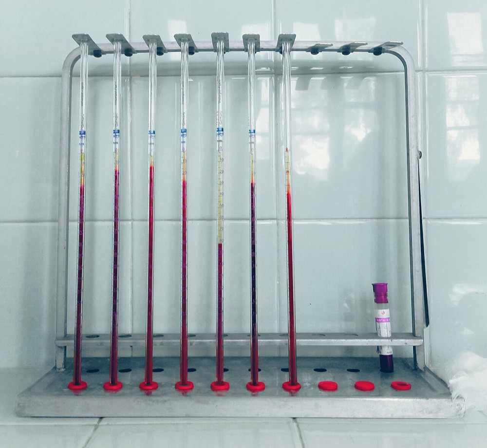
The erythrocyte sedimentation rate measures how far red cells fall in 1 hour. The column is anticoagulated blood, and the distance is read in millimeters. The fall is an indirect sign of inflammation. It reflects plasma proteins, mainly fibrinogen and immunoglobulins, and the red cells themselves. You order it alongside the CBC panel, but it is not a CBC index.
Physiology & regulation
- Rouleaux: Red cells carry a negative charge that keeps them apart. Fibrinogen and immunoglobulins cancel that charge. The cells stack, and heavier stacks fall faster.
- Acute phase: IL-6 pushes the liver to make more fibrinogen, over days. That is why the ESR rises and falls slowly.
- Red cells: Fewer or larger cells settle faster, as in anemia and macrocytosis. More or misshapen cells settle slower, as in polycythemia and sickle cells.
How it is measured
- Specimen: Whole blood in a lavender-top (EDTA) tube, filled and mixed. Some manual methods use a citrate tube instead. No fasting is needed.
- Reference method: Westergren, as defined by the International Council for Standardization in Haematology (ICSH). Diluted blood sits in a pipette on a 200 mm scale, and the reading is taken at 60 minutes.
- Automated methods: Many labs use modified or alternate methods. They are often faster and run from the standard EDTA tube.
- Turnaround: About 1 hour by hand and less on analyzers. A point-of-care ESR is uncommon.
Why it matters
The ESR rarely changes the next minute of emergency care. It does shape high-stakes decisions. Those include steroids for suspected GCA, imaging for epidural abscess or osteomyelitis, and the workup of prolonged fever in a child.
Clinical Pearls
References
- Mukhtyar, C. B., Nikiphorou, E., Bartoletti, A., Dey, M., Ducker, G., Misra, D. P., et al. (2026). 2025 EULAR recommendations for the management of polymyalgia rheumatica and primary large vessel vasculitis. Annals of the Rheumatic Diseases (online ahead of print). https://doi.org/10.1016/j.ard.2026.06.009
- Ponte, C., Grayson, P. C., Robson, J. C., Suppiah, R., Gribbons, K. B., Judge, A., et al. (2022). 2022 American College of Rheumatology/EULAR classification criteria for giant cell arteritis. Annals of the Rheumatic Diseases, 81(12), 1647–1653. https://doi.org/10.1136/ard-2022-223480
- Mackie, S. L., Dejaco, C., Appenzeller, S., Camellino, D., Duftner, C., Gonzalez-Chiappe, S., et al. (2020). British Society for Rheumatology guideline on diagnosis and treatment of giant cell arteritis. Rheumatology, 59(3), e1–e23. https://doi.org/10.1093/rheumatology/kez672
- van der Geest, K. S. M., Sandovici, M., Brouwer, E., & Mackie, S. L. (2020). Diagnostic accuracy of symptoms, physical signs, and laboratory tests for giant cell arteritis: A systematic review and meta-analysis. JAMA Internal Medicine, 180(10), 1295–1304. https://doi.org/10.1001/jamainternmed.2020.3050
- Senneville, É., Albalawi, Z., van Asten, S. A., Abbas, Z. G., Allison, G., Aragón-Sánchez, J., et al. (2023). IWGDF/IDSA guidelines on the diagnosis and treatment of diabetes-related foot infections (IWGDF/IDSA 2023). Clinical Infectious Diseases, ciad527. https://doi.org/10.1093/cid/ciad527
- McCrindle, B. W., Rowley, A. H., Newburger, J. W., Burns, J. C., Bolger, A. F., Gewitz, M., et al. (2017). Diagnosis, treatment, and long-term management of Kawasaki disease: A scientific statement for health professionals from the American Heart Association. Circulation, 135(17), e927–e999. https://doi.org/10.1161/CIR.0000000000000484
- Daniels, L. M., Tosh, P. K., Fiala, J. A., Schleck, C. D., Mandrekar, J. N., & Beckman, T. J. (2017). Extremely elevated erythrocyte sedimentation rates: Associations with patients' diagnoses, demographic characteristics, and comorbidities. Mayo Clinic Proceedings, 92(11), 1636–1643. https://doi.org/10.1016/j.mayocp.2017.07.018
- Kratz, A., Plebani, M., Peng, M., Lee, Y. K., McCafferty, R., Machin, S. J., & International Council for Standardization in Haematology. (2017). ICSH recommendations for modified and alternate methods measuring the erythrocyte sedimentation rate. International Journal of Laboratory Hematology, 39(5), 448–457. https://doi.org/10.1111/ijlh.12693
Medical Disclaimer
- For Educational Purposes Only: This content is intended for educational reference and should not be used for clinical decision-making.
- Not a Substitute for Professional Judgment: Always consult your local protocols, institutional guidelines, and supervising physicians.
- Verify Before Acting: Users are responsible for verifying information through authoritative sources before any clinical application.