

Bedside Snapshot
- Indications
- Suspected pelvic fracture with hypotension or shock, per local protocol
- Suspected unstable pelvic ring injury with hemodynamic compromise (Tile B or C on imaging)
- Selective, not routine: NAEMSP 2025 advises reconsidering routine use
- Contraindications / Abort
- Do not spring the pelvis to test stability: may cause harm
- Loss of distal pulse or sensation: reposition and reassess
- Shock from chest, abdomen or long bones is not treated by a binder
- Critical Steps
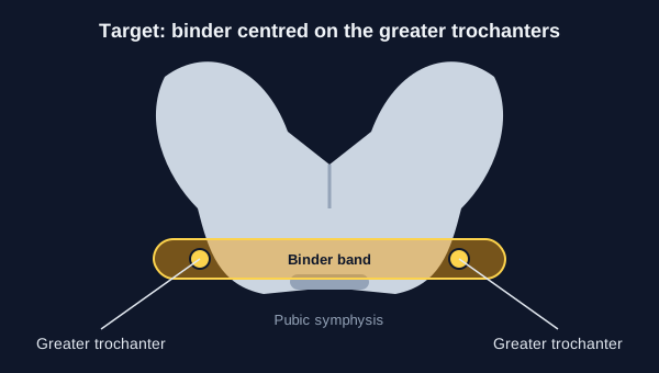
- Centre the band over the greater trochanters
- Secure the feet together so the legs internally rotate
- Tighten per device IFU; sheet binders have no stop point, so do not over-tighten
- Transport to a major trauma center when criteria are met
- Confirm & Monitor
- Distal pulses, sensation and skin at every reassessment
- Re-check placement after every move
- Image per protocol after removal (bladder injury can be masked)
- Document time, device, placement and tension
Key Numbers
Use a pelvic binder selectively, per protocol, for suspected pelvic fracture with hemodynamic compromise. When used, it is placed over the greater trochanters, not the abdomen, and the pelvis is never sprung to test it.
| Parameter | Value |
|---|---|
| Hypotension in the reviewed trauma cohort | Systolic BP < 90 mmHg on admission (TQIP analysis definition; trigger is set by local protocol) |
| Placement | Centred over the greater trochanters (level of both trochanters) |
| Legs | Feet secured together so the legs are internally rotated |
| Tension | Per device instructions for use; sheet binders have no stop point, so follow agency protocol and do not over-tighten. Re-check after every move |
| Manual pelvic stability (springing) | Do not use to rule in or out pelvic injury: neither sensitive nor specific, and may cause harm |
| Routine prehospital use | Reconsider; selective use per protocol, per the NAEMSP 2025 statement |
| Destination | Major trauma center when national triage criteria are met (NAEMSP 2025 statement) |
| Transfusion effect (matched Japan cohort) | No difference in 24 h red cell transfusion volume after matching |
| Angioembolization (matched Japan cohort) | 33.0% with prehospital binder vs 21.2% without (OR 1.82, 95% CI 1.24–2.66; observational association, not proof of harm) |
| Adjusted risks (ACS TQIP, hypotensive patients) | Decubitus ulcer +59%; lower urinary tract injury +34% |
Overview & Principles
A pelvic binder (pelvic circumferential compression device, PCCD) is a strap or sheet wrapped around the pelvis. It shrinks the space inside the pelvis and steadies an unstable pelvic ring fracture while the bleeding is treated. Its job is to tamponade pelvic bleeding and limit fracture movement until definitive care takes over.
How It Works
- Circumferential compression: The band wraps the pelvis at trochanter level. Intrapelvic volume drops and the fracture moves less.
- Correct level matters: Get the level right. The band belongs over the greater trochanters. In a simulation study of doctors, placement accuracy was 83.5% (147 of 176 devices correct).
- Stability is not the same as hemostasis: A steady pelvis is not a stopped bleed. Pooled analyses have not shown a mortality benefit from binder use. Keep looking for the other bleeding sources.
- Imaging still matters: One case report describes a binder masking a bladder injury on CT. Consider repeat imaging after removal per trauma protocol.
Where It Fits
The 2025 NAEMSP compendium on suspected pelvis fractures leans toward caution. Pelvic fracture is hard to identify in the field. Manual stability testing is neither sensitive nor specific, and it may cause harm. Prehospital pelvic circumferential compression devices (PCCDs) are worth reconsidering, because proven benefit in hemorrhage or mortality has not been shown. The statement advises that, if devices are used, care be taken to place them over the trochanters with the legs internally rotated by securing the feet together. Two more analyses add to this. A 2025 systematic review (Surgeon) found no clear superiority of early binder application over no or late application for blood product use, mortality, pain or complications. A 2026 meta-analysis found no association between binder use and mortality in adjusted, unadjusted or pooled analyses. The 2025 German guideline takes a different view. Its expert panel recommends a binder for pelvic ring injury with haemodynamic instability (good practice point, 100% consensus). Local protocol and medical direction decide when a binder is used.
Indications & Contraindications
Indications
- Suspected pelvic fracture with hypotension or signs of shock, when local protocol calls for a binder (the reviewed trauma cohort used systolic BP < 90 mmHg)
- Suspected unstable pelvic ring injury with hemodynamic compromise, per protocol (Tile B or C, classified on imaging)
- Use it selectively. The 2025 NAEMSP statement advises reconsidering routine use for all suspected pelvic fractures
Absolute Contraindications
No absolute contraindication turned up in the sources reviewed for this page. The 2025 German guideline expert panel also sees none for prehospital use. It stresses appropriate indications and correct application, though. Device instructions for use and local protocol set the device-specific limits.
Relative Contraindications & Cautions
- Normal examination: A normal manual pelvic exam does not rule out injury. Springing the pelvis is not a valid test.
- Suspected open or unstable wounds at the trochanter level: Assess and pad per device instructions and local protocol before tightening.
- Suspected hip, proximal femur or acetabular fracture, or a lateral-compression pattern: Internal rotation and tight compression can move fragments. In lateral-compression fractures, binder compression can change displacement. Use the least rotation and tension that still gives placement. Follow the device IFU and local protocol, and involve medical direction.
- Femur fracture needing a splint or traction: Foot securing and the splint or traction can work against each other. Coordinate them per protocol so neither undoes the other.
- Isolated non-pelvic shock: A binder will not stop thoracic, abdominal or limb bleeding. When that bleeding is causing the shock, the binder does not fix it.
Consider an Alternative
Limb and other external bleeding gets direct pressure, packing or a tourniquet per protocol. A binder is not the tool for it. For the general trauma approach, see Trauma Assessment. For vascular access during resuscitation, see IV Insertion and IO Access.
Abort / Stop Criteria
- Loss of distal pulse or sensation: Reassess the placement and tension per the device instructions and local protocol.
- Skin breakdown or pressure injury: Reposition and pad, and document the site.
- Placement not over the trochanters: Reposition before tightening further.
Scope & Team Roles
| Provider Level | Typical Role | Notes |
|---|---|---|
| EMT | Performs (protocol/credentialing required) | Placement only where state protocol includes it; the national scope model placement was not verified |
| AEMT | Performs (protocol/credentialing required) | Same skill as EMT; follow state protocol |
| Paramedic | Performs (protocol/credentialing required) | Selective use with reassessment; transport decision per triage criteria |
| RN (ED/ICU) | Performs | Placement and re-check per facility protocol; post-removal imaging coordination |
| Critical Care Transport (CFRN / FP-C / CCP-C) | Performs (protocol/credentialing required) | Reassess distal perfusion after each move |
| Physician / APP | Performs | Decides on definitive pelvic hemorrhage control and imaging |
Team Roles During the Procedure
- Operator: Positions the device over the greater trochanters and tightens per device instructions (sheet binders have no stop point; follow agency protocol).
- Limb holder: Secures the feet together so the legs are internally rotated.
- Monitor: Checks pulse, perfusion, skin and vital signs during and after placement.
- Recorder: Records time applied, device, tension method, distal checks and transport decision.
Anatomy & Landmarks
| Site | Best For | Landmark | Angle/Depth | Avoid If |
|---|---|---|---|---|
| Greater trochanters (target) | Unstable pelvic ring with hemodynamic compromise | Bony prominence of both greater trochanters | Band level with both trochanters, centred over the pelvis | Not applicable: this is the placement target |
| Above the trochanters (iliac crest or abdomen) | Not a target site | Iliac crests or abdomen | Not applicable | Any placement above the trochanters: reposition |
Greater Trochanters
- Locate the trochanters: Palpate the greater trochanter on each side with the patient supine.
- Centre the band: Place the band so its centre sits between the two trochanters at the level of both.
- Secure the feet: Tie or hold the feet together so the legs are internally rotated.
- Tension and check: Tighten per device instructions and check distal pulses and skin.
Pediatric
The sources reviewed for this page did not verify pediatric landmarks or sizing. Follow local protocol and the device pediatric range.
Equipment & Preparation
Equipment
- Commercial pelvic circumferential compression device (for example the T-POD or SAM Pelvic Sling II, as stocked by the agency), used per device instructions for use
- Improvised sheet binder only where the agency protocol names it; mechanical studies of improvised and proprietary binders are available
- Backup: direct pressure, packing and tourniquet supplies for other bleeding (see Trauma Assessment)
- Monitor with pulse oximetry, blood pressure and capnography per protocol
Sizing
Use a device sized for the adult pelvis per its instructions for use. Pediatric size ranges were not verified in the sources reviewed.
Patient Positioning & Preparation
Keep the patient supine with minimal log-rolling. Expose the pelvis, remove clothing and check the skin before placing the device.
Pre-Procedure Checklist
- Confirm a suspected pelvic fracture per protocol (mechanism, exam, shock)
- Check the device size, labeling and expiry
- Expose the pelvis and check the skin at the trochanters
- Identify the greater trochanters on both sides
- Plan how the feet will be secured
- Confirm baseline pulses, sensation and vital signs
- Confirm transport destination and time of injury
Step-by-Step Procedure
Phase 1: Prepare
- Decide by protocol: Use the binder for suspected pelvic fracture with hemodynamic compromise, not as a reflex for every pelvic complaint.
- Expose and check: Expose the pelvis, record distal pulses and sensation, and check the skin at the trochanters.
Phase 2: Perform
- Find the trochanters: Palpate both greater trochanters with the patient supine.
- Place the band: Centre the device over the greater trochanters, not the abdomen or iliac crests.
- Secure the feet: Tie the feet together so the legs are internally rotated, as the NAEMSP 2025 statement advises when devices are used. Use the least internal rotation that allows placement.
- Tighten and secure: Tighten per device instructions and secure the closure. Sheet binders have no stop point: follow agency protocol and do not over-tighten.
Phase 3: Confirm & Secure
- Re-check distal function: Recheck pulses, sensation and skin at the site (see Confirmation & Monitoring).
- Reassess perfusion: Re-check vitals and look for other sources of bleeding.
- Document and hand off: Record time, device, placement and the transport destination.
Alternative Technique: Sheet Binder
The same landmarks apply when the agency protocol names a sheet binder. In one simulation study of doctors, placement accuracy with sheet binders was 83.5%. Body mass index did not change that accuracy in the study.
Confirmation & Monitoring
Primary Confirmation
Confirm by inspection. A good placement shows the band centred over the greater trochanters, legs internally rotated with the feet secured, tension set per the device instructions, and distal pulses and sensation present.
Secondary Confirmation
- Skin under and around the device is intact at the time of placement
- Distal pulses and capillary refill are unchanged or improved
- Vital signs and mental status are recorded before and after placement
- The device has not shifted up the abdomen after transfer
| Finding | Success | Failure / Malposition |
|---|---|---|
| Band level | Centred over the greater trochanters | Above the trochanters or on the abdomen |
| Leg position | Feet secured, legs internally rotated | Legs free to rotate outward |
| Distal perfusion | Pulses and sensation present | Absent pulse or new numbness: reposition |
| Skin | Intact; pads in place | Pressure marks or breakdown at the trochanters |
| Hemodynamics | Improvement with other bleeding treated | Persistent shock: look for other sources |
Ongoing Monitoring
- Every reassessment: pulses, sensation, skin and vital signs
- Every move: check placement and tension, since the device can shift
- After removal: imaging per trauma protocol to look for pelvic and bladder injury
Troubleshooting
| Problem | Likely Cause | Fix |
|---|---|---|
| Band rides up over the abdomen | Placed above the trochanters | Reposition over the trochanters before further tightening |
| Legs rotate outward | Feet not secured | Secure the feet together so the legs are internally rotated |
| Pressure marks or skin breakdown | Excess tension or no padding | Reposition and pad per device instructions; document the site |
| Pulse or sensation lost in the leg | Too tight or limb compression | Loosen and reassess the tension per instructions and protocol; re-check the distal pulse |
| Still in shock after placement | Bleeding from another site | Treat chest, abdominal, long-bone or external bleeding per protocol |
| Device will not stay on | Wrong device or sizing | Use a device sized for the adult pelvis; re-apply per instructions |
If Shock Persists
- Look for other sources: chest, abdomen, long bones and external wounds.
- Resuscitate per protocol: Hemorrhagic shock care and transport to a major trauma center.
- Keep the pelvis still: Avoid log-rolling and springing; do not add more compression as the only fix.
- Hemorrhage control adjuncts: Tranexamic acid and blood products per protocol; see Tranexamic Acid.
Complications
| Complication | Recognition | Prevention | Management |
|---|---|---|---|
| Immediate iatrogenic injury | Pain or new findings after placement; device-related harm noted in the NAEMSP 2025 statement | Correct placement; no manual springing | Reassess and reposition per protocol; escalate care |
| Delayed pressure injury (decubitus ulcer) | Skin marks or breakdown under the device | Padding and tension per instructions; re-check at each move | Reposition, pad and document; wound care per facility |
| Delayed lower urinary tract injury | Blood at the urethral meatus or hematuria, per local protocol | Correct placement; post-removal imaging | Urology and imaging per trauma protocol |
| Delayed missed bladder injury | Case report: bladder injury masked by a binder on CT; contrast extravasation seen on post-removal X-ray | Consider repeat imaging after removal if hematuria or clinical suspicion, per trauma protocol | Urological evaluation per protocol |
In the ACS TQIP hypotensive cohort, binder use came with adjusted risks 59% higher for decubitus ulcer and 34% higher for lower urinary tract injury. A matched observational cohort from Japan compared 182 binder patients with 546 controls. Pelvic angioembolization was more frequent with prehospital binders: 33.0% vs 21.2% (OR 1.82, 95% CI 1.24–2.66). That is an association. It is not proof of harm or benefit.
Special Populations
Pediatrics
The sources reviewed for this page did not verify pediatric binder sizes, indications or evidence. Follow local protocol and the device pediatric range. Where a size or dose is needed, go to the weight-based references: Equipment Sizing, Weight-Based Dosing, Pediatric Vital Signs, and Pediatric Shock.
Pregnancy
Pelvic trauma in a pregnant patient needs obstetric assessment and fetal evaluation per protocol. A binder does not replace that evaluation. Placement does not change, though: it still goes on the greater trochanters.
Obesity
- Body mass index did not change placement accuracy in a simulation study (83.5% overall).
Post-Procedure Care & Transport
Immediate Aftercare
- Recheck distal pulses, sensation and skin after placement and after any move.
- Keep the feet secured and the tension as set per instructions.
- Do not spring the pelvis for a repeat examination.
Ongoing Maintenance
- Re-check tension and placement at each handoff.
- Maintain pad and skin care per facility protocol.
- Plan removal only in the hospital, after imaging per trauma protocol.
Transport Considerations
- Destination: Transport to a major trauma center when triage criteria are met, per the NAEMSP 2025 statement. Air-based EMS may be appropriate in select cases.
- Reassessment: Repeat the distal check and vitals after every move and before departure.
- Environment: Confirm distal perfusion by pulse and skin checks, not only by monitor readings.
- Dislodgement: Re-check placement after loading and during transport.
Document & Hand Off
- Time of injury and time of binder placement
- Device type and placement relative to the greater trochanters
- Foot securing method and tension applied
- Distal pulses, sensation and skin before and after placement
- Vital signs and any response to treatment
- Other bleeding sources and interventions given
- Transport destination and reason
Clinical Pearls & Pitfalls
Pearls
- Trochanters, not umbilicus: Find the greater trochanters first. The band goes there, not at the belly button.
- Feet together: Securing the feet internally rotates the legs. The NAEMSP 2025 statement advises this.
- Exam does not rule out: Normal on manual exam? You still cannot exclude a pelvic fracture.
- Other bleeding first: In pelvic trauma, chest, abdomen and long bones often cause the shock. Find those bleeds too.
- Image after removal: One case report describes a binder masking a bladder injury on CT. Consider repeat imaging per trauma protocol.
Common Pitfalls
- Pitfall: Springing the pelvis to test stability → Do not spring. It is neither sensitive nor specific, and it may cause harm.
- Pitfall: Placing the band too high → Re-centre on the trochanters.
- Pitfall: Leaving the legs free → Secure the feet together.
- Pitfall: Treating the binder as definitive for shock → Keep looking for other sources.
Education & Skill Maintenance
Skill Maintenance
- Practice trochanter identification and binder placement in simulation at the intervals set by agency policy.
- Use a checklist covering placement, foot securing, distal checks and documentation.
- Debrief each use, including whether the binder was indicated under protocol.
- Include recognition of pelvic fracture and training on when not to use a binder, as the NAEMSP 2025 statement advises.
References
- National Association of EMS Physicians (NAEMSP). Lyng JW, Corsa JG, Raetzke BD, Wilson BR, McCoy A, Patterson SC, Rose A, Kraft E, Bosson N. Prehospital Trauma Compendium: Evaluation and Management of Suspected Pelvis Fractures — An NAEMSP Position Statement and Resource Document. Prehosp Emerg Care. 2025 (published online August 2025). https://doi.org/10.1080/10903127.2025.2540420
- Papaleontiou A, Poupard AM, Parker P. Are pelvic binders an effective measure to lower mortality and decrease blood loss after high energy pelvic ring injuries? A systematic review. Surgeon. 2025;23:371–380. https://doi.org/10.1016/j.surge.2025.08.002
- Forssten MP, Forssten SP, Westas Ismail A, Marie H, AlHussaini Y, Cao Y, Mohseni S. Pelvic Binders in Pelvic Ring Trauma: Reassessing Mortality as an Endpoint in a Systematic Review and Meta-Analysis. Am Surg. 2026. https://doi.org/10.1177/00031348261493537
- Forssten MP, Westas Ismail A, Ekestubbe L, AlHussaini Y, Sarani B, Mohseni S. Outcomes Associated With Pelvic Binder Use in Hypotensive Patients With Unstable Pelvic Fractures. Am Surg. 2026. https://doi.org/10.1177/00031348261480824
- Matsumoto S, Aoki M, Shimizu M. Prehospital pelvic binder use and transfusion requirements in patients with pelvic fractures: A nationwide propensity-matched cohort study. Injury. 2026;57:113501. https://doi.org/10.1016/j.injury.2026.113501
- Trentzsch H, Goossen K, Prediger B, Schweigkofler U, Hilbert-Carius P, Hanken H, et al. "Stop the Bleed" – Prehospital bleeding control in patients with multiple and/or severe injuries – A systematic review and clinical practice guideline. Eur J Trauma Emerg Surg. 2025;51:92. https://doi.org/10.1007/s00068-024-02726-1
- Kruger MW, Plessis JD, Moodley P. Pelvic sheet binders: Are doctors placing them in the correct position? Afr J Emerg Med. 2024;14:512–517. https://doi.org/10.1016/j.afjem.2024.12.001
- Heathcote C, Vincent CN, Alsousou J. Occult bladder wall injury with pelvic binder. BMJ Case Rep. 2025;18. https://doi.org/10.1136/bcr-2024-264074
Medical Disclaimer
- For Educational Purposes Only: This content is intended for educational reference and should not be used for clinical decision-making.
- Not a Substitute for Professional Judgment: Always consult your local protocols, institutional guidelines, and supervising physicians.
- Verify Before Acting: Users are responsible for verifying information through authoritative sources before any clinical application.