

Bedside Topline
- What It Does
- A ventilator delivers breaths through a tube in the trachea, takes over some or all of the work of breathing, and sets the oxygen and CO₂ the lungs must handle.
- Key Settings & Targets
- Tidal volume 4–8 mL/kg PBW in ARDS (ARDS Network target 6)
- Plateau pressure ≤30 cmH₂O
- PEEP at least 5 cmH₂O; ARDS per the ARDS Network table
- pH 7.30–7.45
- Check Every Hour
- Chest rise, breath sounds, SpO₂ and EtCO₂, peak and plateau pressure, tube depth, sedation score (RASS) and alarm history. Plateau at least every 4 hours.
- Top Alarm → First Move
- High peak pressure: suction and check for kinks or coughing. If the patient is unstable, disconnect and bag with 100% oxygen while you fix the cause.
- Never
- Paralyze without sedation
- Silence an alarm without finding its cause
- Raise tidal volume routinely to clear CO₂ in ARDS (protocol allows it only at pH below 7.15 with RR 35)
- Leave a disconnected patient without a bag
- Escalate Now If
- SpO₂ falls on FiO₂ 1.0 despite a tube check and PEEP
- Plateau above 30 cmH₂O that does not fall after VT cuts
- pH below 7.15 with the rate already at 35
- Falling BP with suspected auto-PEEP or pneumothorax
Overview & How It Works
Mechanical ventilation (MV) is a machine that pushes gas into the lungs and pulls it back out, through an endotracheal tube or tracheostomy. It takes over some or all of the work of breathing. You set the oxygenation and the CO₂ removal. Every setting is a trade: you gain gas exchange and pay for it in lung and circulatory stress.
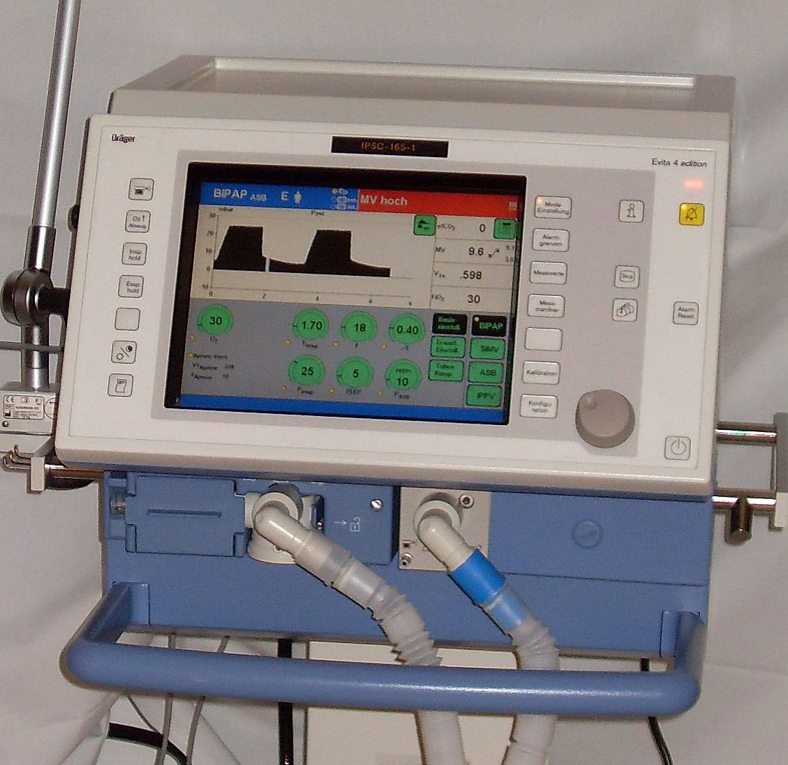
Key Components
| Component | Where It Sits | What It Does |
|---|---|---|
| Ventilator (console) | Bedside, ICU stand or transport unit | Delivers breaths by volume or pressure; sets rate, PEEP, FiO₂ and alarm limits |
| Gas supply | Wall oxygen and air, or cylinders | Supplies the set oxygen fraction; loss of supply stops delivery |
| Breathing circuit | Between the ventilator and the airway | Inspiratory and expiratory limbs carry gas; the exhalation valve releases it |
| Humidifier or heat-moisture exchanger | In-line near the patient | Conditions inspired gas and limits drying of secretions |
| Endotracheal tube or tracheostomy | Trachea | Connects the circuit to the airway; the cuff seals it |
| Capnography and pressure sensors | Y-piece or ventilator | Show EtCO₂ and airway pressure breath by breath |
Flow Path
- On inspiration the ventilator pushes gas in, by set volume or by set pressure.
- Gas runs down the inspiratory limb and through the humidifier to the Y-piece.
- Gas goes down the tube into the lungs. The chest rises as they fill.
- Exhaled gas comes back up the expiratory limb and out the exhalation valve. A PEEP valve or setting holds a baseline pressure in place.
- The sensors log flow, volume, pressure and EtCO₂ every cycle. A mismatch sets off the alarm.
Physiology & Hemodynamic Effects
Every positive-pressure breath pushes up the pressure in the chest. Venous return drops as a result, and the lung gets stretched. Positive end-expiratory pressure (PEEP) holds the alveoli open at end-exhalation. Plateau pressure (Pplat) is the static pressure at end-inspiration, read after a short pause. It tells you how elastic the lung and chest wall are.
What It Does
| Parameter | Effect | Why It Matters |
|---|---|---|
| Intrathoracic pressure | ↑ during inflation | Reduces venous return and can lower blood pressure, especially after intubation |
| Tidal volume (VT) | ↑ with larger breaths | Stretches lung tissue; high VT and high plateau pressure drive ventilator-induced lung injury |
| PEEP | ↑ end-expiratory lung volume | Recruits collapsed alveoli and can improve oxygenation; too much overdistends healthy lung and reduces venous return |
| Minute ventilation (VE = VT × rate) | ↑ with rate or VT | Main lever for PaCO₂ and pH; lowers PaCO₂ when raised |
| Auto-PEEP (air trapping) | ↑ end-expiratory pressure | Raises intrathoracic pressure without a PEEP setting; reduces preload and can cause hypotension |
| Peak pressure | ↑ with airway resistance | Rises with secretions, bronchospasm or a kink even when lung stiffness is normal |
What It Depends On
- Compliance and resistance: stiff lungs (ARDS, pulmonary edema) push both plateau and peak pressure up. A narrowed airway pushes peak up and leaves plateau alone.
- Chest wall: abdominal distension, obesity and chest wall edema all take more pressure to deliver the same volume.
- Patient effort: the patient's own breaths either add to the set breath or fight it. Either way, dyssynchrony changes every pressure reading you see.
- Preload and rhythm: cardiac output moves with every change in intrathoracic pressure.
- Pleural space: a pneumothorax skews the pressure readings, and it can turn into a tension pneumothorax.
Types, Modes & Configurations
Every manufacturer names its modes differently, so ignore the brand name. The table sorts each mode by what controls the breath, volume or pressure, and by what the patient still decides.
| Mode | Control Variable | What Each Breath Does | Best For | Key Risk |
|---|---|---|---|---|
| AC-VC (assist-control, volume control) | Volume | Delivers the set VT on every breath, triggered by patient or machine; pressure varies with compliance | Lung-protective ventilation for ARDS and most ICU settings | Peak pressure can rise unseen as compliance falls; check plateau pressure |
| AC-PC (assist-control, pressure control) | Pressure and inspiratory time | Delivers the set pressure on every breath; VT varies with compliance | Limiting peak pressure in selected patients | VT falls when the lungs stiffen; watch VT and minute ventilation |
| PRVC (pressure-regulated volume control) | Volume target, pressure adjusted | Adjusts pressure breath to breath to reach the set VT | Volume targeting with changing lung mechanics | Pressure changes between breaths; confirm the delivered VT |
| SIMV (synchronized intermittent mandatory ventilation) | Volume or pressure on mandatory breaths | Mandatory breaths are synchronized; spontaneous breaths occur between them | Older weaning approach; still in use | Spontaneous breaths can add large volumes or extra work at low set rates |
| PSV (pressure support) | Pressure; patient sets rate and VT | Assists each spontaneous breath to a set pressure | Spontaneous breathing and weaning | Apnea without a backup rate; large VT if effort is strong |
| APRV (airway pressure release) | Pressure (high CPAP with brief release) | Long high-pressure phase with a brief release; spontaneous breathing throughout | Selected severe ARDS in experienced units | CO₂ clearance depends on the release phase; needs experienced practice |
Indications, Contraindications & Evidence
Indications
- Failure to oxygenate or ventilate: hypoxemia or hypercapnic acidosis that persists despite non-invasive support and maximal oxygen therapy.
- Airway protection: loss of protective reflexes or inability to protect the airway. Severe TBI needs definitive airway protection (see Special Populations).
- ARDS: PaO₂/FiO₂ of 300 or lower with bilateral opacities, not explained by left atrial hypertension (ARDS Network inclusion criteria; Berlin Definition).
- Post-arrest or coma: control of airway and PaCO₂ under the post-resuscitation protocol.
- Transport: patients who need controlled ventilation for the trip, under protocol and medical direction.
Contraindications
| Absolute | Relative |
|---|---|
| None when ventilation is required to keep the patient alive | Untreated tension pneumothorax: decompress before positive pressure |
Uncontrolled bronchospasm: treat before high-pressure ventilation | |
Severe hypotension or preload dependence: positive pressure can lower blood pressure further; resuscitate first | |
Raised intracranial pressure: set ventilation to a PaCO₂ target and avoid unsafe hyperventilation |
Limitations
- Positive pressure cuts venous return. Blood pressure can fall because of it.
- Big tidal volumes and high plateau pressures hurt healthy lung, not only diseased lung (2017 ATS guideline).
- Ventilator-associated pneumonia is strongly tied to time on the ventilator (BTF 4th edition).
- Sedation and paralysis hide effort and discomfort. PADIS suggests light sedation over deep (conditional).
Key Evidence
| Trial / Guideline (Year) | Population | Bottom Line |
|---|---|---|
| ARDS Network ARMA trial (2000) | 861 patients with acute lung injury or ARDS; VT 6 vs 12 mL/kg PBW, plateau 30 vs 50 cmH₂O | Mortality 31.0% vs 39.8% (P=0.007) with the lower VT and plateau limit |
| Amato driving-pressure analysis (2015) | Individual data from 3,562 ARDS patients in nine randomized trials | Driving pressure was the strongest ventilator variable linked to survival; each 1-SD rise (about 7 cmH₂O) raised mortality (RR 1.41) |
| ATS/ESICM/SCCM guideline (2017) | Adults with ARDS | Strong: VT 4–8 mL/kg PBW and plateau pressure below 30 cmH₂O (the ARDS Network protocol uses at or below 30); prone positioning over 12 h/day in severe ARDS; against routine high-frequency oscillation |
| ATS guideline update (2024) | Adults with ARDS | Conditional: neuromuscular blockers in early severe ARDS (PaO₂/FiO₂ 100 or lower, less than 48 h from onset), higher PEEP without recruitment maneuvers, corticosteroids, selected VV-ECMO; strong against prolonged recruitment maneuvers |
| PALICC-2 (2023) | Children with pediatric ARDS | VT 6–8 mL/kg, plateau at or below 28 cmH₂O and driving pressure at or below 15 cmH₂O (conditional) |
Setup, Settings & Targets
Targets
| Parameter | Typical Target | If Below | If Above |
|---|---|---|---|
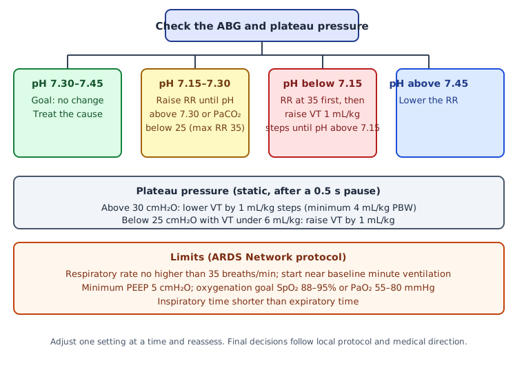
| Tidal volume | ARDS: 4–8 mL/kg PBW (ARDS Network target 6 mL/kg PBW) | Find the cause first (effort, sedation, pain); do not raise VT routinely to chase CO₂. A rise is a last protocol step at pH below 7.15 (see pH row) | Reduce VT by 1 mL/kg steps; minimum 4 mL/kg PBW |
| Plateau pressure | At or below 30 cmH₂O (static, after a 0.5 s pause); pediatric at or below 28 cmH₂O | Below 25 cmH₂O with VT under 6 mL/kg: raise VT by 1 mL/kg | Above 30 cmH₂O: reduce VT by 1 mL/kg steps |
| PEEP | At least 5 cmH₂O; ARDS: at or above the ARDS Network low-PEEP/high-FiO₂ table | Hypoxemia: raise FiO₂ and PEEP together per the table | Hypotension or overdistension: lower PEEP stepwise with reassessment |
| Driving pressure (Pplat − PEEP) | At or below 15 cmH₂O (PALICC-2, children); adult limit per protocol | Not a lower-limit target | Reduce VT first, then review PEEP per protocol |
| pH | 7.30–7.45 (ARDS Network). Pediatric PARDS: permissive floor of 7.20 (PALICC-2; see Special Populations) | 7.15–7.30: raise RR, up to 35, until pH above 7.30 or PaCO₂ below 25 | Above 7.45: lower the RR |
| pH below 7.15 (last step) | Not a routine target. Protocol step under physician direction: RR already at 35; sodium bicarbonate may be given per protocol | Raise VT in 1 mL/kg steps while pH stays below 7.15; the plateau pressure goal of 30 may be exceeded in this step. Not with raised intracranial pressure (see Special Populations) | Stop raising VT once pH is above 7.15 |
| Respiratory rate | Start near baseline minute ventilation; no higher than 35 breaths/min | Check sedation and effort; a falling rate can raise PaCO₂ | Reduce only when pH is high (see pH row) |
| SpO₂ | ARDS: 88–95% (PaO₂ 55–80 mmHg). Pediatric PARDS, mild or moderate: 92–97% | Raise FiO₂, then PEEP, per the table; in severe pediatric PARDS, accept below 92% once PEEP is optimized | Lower FiO₂ to the lowest level that meets target; avoid prolonged values above 97% in children |
| Inspiratory : expiratory time | Inspiration shorter than expiration (ARDS Network goal) | Not a lower-limit target | Long inspiration leaves less time to exhale; shorten it to reduce air trapping |
Initial Settings
- Mode: per protocol. Name the mode and its control variable in the chart.
- Tidal volume (ARDS): start at 8 mL/kg PBW, then reduce by 1 mL/kg at intervals of 2 hours or less until 6 mL/kg PBW is reached (ARDS Network).
- Rate: start near baseline minute ventilation, not above 35 breaths/min.
- PEEP: minimum 5 cmH₂O; in ARDS, use the ARDS Network low-PEEP/high-FiO₂ table.
- FiO₂: start high enough to reach the SpO₂ goal, then wean to the lowest level that meets it.
- Plateau pressure: check at least every 4 hours and after each change in PEEP or VT.
- Non-ARDS adults: this page only gives ARDS starting settings. None of the sources used here sets a starting volume or rate for other adults. Your ICU or transport protocol decides those.
| FiO₂ | 0.3 | 0.4 | 0.5 | 0.6 | 0.7 | 0.8 | 0.9 | 1.0 |
|---|---|---|---|---|---|---|---|---|
| PEEP (cmH₂O) | 5 | 5–8 | 8–10 | 10 | 10–14 | 14 | 14–18 | 18–24 |
Sizing & Positioning
Female: 45.5 + 2.3 × (height in inches − 60)
Height and sex set PBW, never actual weight. Take a 70-inch male: his PBW is 73 kg, so 6 mL/kg PBW is 438 mL. Confirm tube depth and position before you set the ventilator (see the intubation page).
Calculations & Derived Values
Raw pressures mean little until you turn them into lung mechanics. Run these numbers every time you measure a plateau and every time you draw an ABG.
| Variable | Formula | Normal or Target | Why You Care |
|---|---|---|---|
| Driving pressure | Pplat − PEEP | At or below 15 cmH₂O (PALICC-2, children); adult limit per protocol | Strongest ventilator variable linked to survival in the Amato analysis |
| Compliance (static) | VT ÷ (Pplat − PEEP) | Trend, not a fixed target | Falling compliance means stiffer lung or overdistension; recheck after PEEP changes |
| Minute ventilation | VT × rate | Set near baseline to start | Main lever for PaCO₂; compare with the measured exhaled volume |
| PaO₂/FiO₂ ratio | PaO₂ ÷ FiO₂ (fraction, for example 0.4) | Above 300: not ARDS; 200–300 mild; 100–200 moderate; 100 or lower severe (Berlin) | Defines ARDS and its severity; read it with the PEEP in use |
Circuit Pressures & Console
Normal
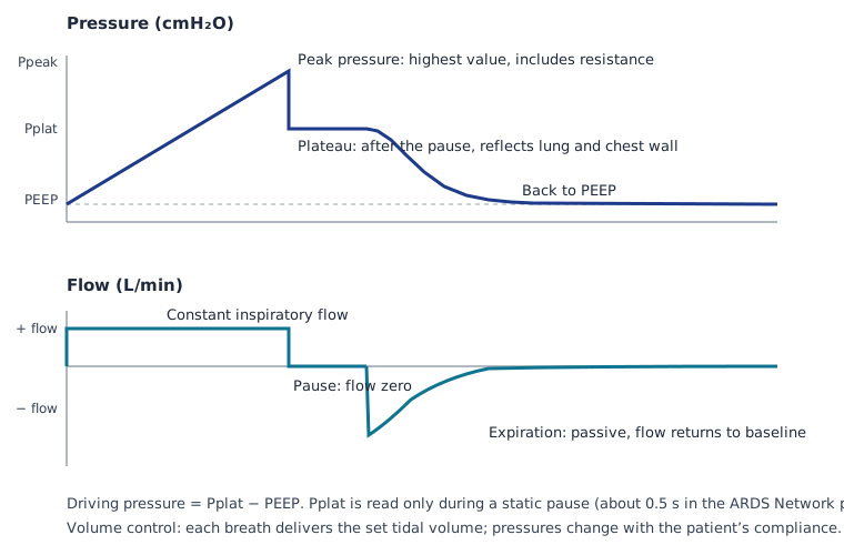
- Volume control: flow climbs to a set constant value during inspiration. Plot that and you get a square wave.
- Pressure: climbs to peak during inspiration. Add an inspiratory pause and it drops to a plateau and holds there.
- Expiration: flow falls back to baseline before the next breath. Pressure goes back down to PEEP.
- Loops and curves: pressure-time and flow-time curves repeat breath to breath when the patient is passive.
Console Checklist
- Mode, control variable, set VT, rate, PEEP, FiO₂ and inspiratory time match the chart.
- Trigger sensitivity is set so the patient can start a breath without auto-triggering.
- Alarm limits are set for this patient, not left at defaults.
Abnormal Patterns
| Pattern | What You See | Cause | Fix |
|---|---|---|---|
| Auto-PEEP (air trapping) | Expiratory flow does not return to zero before the next breath; end-expiratory pressure sits above set PEEP | Rate or inspiratory time too high for the expiratory time; airway obstruction | Lower the rate or shorten inspiration; check for obstruction; if unstable, disconnect briefly to let the chest empty |
| Peak up, plateau normal | Peak pressure rises; plateau stays in range | Airway resistance: secretions, kinked tube, bronchospasm | Suction, check tube and circuit, bronchodilator per protocol |
| Peak and plateau both up | Both pressures rise together | Stiffer lung, overdistension, chest wall pressure or pneumothorax | Check breath sounds and chest movement; reduce VT; look for pneumothorax |
| Patient effort or dyssynchrony | Negative deflection before a breath; double breaths; flow notches | Support mismatched to effort, trigger set too sensitive or too insensitive, or sedation mismatch | Adjust trigger and support per protocol; reassess pain and sedation (PADIS) |
| Leak | Exhaled VT lower than inspired VT; pressure does not reach set level | Cuff leak, disconnect, tube out of position | Check connections and cuff; confirm tube depth with EtCO₂ |
Monitoring & Ongoing Care
| Frequency | What to Check | Action |
|---|---|---|
| Continuous | SpO₂, EtCO₂ waveform, ECG, arterial pressure, ventilator alarms | Respond to every alarm; find the cause before silencing it |
| Hourly | Chest rise, breath sounds, peak pressure, delivered VT, SpO₂, sedation score (RASS) | Reassess after any change; adjust sedation to the goal |
| At least every 4 h, and after each PEEP or VT change | Plateau pressure (static pause); driving pressure | Adjust VT or PEEP per the ARDS protocol |
| After each major change | ABG: pH, PaCO₂, PaO₂; FiO₂ in use | Use the pH goals in the Setup table; wean FiO₂ to target |
| Daily | Sedation interruption and spontaneous breathing trial readiness; tube depth and cuff; oral care; head of bed | Pair daily sedation interruption with a trial of breathing (see Weaning) |
Positioning & Prone Ventilation
- Prone positioning: for more than 12 hours a day in severe ARDS (ATS 2017, strong recommendation).
- Head of bed and oral care: per the unit ventilator-associated pneumonia protocol.
- Early mobility: when the patient is stable and per protocol.
If prone positioning fails or ARDS is severe, consider the ECMO page. ATS 2024 suggests VV-ECMO only in selected severe ARDS.
Anticoagulation & Hematology
Ventilation does not need anticoagulation on its own. The bleeding risks on this page come from airway procedures and from the prophylaxis ventilated patients get under the unit protocol.
- Before a procedure: confirm the platelet count, coagulation tests and any anticoagulant on board, per the procedure protocol.
- Heparin exposure: when platelets fall on heparin, consider heparin-induced thrombocytopenia (see the heparin page).
- Bleeding: new bleeding from the tube, the tracheostomy or a line site needs the team and a fresh coagulation check.
| Issue | Typical Target | Monitoring | Watch For |
|---|---|---|---|
| VTE prophylaxis (heparin) High-alert | Per unit protocol | Per protocol; platelet count (see platelets) | Bleeding at tube, line or tracheostomy sites; heparin-induced thrombocytopenia |
| Bronchoscopy or tracheostomy | Anticoagulant held or reversed per the proceduralist | Coagulation tests per team (see aPTT) | Bleeding during or after the procedure; blood in secretions |
| Tracheal suction | No bleeding beyond streaks | Secretion color and volume each shift | Persistent blood: check platelets and coagulation, and notify the team |
Key Medications & Drug Considerations
| Medication | Role Here | Typical Adult Dose / Target | Ventilator-Specific Notes |
|---|---|---|---|
| Propofol | Sedation in ventilated adults | Start 5 mcg/kg/min IV infusion; increase by 5–10 mcg/kg/min to the sedation goal; usual 5–50 mcg/kg/min (dose per propofol page) | High-alert Hypotension; a bolus can cause apnea. PADIS suggests propofol or dexmedetomidine over benzodiazepines (conditional). |
| Dexmedetomidine | Sedation alternative to propofol | 0.2–0.7 mcg/kg/hr IV infusion (label); load often omitted in the ICU | High-alert Bradycardia and hypotension. Pair with analgesia; PADIS treats pain before a sedative is considered. |
| Midazolam | Second-line sedation when propofol or dexmedetomidine is unsuitable | 0.02–0.1 mg/kg/hr (1–7 mg/hr) IV infusion; optional load per midazolam page | High-alert PADIS suggests propofol or dexmedetomidine over benzodiazepines. |
| Fentanyl | Analgesia before sedation | 0.35–0.5 mcg/kg IV every 30–60 min as needed; infusion per fentanyl page | High-alert PADIS: treat pain before a sedative is considered. |
| Rocuronium | Neuromuscular blockade (ICU or transport) | 0.6 mg/kg IV loading dose after deep sedation is confirmed; infusion 10–12 mcg/kg/min at early recovery (per rocuronium page) | High-alert Never give without sedation. ATS 2024 conditionally suggests neuromuscular blockers in early severe ARDS (PaO₂/FiO₂ 100 or lower, less than 48 h from ARDS onset) and limits use to 48 h where possible. The guideline names no agent; cisatracurium was used in the largest trials, and this page does not cover it. |
| Norepinephrine | Hypotension after positive pressure or induction | 0.05–0.5 mcg/kg/min IV infusion, titrated to the MAP target (per norepinephrine page) | High-alert Confirm mcg/min versus mcg/kg/min before each change. Fix preload and auto-PEEP first where possible. |
| Albuterol | Bronchospasm on the ventilator | Nebulized 2.5–5 mg, repeated per protocol; continuous nebulization in severe asthma per albuterol page | Does not fix auto-PEEP alone; also lower the rate and lengthen expiration. |
| Sodium bicarbonate | Metabolic acidemia not corrected by ventilator changes | Per sodium bicarbonate page; pH endpoint per that page | Not a ventilator fix. The ARDS Network protocol lists it only after rate changes and within its acidosis steps. |
Doses vary by protocol; follow local protocols and medical direction.
Alarms & Troubleshooting
| Alarm / Problem | What You See | Likely Causes | First Actions |
|---|---|---|---|
| High peak pressure | Peak alarm; plateau normal or high | Secretions, kinked tube, bronchospasm, coughing | 1) Look at the patient. 2) Suction. 3) Check the tube and circuit. 4) Bronchodilator per protocol. |
| Low pressure or low VT | Low-pressure or low-volume alarm; EtCO₂ falls | Disconnect, cuff leak, tube out of position, circuit leak | 1) Check every connection. 2) Bag with 100% oxygen if SpO₂ falls. 3) Check tube depth and cuff. |
| Auto-PEEP (air trapping) | Expiratory flow does not return to zero; blood pressure falls | Rate too high, short expiratory time, obstruction | 1) Disconnect briefly if unstable. 2) Lower the rate. 3) Bronchodilator per protocol. 4) Recheck flow. |
| Waveform lost or EtCO₂ flat | Capnogram flat or falling | Tube out or esophageal, circulatory collapse, disconnect | 1) Bag with oxygen. 2) Run DOPE (displacement, obstruction, pneumothorax, equipment). 3) Check tube depth (see intubation page). |
| Hypoxemia | SpO₂ falling | Mucus plug, PEEP too low, pneumothorax, shunt | 1) Bag with 100% oxygen and check the tube. 2) Raise FiO₂, then PEEP, per the table. 3) Look for pneumothorax. |
| Hypercapnia or acidemia | pH falls; PaCO₂ rises | Rate too low, dead space, low VT, sedation reducing effort | 1) Raise the rate in steps toward 35 per the pH steps. 2) Check delivered VT. 3) Check the circuit. |
| Hypotension after positive pressure | MAP falls soon after induction or a PEEP increase | Intrathoracic pressure, hypovolemia, auto-PEEP, sedation | 1) Check for auto-PEEP. 2) Volume or vasopressor per protocol. 3) Lower PEEP or rate if preload is limited. |
| Patient-ventilator dyssynchrony | Patient fights the ventilator; double breaths | Sedation mismatch, trigger setting, too little support, pain or anxiety | 1) Check trigger and support. 2) Assess pain, sedation and anxiety. 3) Adjust support per protocol. |
Complications & Red Flags
| Complication | How You Recognize It | Immediate Action | Prevention |
|---|---|---|---|
| Barotrauma (pneumothorax) | Sudden fall in SpO₂ and BP; rising peak pressure; absent breath sounds on one side | Treat as tension until proven otherwise; decompress per protocol | Lung-protective VT and plateau limits |
| Ventilator-induced lung injury | Rising plateau pressure; stiffer lungs; new bilateral opacities | Reduce VT by 1 mL/kg steps; check plateau | VT 4–8 mL/kg PBW and plateau below 30 cmH₂O (2017 ATS guideline) |
| Auto-PEEP (dynamic hyperinflation) | Hypotension; rising pressures; expiratory flow fails to return to zero | Disconnect briefly to let the chest empty; lower the rate | Shorten inspiration and lengthen expiration in obstructive disease |
| Hypotension | MAP falls after induction or a PEEP increase | Volume, vasopressor or lower PEEP per protocol | Resuscitate before induction (see the RSI page) |
| Tube displacement or mainstem intubation | Asymmetric chest rise; one-sided breath sounds; EtCO₂ change | Check depth; pull back and re-auscultate | Secure the tube; check depth after every move |
| Ventilator-associated pneumonia | New fever; purulent secretions; rising oxygen need | Cultures and treatment per protocol | Head of bed, oral care and daily sedation interruption per unit protocol |
| Dyssynchrony and agitation | Fighting the ventilator; double triggering | Check settings, pain and sedation | Analgesia first, then light sedation (PADIS) |
- SpO₂ falling after a disconnect or alarm and not recovering with a bag and oxygen
- Sudden hypotension with rising peak pressure (possible tension pneumothorax)
- Flat or lost EtCO₂ waveform in a patient who was ventilating well
- New one-sided absent breath sounds with rising pressures
Emergencies: Arrest, Power Loss & Device Failure
Cardiac Arrest on the Ventilator
- Start CPR if the patient is pulseless, per your ACLS protocol.
- Stop the ventilator and ventilate with a bag-mask and 100% oxygen, or an advanced airway if the team has one.
- Set the ventilation rate with an advanced airway per your ACLS protocol (AHA 2025 Part 9 covers advanced airway management in arrest).
- Confirm the tube with waveform EtCO₂ (see the intubation page).
- Look for reversible causes: tension pneumothorax, severe auto-PEEP, tube displacement.
- Resume ventilation per the post-arrest protocol once the team has a plan.
Ventilator Failure or Power Loss
- Disconnect the failed unit from the tube and bag with 100% oxygen. Use a PEEP valve if available.
- Check the tube and connections while you bag, and confirm EtCO₂.
- Switch to battery or the backup unit if one is available.
- Watch SpO₂ and blood pressure and call for help early (critical-care transport or the ICU team).
Suspected Tension Pneumothorax on Positive Pressure
- Stop positive pressure: disconnect and bag if the patient is unstable.
- Decompress per protocol (needle or finger thoracostomy; procedure scope per team and protocol).
- Reassess breath sounds, BP and SpO₂ after decompression.
- Chest tube per the physician or APP.
Tube Dislodged or Extubated
- Stop ventilating through a displaced tube. Bag-mask with 100% oxygen.
- Call for help and assess the airway.
- Reintubate or place a supraglottic airway per the airway plan (Paramedic, RN or CCT per protocol).
- Confirm with waveform EtCO₂ and reassess SpO₂.
Weaning & Discontinuation
Readiness Criteria
- Cause treated: the reason for ventilation is improving.
- Oxygenation on low support: FiO₂ at or below 0.40 with PEEP at or below 8 cmH₂O, or FiO₂ below 0.50 with PEEP below 5 cmH₂O (ARDS Network).
- Stable circulation: systolic BP at least 90 mmHg without vasopressors; no neuromuscular blocker on board.
- Effort and settings: acceptable spontaneous efforts; PEEP and FiO₂ no higher than the previous day.
Weaning Steps
- Screen daily for readiness, and interrupt sedation where safe (the ABC trial paired a daily awakening trial with a breathing trial).
- Run the trial for up to 120 minutes on a T-piece, trach collar, or CPAP at or below 5 cmH₂O with pressure support below 5 cmH₂O (FiO₂ below 0.5 and PEEP below 5).
- Check tolerance during the trial: SpO₂ at least 90% and/or PaO₂ at least 60 mmHg; spontaneous VT at least 4 mL/kg PBW; rate 35 or less; pH at least 7.30; no distress.
- If tolerated for at least 30 minutes, consider extubation (ARDS Network).
- If not tolerated, return to the settings used before the trial.
Failure Criteria: Step Back Up
- Heart rate above 120% of baseline
- Marked accessory muscle use or abdominal paradox
- Diaphoresis or marked dyspnea
- SpO₂ below target during the trial
Removal & Post-Removal Care
- Check sedation and paralysis status before extubation.
- Watch for upper airway obstruction, secretions and failure to protect the airway after extubation, per protocol.
Transport Considerations
Before You Move
- Check power: battery minutes and backup power. Confirm the ventilator runs on battery before you disconnect from wall power.
- Check oxygen: cylinder content and duration for the trip, with a spare.
- Secure the airway: tube, circuit and EtCO₂ sensor. Document tube depth.
- Carry spares: spare tube, suction, bag-mask-valve with PEEP, and a backup ventilator if available.
- Brief the receiving team on mode, settings, last plateau pressure and sedation.
If You Are Not the Ventilator Specialist
- Watch: SpO₂, EtCO₂ waveform, peak and plateau pressure, rate and chest rise.
- Never: change the mode or remove PEEP to silence an alarm. Disconnect only when the patient is in danger and bag at once.
- Call: the critical-care team or medical control for any change beyond protocol.
During Transport
- Recheck tube depth and cuff after every move: stretcher, ambulance, elevator and stairs.
- Expect sedation and paralysis to need checks at each handoff and after each drug change.
Air Medical & Altitude
As you climb, ambient pressure falls and the gas in the cuff and circuit expands (Boyle's law). Cuff pressure rises on ascent and falls on descent. Recheck the cuff at every altitude change. Transport ventilators do not all compensate the same way, so the device manual governs altitude.
Handoff
- Mode, settings, FiO₂ and PEEP
- Last ABG, plateau pressure and driving pressure
- Sedation score, analgesia and any paralysis
- Recent alarms, complications and tube depth with cuff status
- Goals of care and the next planned step (trial of breathing, procedure or transfer)
Special Populations
Pediatric & Neonatal
- Tidal volume: 6–8 mL/kg of the lesser of predicted or actual body weight. Go below 6 mL/kg only if needed to stay within plateau and driving-pressure limits, and use caution below 4 mL/kg (PALICC-2, conditional).
- Plateau pressure: at or below 28 cmH₂O (29–32 cmH₂O may be accepted with reduced chest wall compliance).
- Driving pressure: at or below 15 cmH₂O (static).
- PEEP: at or above the ARDS Network low-PEEP/high-FiO₂ table.
- SpO₂: 92–97% for mild or moderate PARDS; in severe PARDS, accept below 92% after PEEP is optimized.
- Permissive hypercapnia: to a pH floor of 7.20, within the pressure and volume limits above. Not suggested with intracranial hypertension, severe pulmonary hypertension, select congenital heart lesions, hemodynamic instability or significant ventricular dysfunction (PALICC-2, conditional).
- Recruitment maneuvers and high-frequency oscillation: PALICC-2 offers no recommendation for or against either one.
- Neonates: This page does not cover neonates. Follow the neonatal resuscitation protocol and the neonatal transport team.
Pediatric sedation runs under PICU protocol. Start at the midazolam page and weight-based dosing.
Acute Brain Injury (TBI)
- Goal: normal ventilation with PaCO₂ 35–45 mmHg when there is no herniation (BTF 4th edition).
- Prophylactic hyperventilation: prolonged hyperventilation to PaCO₂ 25 mmHg or less is not recommended.
- Temporizing hyperventilation: the 3rd-edition statements were restated for herniation. If you use it, watch oxygen delivery through jugular venous saturation or brain tissue oxygen.
- Oxygenation: avoid hypoxia. Hypoxia caught on the monitor goes with worse outcomes.
Obstructive Lung Disease (COPD and Asthma)
- Expiratory time: the rate has to be low enough that the chest empties before the next breath. Look for auto-PEEP at each change.
- Hypercapnia: accept permissive hypercapnia within the pH floor protocol sets. Keep it away from raised intracranial pressure (see Acute Brain Injury), severe pulmonary hypertension, hemodynamic instability or significant ventricular dysfunction. PALICC-2 wrote those exceptions for children, so apply the same caution to adults under medical direction.
- Oxygen: aim for the lower SpO₂ target that protocol sets for hypercapnic COPD.
- PEEP: PEEP goes on only as protocol directs. Auto-PEEP stacks onto the load already there.
Pregnancy & Obesity
In pregnancy and obesity, the same volume takes more pressure. A given plateau may reflect a stiffer chest wall, not a stiffer lung. This page gives no numeric targets for either. Follow obstetric or ICU protocol and medical direction.
Bedside Pearls & Teaching Script
Systematic Walk-Through
- Indication: why does this patient need support, and what is the goal (oxygen, CO₂, rest or airway)?
- Mode and control: name the mode and whether volume or pressure controls each breath.
- Settings: VT by PBW, rate, PEEP, FiO₂ and inspiratory time.
- Pressures and ABG: check plateau and driving pressure; read the pH and PaO₂/FiO₂.
- Comfort: treat pain first, then use light sedation; paralysis only with sedation.
- Complications and goal: check for pneumothorax, auto-PEEP and hypotension, then set today's goal (trial of breathing, extubation or transport).
Pearls
- Build tidal volume from predicted body weight. Actual weight does not enter into it.
- Peak pressure tells you about the airway. Plateau tells you about the lung.
- For hypoxemia, check the tube first, then FiO₂, then PEEP.
- Auto-PEEP shows up as expiratory flow that never gets back to zero. Check for it before you add PEEP.
- Pain before sedation, and light sedation when safe.
- Change one setting at a time and reassess after each change.
References
- The Acute Respiratory Distress Syndrome Network. (2000). Ventilation with lower tidal volumes as compared with traditional tidal volumes for acute lung injury and the acute respiratory distress syndrome. New England Journal of Medicine, 342(18), 1301–1308. https://doi.org/10.1056/NEJM200005043421801
- Amato, M. B. P., Meade, M. O., Slutsky, A. S., et al. (2015). Driving pressure and survival in the acute respiratory distress syndrome. New England Journal of Medicine, 372(8), 747–755. https://doi.org/10.1056/NEJMsa1410639
- Fan, E., Del Sorbo, L., Goligher, E. C., et al. (2017). An official American Thoracic Society/European Society of Intensive Care Medicine/Society of Critical Care Medicine clinical practice guideline: Mechanical ventilation in adult patients with acute respiratory distress syndrome. American Journal of Respiratory and Critical Care Medicine, 195(9), 1253–1263. https://doi.org/10.1164/rccm.201703-0548ST
- Qadir, N., Sahetya, S., Munshi, L., et al. (2024). An update on management of adult patients with acute respiratory distress syndrome: An official American Thoracic Society clinical practice guideline. American Journal of Respiratory and Critical Care Medicine, 209(1), 24–36. https://doi.org/10.1164/rccm.202311-2011ST
- Emeriaud, G., López-Fernández, Y. M., Iyer, N. P., et al. (2023). Executive summary of the second international guidelines for the diagnosis and management of pediatric acute respiratory distress syndrome (PALICC-2). Pediatric Critical Care Medicine, 24(2), 143–168. https://doi.org/10.1097/PCC.0000000000003147
- Girard, T. D., Kress, J. P., Fuchs, B. D., et al. (2008). Efficacy and safety of a paired sedation and ventilator weaning protocol for mechanically ventilated patients in intensive care (Awakening and Breathing Controlled trial): A randomised controlled trial. The Lancet, 371(9607), 126–134. https://doi.org/10.1016/S0140-6736(08)60105-1
- Devlin, J. W., Skrobik, Y., Gélinas, C., et al. (2018). Clinical practice guidelines for the prevention and management of pain, agitation/sedation, delirium, immobility, and sleep disruption in adult patients in the ICU. Critical Care Medicine, 46(9), e825–e873. https://doi.org/10.1097/CCM.0000000000003299
- Carney, N., Totten, A. M., O'Reilly, C., et al. (2017). Guidelines for the management of severe traumatic brain injury, fourth edition. Neurosurgery, 80(1), 6–15. https://doi.org/10.1227/NEU.0000000000001432
- ARDS Definition Task Force. (2012). Acute respiratory distress syndrome: The Berlin Definition. JAMA, 307(23). https://doi.org/10.1001/jama.2012.5669
- NHLBI ARDS Clinical Network. (2008). Mechanical ventilation protocol summary. National Heart, Lung, and Blood Institute. https://www.ardsnet.org/files/ventilator_protocol_2008-07.pdf
- Roginski, M. A., Carroll, M. C., Trautwein, M. L., Watkins, E. D., and Esteves, A. M. (2023). Sedation practices of mechanically ventilated patients during critical care transport. Air Medical Journal, 42(5), 343–347. https://doi.org/10.1016/j.amj.2023.05.002
- Wigginton, J. G., Agarwal, S., Bartos, J. A., Coute, R. A., Drennan, I. R., Haamid, A., Kudenchuk, P. J., Link, M. S., et al. (2025). Part 9: Adult advanced life support: 2025 American Heart Association guidelines for cardiopulmonary resuscitation and emergency cardiovascular care. Circulation, 152(16 suppl 2), S538–S577. https://doi.org/10.1161/CIR.0000000000001376
Medical Disclaimer
- For Educational Purposes Only: This content is intended for educational reference and should not be used for clinical decision-making.
- Not a Substitute for Professional Judgment: Always consult your local protocols, institutional guidelines, and supervising physicians.
- Verify Before Acting: Users are responsible for verifying information through authoritative sources before any clinical application.