

At a Glance
- Covers
- First-degree AV block, Mobitz I, Mobitz II, 2:1 and high-grade AV block, complete heart block, bundle branch and fascicular blocks.
- Recognize It
- PR 120–200 ms normal. Mobitz I: PR lengthens, then a QRS drops. Mobitz II: constant PR, sudden drop. Complete block: P waves and QRS unrelated.
- Most Dangerous
- Mobitz II and complete heart block below the AV node: slow, wide escape and a risk of sudden asystole.
- Unstable Patient
- Hypotension, altered mental status, ischemic chest pain or shock: atropine 1 mg IV/IO, apply pads, then transcutaneous pacing.
- Stable Patient
- Monitor and get a 12-lead. Treat reversible causes. Mobitz I often needs only monitoring; Mobitz II and complete block need cardiology review.
- Do Not
- Do not expect atropine to fix infranodal block. Do not discharge Mobitz II or complete heart block without a pacing plan.
Physiology & What It Represents
Start at the sinus node. The beat crosses the atria, then arrives at the atrioventricular (AV) node, where it is held up briefly. From there it runs through the His bundle, the bundle branches and the Purkinje network out to the ventricular myocardium. Normally the AV node is the only electrical bridge between atria and ventricles. The hold-up is what the PR interval measures. A block either stretches it or cuts the signal altogether.
Where the block sits tells you what backs it up. Above the His bundle (AV nodal, supra-Hisian), the QRS stays narrow and a junctional escape at about 40–60 bpm takes over. That escape is often transient. Below the His bundle (infranodal), the QRS goes wide and the ventricle has to supply a slow beat, about 20–40 bpm. Asystole can arrive without warning.
- Automatic pacemakers, fastest to slowest: sinus node (60–100 bpm), AV junction (40–60 bpm), ventricle (20–40 bpm)
Recognition Approach
Work through rate, regularity and the P-to-QRS relationship, in that order. Width comes next. The QRS tells you where the block sits.
| Rhythm | Rate (bpm) | Regularity | P Waves | PR | QRS | First-Line Action |
|---|---|---|---|---|---|---|
| First-degree AV block | Sinus rate | Regular | Every P conducted | >200 ms, constant | Narrow unless BBB | Monitor; treat reversible causes |
| Mobitz I (Wenckebach) | Sinus rate, grouped | Irregular in groups | Regular march; one blocked | Lengthens, then a dropped QRS | Narrow | Monitor; atropine if symptomatic; pace if unstable |
| Mobitz II | Sinus rate | Irregular (dropped beats) | Regular; some not conducted | Constant before a drop | Often wide | Unstable: atropine, then pace; cardiology for pacemaker |
| 2:1 AV block | Half the sinus rate | Regular | Every other P conducted | Cannot be typed | Narrow or wide | Wide QRS: treat as Mobitz II |
| High-grade AV block | Below sinus rate | Irregular | Two or more blocked in a row | Constant on conducted beats | Narrow or wide | Unstable: atropine, then pace |
| Third-degree (complete) | Atrial rate above ventricular | Atrial and ventricular each regular | Independent of QRS | Varies (no relationship) | Narrow (junctional) or wide (ventricular) | Narrow: atropine trial; unstable: pace |
| Bundle branch block | Sinus rate | Regular | Every P conducted | Normal | 120 ms or more, typical shape | Treat the cause; assess for ischemia |
| Fascicular block | Sinus rate | Regular | Every P conducted | Normal | Normal or under 120 ms; axis shift | Usually none; check for bifascicular block |
- Rate: Count the ventricular rate and compare it with the atrial (P-wave) rate.
- Regularity: Decide whether the P-P and R-R intervals are constant or change in a pattern.
- P waves: Confirm a P wave comes before each QRS, and find any P waves with no QRS.
- PR and P:QRS relationship: Measure the PR before each conducted beat. Note whether it lengthens, stays constant, or the P wave is blocked.
- QRS width: Measure the QRS in the widest lead. Narrow (under 120 ms; 110–119 ms is borderline) suggests a nodal site; wide suggests infranodal.
- Stability: Decide stable or unstable before any drug. Unstable means hypotension, altered mental status, ischemic chest pain, shock or heart failure.
| Parameter | Normal Value | Notes |
|---|---|---|
| PR interval | 120–200 ms (0.12–0.20 s; 3–5 small boxes) | From P-wave onset to QRS onset |
| QRS duration | 110 ms or less | 110–119 ms is borderline: incomplete BBB or nonspecific IVCD; 120 ms or more: wide |
| Junctional escape | 40–60 bpm | Narrow QRS; more reliable |
| Ventricular escape | 20–40 bpm | Wide QRS; less reliable |
First-Degree AV Block
First-degree AV block is a steady conduction delay. Every P wave gets a QRS, but the PR runs longer than normal. The full PR-interval teaching is on the PR Interval page.
ECG Criteria
- PR interval: More than 200 ms (0.20 s; more than 5 small boxes), constant beat to beat
- Conduction: 1:1; every P wave followed by a QRS
- QRS: Narrow unless a bundle branch block coexists
Causes
- Increased vagal tone, athletic conditioning, sleep
- Drugs: beta-blockers, calcium channel blockers, digoxin, amiodarone
- Inferior MI (AV nodal ischemia), myocarditis, Lyme carditis
Clinical Significance
- Usually benign. On its own it needs no specific treatment
- A very long PR (commonly above 300 ms) can cause symptoms. Loss of AV synchrony means the atria contract against closed AV valves
- It can progress to higher-grade block. Review the drugs, and repeat the ECG if the clinical picture changes
Second-Degree Mobitz I (Wenckebach) AV Block
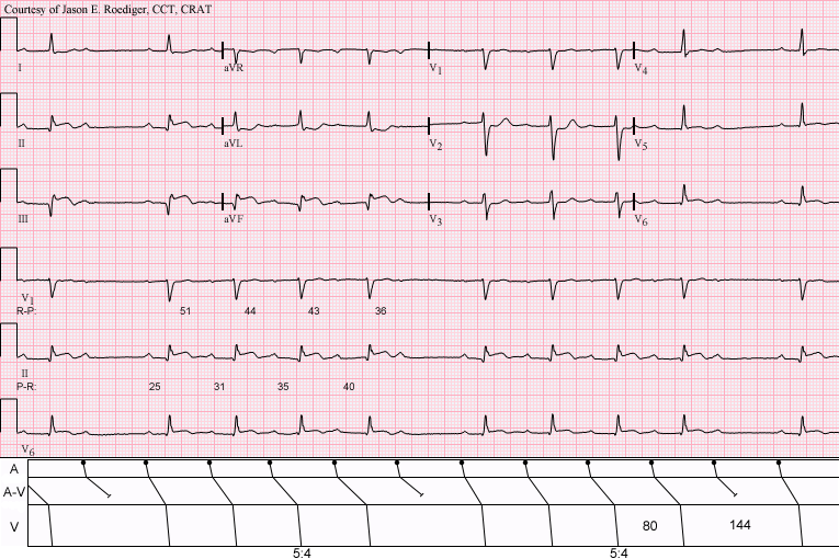
Mobitz I is a nodal block, and the PR tells the story. It lengthens with every beat until a P wave fails to conduct. Then the cycle starts over. The pause lets conduction recover, so the QRS stays narrow.
ECG Criteria
- PR: Lengthens progressively across the group, with shrinking increments before the drop
- Dropped QRS: One P wave with no QRS ends each group
- R-R: Shortens across the group; the R-R interval that contains the dropped beat is less than twice the shortest R-R before it
- QRS: Narrow, because the block is above the His bundle
Causes
- Increased vagal tone, sleep, athletic training
- Inferior MI (usually transient), beta-blockers, calcium channel blockers, digoxin
- Myocarditis and Lyme carditis
Clinical Significance
- Usually benign, and the chance of sliding into complete block is low
- You can count on the escape rhythm here. The block sits in the node
- Pacing is rarely the answer. Reach for it only when the patient is symptomatic or hemodynamically unstable
Look-Alikes
- Sinus arrest or exit block: a pause with no P wave in front of it (see Differential Diagnosis)
Second-Degree Mobitz II AV Block
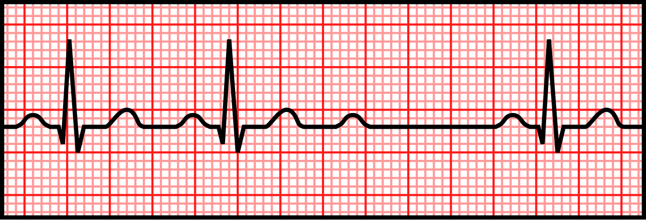
Mobitz II is an infranodal block. Conducted beats keep a constant PR interval. Then some P waves fail to conduct, with no warning. The block lies in the His–Purkinje system.
ECG Criteria
- PR: Constant for conducted beats; no lengthening before the drop
- Dropped QRS: Sudden and unpredictable; often 2:1 or 3:1, or variable
- P waves: The sinus rhythm continues regularly through the dropped beats
- QRS: Often wide, because the block is below the AV node
Causes
- Anterior MI with septal involvement (LAD territory)
- Degenerative conduction disease and fibrosis
- Infiltrative disease, cardiac surgery, drug or electrolyte toxicity
Clinical Significance
- The risk of progressing to complete heart block is high. It often comes without warning
- Escape rhythms are slow, about 20–40 bpm. They may be wide, and you can't count on them
- It needs a permanent pacing evaluation. Do not discharge without cardiology review
2:1 and High-Grade AV Block
In 2:1 AV block, every other P wave conducts. High-grade AV block has two or more non-conducted P waves in a row, with some conduction preserved. You can't classify either one as Mobitz I or Mobitz II from the 2:1 segment alone. Only one PR interval is visible at a time.
ECG Criteria
- 2:1: Two P waves for each QRS; the PR of the one conducted beat cannot show progression
- High-grade: Two or more consecutive non-conducted P waves
- QRS width: The main clue. Narrow suggests a nodal site; wide suggests infranodal disease
Clinical Significance
- Manage a wide QRS with 2:1 or high-grade block as infranodal. Stay with that until a longer strip shows otherwise
- Record a longer rhythm strip. A stretch with more than one conducted beat shows whether the PR lengthens
- A change in conduction ratio with a wider QRS is a warning sign. It points to progressive infranodal disease
Third-Degree (Complete) AV Block
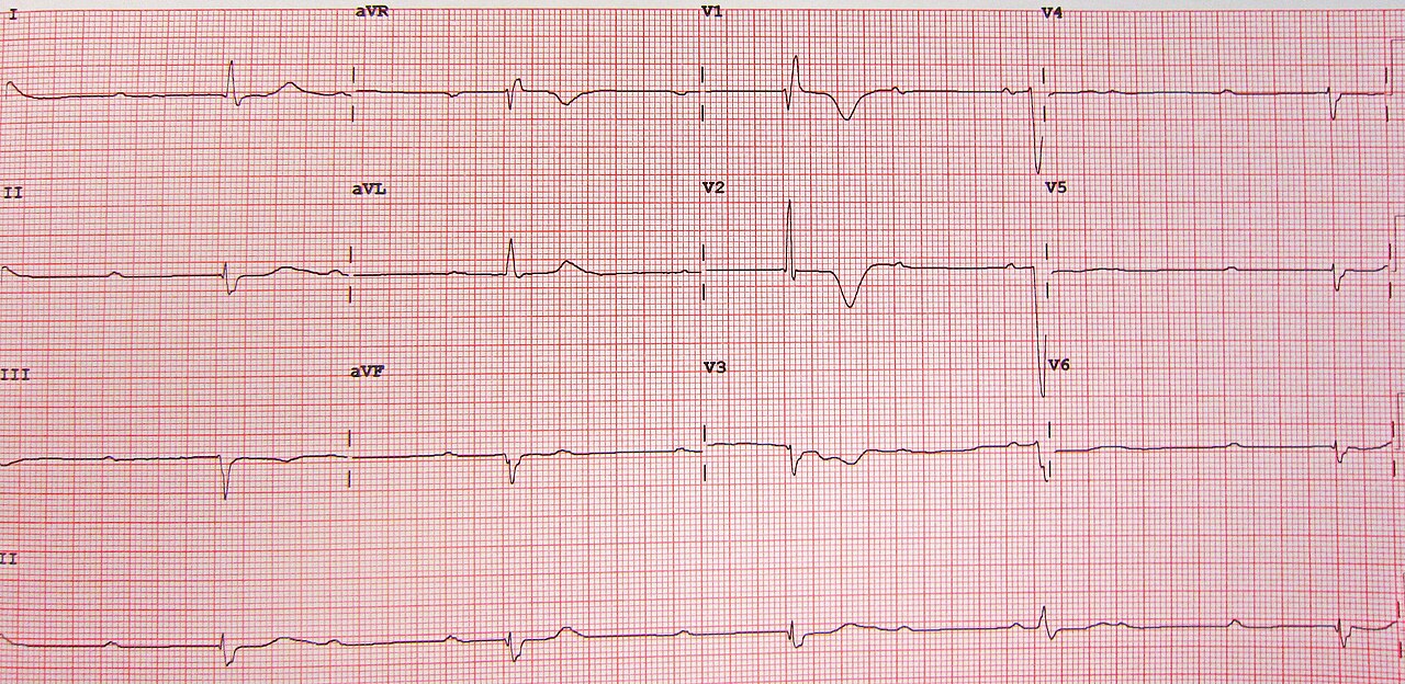
Nothing from the atria gets through in complete AV block. Each chamber keeps its own timing. The ventricles run on an escape rhythm generated below the block.
ECG Criteria
- AV dissociation: P waves and QRS complexes occur independently; the PR interval has no consistent value
- Atrial rate: Regular P-P intervals, usually at the sinus rate
- Ventricular rate: Regular R-R intervals, slower than the atrial rate
- QRS: Narrow with a junctional escape, or wide with a ventricular escape
Junctional vs. Ventricular Escape
| Feature | Junctional Escape (Nodal Block) | Ventricular Escape (Infranodal Block) |
|---|---|---|
| QRS width | Narrow (under 120 ms) | Wide (120 ms or more) |
| Escape rate | 40–60 bpm | 20–40 bpm |
| Reliability | More reliable | Less reliable; risk of asystole |
| Typical setting | Inferior MI, drugs, transient block | Anterior MI, degenerative disease |
| Atropine response | Sometimes improves the rate | Often ineffective |
| Prognosis | Often transient | Often persistent; higher risk |
Causes
- Acute inferior MI (usually nodal, often transient) or anterior MI (infranodal, serious)
- Drugs (for example beta-blockers, calcium channel blockers, digoxin), electrolyte disturbance, Lyme carditis
- Degenerative disease, congenital block, infiltrative disease, cardiac surgery
Clinical Significance
- An anterior MI that causes complete block has done extensive septal damage, and mortality is high. Immediate pacing and urgent cardiology review are both needed
- In inferior MI, complete block is often nodal. Recovery within days is possible
- When acquired complete block refuses to resolve, it needs a permanent pacemaker evaluation
Bundle Branch Blocks (RBBB, LBBB and IVCD)
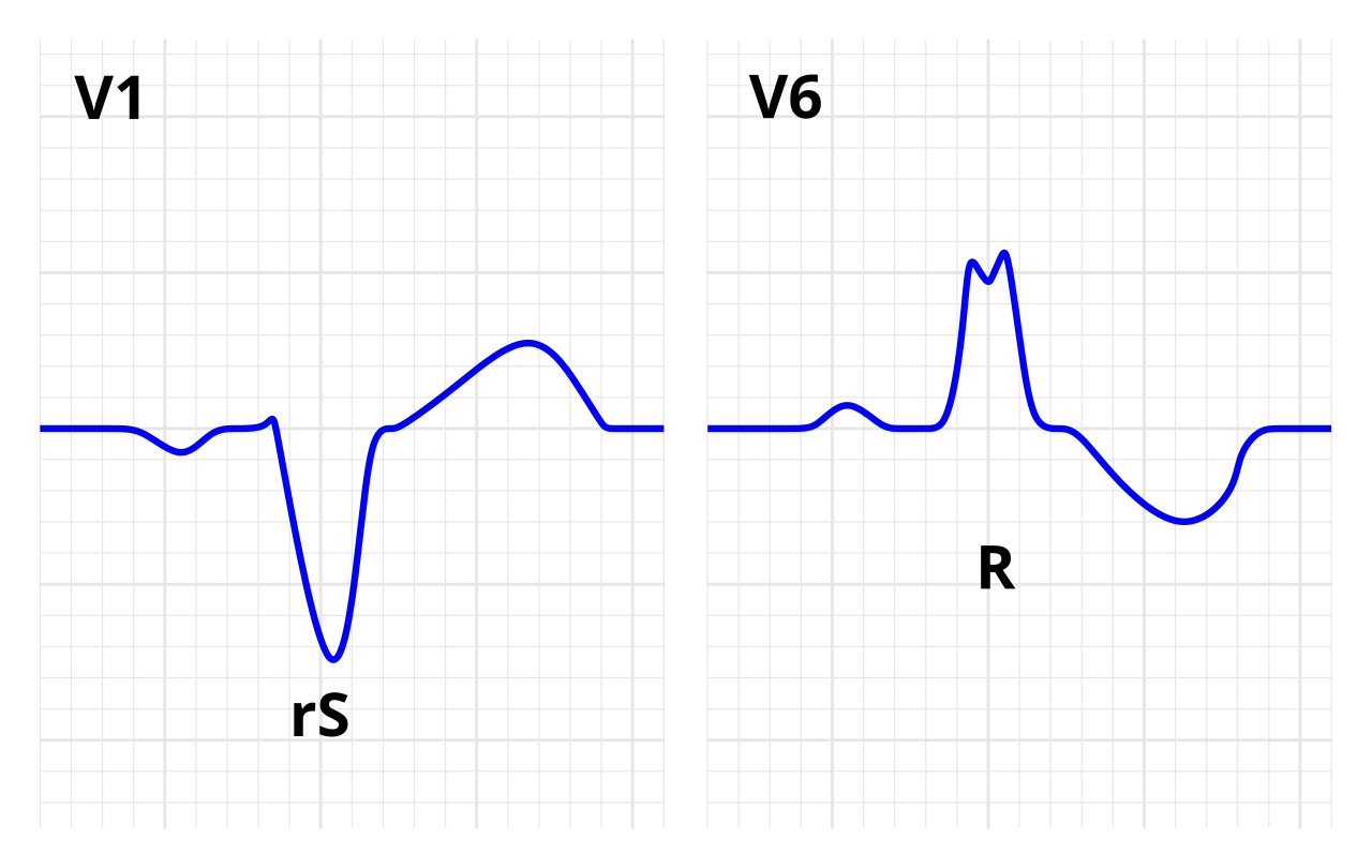
One ventricle gets its signal late. That delay widens the QRS, and the ST-T waves swing the other way. The full criteria, lead patterns and mnemonic live on the QRS Complex page. Here you get just enough criteria to classify a block.
ECG Criteria
- RBBB: QRS 120 ms or more (complete), 110–119 ms (incomplete); rsR′ in V1–V2 and a wide, slurred S wave in I, V5–V6
- LBBB: QRS 120 ms or more; V1 QS or rS, broad notched R in I, V5–V6, no septal Q waves
- Strict LBBB (Strauss criteria, 2011): QRS 140 ms or more in men or 130 ms or more in women, with mid-QRS notching or slurring in 2 or more contiguous leads
- Nonspecific IVCD: QRS 110 ms or more without a complete RBBB or LBBB pattern
Clinical Significance
- Alone, RBBB can be a normal variant. A new one can mean right heart strain, anterior MI or myocarditis
- Nearly every LBBB has structural heart disease behind it. Hypertensive or ischemic heart disease, cardiomyopathy and aortic valve disease are the usual suspects. It also masks ischemia on the ECG
- Rate-related BBB: Aberrant conduction that appears only at fast rates or after a long pause (Ashman phenomenon). It is not a permanent block. Compare it with a slower rhythm
Look-Alikes
- Wide-complex tachycardia with aberrancy (see Differential Diagnosis)
Fascicular, Bifascicular and Trifascicular Block
The left bundle branches into two fascicles. The anterior one is thin and relies mainly on the LAD. The posterior one is thick and has dual LAD and RCA supply. Block either one and the frontal-plane axis shifts, without major QRS widening.
Left Anterior Fascicular Block (LAFB)
- Axis: −45° to −90°; QRS under 120 ms
- Pattern: qR in I and aVL; rS in II, III and aVF
- Significance: The one you will see most often. Frequently benign, but it points to underlying disease
Left Posterior Fascicular Block (LPFB)
- Axis: More than +90°; QRS under 120 ms
- Pattern: rS in I and aVL; qR in III and aVF
- Significance: Rare. You arrive here only after ruling out RVH, pulmonary embolism and lateral MI (see Cardiac Axis)
Bifascicular and Trifascicular Block
- Bifascicular: RBBB plus LAFB (the most common combination) or RBBB plus LPFB
- Trifascicular: bifascicular block plus first-degree AV block. The third pathway is in danger, but that does not mean it is blocked
Differential Diagnosis & Mimics
Separate the common block patterns by PR interval, QRS width and escape rhythm. These features matter more than how many P waves you see on one strip.
| Feature | Mobitz I (Wenckebach) | Mobitz II | 2:1 AV Block |
|---|---|---|---|
| PR before the drop | Lengthens progressively | Constant | Cannot be determined from one conducted beat |
| QRS width | Usually narrow | Often wide | Narrow or wide; check the strip |
| Site | AV node | His–Purkinje system | Node or infranodal |
| Escape and risk | Reliable; low risk | Unreliable; high risk | Depends on the site |
| Bedside direction | Monitor; atropine if symptomatic | Pacing evaluation | Pace if wide QRS or unstable |
- LAFB vs. inferior MI: LAFB gives a left axis without inferior ST elevation. Inferior MI gives inferior ST changes with reciprocal changes in aVL. Lean on the 12-lead and serial ECGs.
Artifact & Lead Misplacement
- Limb lead reversal: A reversed pair of arm electrodes (for example RA and LA) inverts lead I. That can mimic an axis shift or a block pattern
- Precordial misplacement: V1 or V2 placed too high can produce an rSR′ pattern. That mimics RBBB
- Tremor, shivering or transport vibration: They can hide P waves and mimic a dropped beat. Check a second lead before you call a block
Normal Variants
- Isolated RBBB and first-degree AV block in young and athletic patients
- Wenckebach pattern during sleep or with high vagal tone
Bedside Management & Escalation
Doses below come from the 2025 AHA adult bradycardia algorithm (symptomatic bradycardia with a pulse) and the 2025 PALS pediatric bradycardia algorithm, and match the E3 atropine, epinephrine and digoxin-immune-fab pages. Follow local protocols and medical direction.
| Situation | BLS (EMT) | ALS (AEMT / Paramedic / RN) | Critical Care / Transport |
|---|---|---|---|
| Unstable (hypotension, altered mental status, ischemic chest pain, shock, heart failure) | Oxygen if SpO₂ under 94%; monitor; 12-lead if available; call ALS early | Atropine 1 mg IV/IO; apply pacing pads now; start transcutaneous pacing early in acute MI or if atropine fails | Transcutaneous or transvenous pacing; dopamine or epinephrine infusion as a bridge; notify cardiology |
| Stable (no symptoms or minor symptoms) | Monitor; 12-lead; transport | Monitor; 12-lead; check drugs and electrolytes; Mobitz I often needs only monitoring. Apply pacing pads early for Mobitz II, high-grade or complete block, which can deteriorate suddenly | Monitor; cardiology review; plan for pacing if the block worsens |
| Pulseless | CPR and AED per the BLS sequence | Cardiac arrest algorithm; epinephrine; treat reversible causes | Arrest algorithm; pacing does not replace compressions |
| Therapy | Adult | Pediatric | Key Notes |
|---|---|---|---|
| Atropine | 1 mg IV/IO, every 3–5 min; max 3 mg total |
0.02 mg/kg IV/IO, minimum 0.1 mg, maximum 0.5 mg per dose; may repeat once. For bradycardia with compromise from increased vagal tone or AV block, not from hypoxia (2025 AHA Part 8, COR 1, LOE C-LD) |
Adult first drug for symptomatic bradycardia with a pulse (2025 AHA Part 9: reasonable with hemodynamic compromise, COR 2a, LOE B-NR). Unreliable in infranodal block; do not delay pacing while waiting for a response. Raises heart rate and myocardial oxygen demand, so in acute MI favor early pacing over repeated doses. Pediatric: not first-line (see Pediatrics). |
| Transcutaneous pacing | Pacing rate 60–80/min per protocol; start at the lowest output and increase until electrical, then mechanical, capture; add the safety margin set by the device or protocol (often about 10% above threshold). Provide analgesia or sedation per protocol for the awake patient |
No sourced pediatric pacing parameters exist (AHA 2025 Part 8, rec 4, COR 2b, C-EO: emergency transcutaneous pacing may be considered for complete block or sinus node dysfunction unresponsive to CPR). Use smaller pads and the pediatric protocol |
Confirm mechanical capture: a pulse with each paced beat. An ECG spike without a pulse is not capture. |
| Epinephrine | 2–10 mcg/min IV infusion, titrated to response |
0.01 mg/kg (0.1 mL/kg of 0.1 mg/mL) IV/IO every 3–5 min, max 1 mg per dose; no sourced pediatric infusion dose exists (the 2025 PALS bradycardia algorithm gives none), so use the pediatric protocol and medical direction. First-line drug for pediatric bradycardia with poor perfusion that persists after oxygenation, ventilation and correction of other causes (2025 AHA Part 8, COR 1, LOE C-EO; see Pediatrics) |
High-alert Infusion is a bridge to pacing, used when atropine is ineffective (2025 AHA Part 9, COR 2b, LOE C-LD). Check mcg/min versus mcg/kg/min on the pump. |
| Dopamine | 5–20 mcg/kg/min IV infusion, titrated to heart rate and blood pressure |
No sourced pediatric infusion dose exists (the 2025 PALS bradycardia algorithm gives none); use the pediatric protocol and medical direction |
High-alert Infusion only, used when atropine is ineffective (2025 AHA Part 9, COR 2b, LOE C-LD). Confirm mcg/kg/min and the bag concentration. |
- Hyperkalemia: peaked T waves, a wide QRS and flattened or absent P waves. Give calcium gluconate 10% 30 mL (3 g) IV over 10 min for ECG changes, or calcium chloride 10% 10 mL (1 g) over 5 min in peri-arrest or with central access; repeat calcium if ECG changes persist. Then insulin 10 units IV mixed with dextrose 25 g, given over 5–15 min into a large vein; add D10 at 50 mL/hr for 5 hr if pre-dose glucose is under 126 mg/dL, and check glucose frequently for at least 6 hr after insulin. Outside arrest, nebulized albuterol 10–20 mg is an adjunct, never monotherapy; sodium bicarbonate is not routine outside arrest. Differs from FDA label: the gluconate label limits the rate to 200 mg/min (3 g over at least 15 min), and the chloride label limits it to 1 mL/min; the faster infusions here are used for ECG changes (UKKA 2026, Guidelines 16.2a–b, 16.3 and 16.4). In hyperkalemic cardiac arrest, AHA 2025 rates calcium, sodium bicarbonate and insulin-glucose 2b (effectiveness not well established) and inhaled albuterol 3: No Benefit. Check the potassium.
- Beta-blocker or calcium channel blocker toxicity: atropine is not in the AHA 2025 recommendations for this poisoning (standard ALS applies). Vasopressors are COR 1 for hypotension. High-dose insulin is COR 1 (refractory to vasopressors in beta-blocker poisoning; for hypotension in CCB poisoning). Glucagon is 2a for symptomatic bradycardia or hypotension in beta-blocker poisoning, and 2b (uncertain) for CCB poisoning. Calcium is 2b in beta-blocker and 2a in CCB poisoning. Call the poison center or medical direction early.
- Digoxin toxicity: digoxin-specific Fab is COR 1, LOE B-NR for life-threatening poisoning (AHA 2025 Part 10). Arrest or arrest imminent: 10 vials IV push, repeat 10 if no response. Toxicity without arrest: 1–2 vials IV, titrated to response. Differs from FDA label: the label gives a calculated dose; the titrated 1–2 vial dose is used outside arrest. Dialysis is not recommended (COR 3: No Benefit).
- Other causes to correct: hypothyroidism, hypothermia, hypoxia, and increased intracranial pressure (the Cushing response with hypertension).
Follow local protocols and online medical direction.
Pediatric & Special Populations
Pediatrics
- Heart rate: Age-based norms are on the Vital Signs page. In PALS, you treat bradycardia when the heart rate is under 60/min with poor perfusion despite oxygenation and ventilation. If it stays under 60/min with poor perfusion despite that support, start CPR
- Usual cause: Hypoxia or increased vagal tone. Support oxygenation and ventilation first. If the heart rate stays under 60/min with poor perfusion, start CPR. Then give epinephrine 0.01 mg/kg IV/IO every 3–5 min as the first drug (see the table above). Atropine (0.02 mg/kg) is for bradycardia with compromise from increased vagal tone or AV conduction block not caused by hypoxia (2025 AHA Part 8, COR 1). Pacing is considered for block that does not respond to drugs
- Congenital heart disease: After surgery for congenital heart disease, complete block can follow. Sometimes epicardial pacing wires are in place. Check wire capture and sensing with each change
- Infants: The PR interval is shorter in infants. Do not apply adult PR thresholds without age context
- Dosing: Weight-based doses come from the Weight-Based Dosing page
Other Populations
- Pregnancy: Check electrolytes and drug effects first. Magnesium is on that list, because it can cause bradycardia and AV block. For pacing, follow the obstetric protocol
- Older adults: Degenerative conduction disease is the most common cause of new block. Symptoms can be subtle
- Athletes: First-degree AV block and Wenckebach can be normal in athletes. Assess for symptoms
- Heart transplant recipients: The sinus node is denervated. That makes atropine ineffective, and the heart rate is often fixed. If the patient is unstable, pacing is the bedside option
- Pacemaker or ICD patients: Confirm the pacing mode. A missing paced beat can look like block. Check for a pacing spike before each QRS
Monitoring & Transport Considerations
| Issue | What to Do |
|---|---|
| Monitoring lead | Lead II to see P waves; V1 or MCL1 for bundle branch and QRS shape |
| Serial 12-leads | Repeat every 15–30 min during ongoing chest pain or after any new block |
| Right-sided and posterior leads | Add V4R and V7–V9 when inferior ischemia, RV involvement or posterior involvement is suspected |
| Artifact | Vibration, shivering and movement can hide P waves; check a second lead before calling a block |
| Pacing pads | Apply anterior–posterior pads before moving an unstable bradycardic patient |
| Pacing capture | Check the pulse and the SpO₂ waveform with every paced beat; recheck after every move |
| Transport | Hand off the rhythm, pacing settings and drug doses; recheck the pulse and capture after each transfer |
Step-by-Step Approach & Pitfalls
- Decide whether the patient is stable or unstable before reading the strip in detail.
- Measure the rate and check whether the P-P and R-R intervals are regular.
- Confirm each QRS has a P wave in front of it, and find the P waves that do not conduct.
- Measure the PR before each conducted beat and look for lengthening before the dropped beat.
- Measure the QRS width in the widest lead.
- Name the block, its site, and the escape rhythm and rate.
- Act: treat an unstable patient per the table above, apply pads for unstable high-grade block, and notify cardiology for Mobitz II or complete block.
Common Pitfalls
- Calling a 2:1 block Wenckebach or Mobitz II from one conducted beat
- Waiting for atropine to work when an infranodal block needs pacing
- Giving lidocaine or another antiarrhythmic to a patient whose wide escape rhythm is keeping them alive. Lidocaine can suppress that escape. The patient then drops into asystole
- Treating electrical capture on the monitor as successful pacing when there is no pulse
- Missing a sinus pause or exit block that has no P wave in front of the pause
- Discharging a patient with Mobitz II or complete heart block without a pacing plan
ECG Examples
Clinical Pearls
References
- Kusumoto FM, Schoenfeld MH, Barrett C, et al. 2018 ACC/AHA/HRS guideline on the evaluation and management of patients with bradycardia and cardiac conduction delay. Circulation. 2019;140(8):e382-e482. https://doi.org/10.1161/CIR.0000000000000628
- Wigginton JG, Agarwal S, Bartos JA, et al. Part 9: Adult advanced life support: 2025 American Heart Association guidelines for cardiopulmonary resuscitation and emergency cardiovascular care. Circulation. 2025;152(16_suppl_2). https://doi.org/10.1161/CIR.0000000000001376 (Initial Management of Bradycardia: atropine 2a, B-NR; epinephrine or dopamine 2b, C-LD; transvenous pacing 2a, C-LD)
- Lasa JJ, Dhillon GS, et al. Part 8: Pediatric advanced life support: 2025 American Heart Association and American Academy of Pediatrics guidelines for cardiopulmonary resuscitation and emergency cardiovascular care. Circulation. 2025;152(16_suppl_2). https://doi.org/10.1161/CIR.0000000000001368 (Management of Bradycardia: atropine 1, C-LD for vagal or AV-block bradycardia not due to hypoxia; epinephrine 1, C-EO after correction of other factors; transcutaneous pacing 2b, C-EO)
- Sgarbossa EB, Pinski SL, Barbagelata A, et al. Electrocardiographic diagnosis of evolving acute myocardial infarction in the presence of left bundle-branch block. N Engl J Med. 1996;334:481-487. (landmark derivation study) https://doi.org/10.1056/NEJM199602223340801
- Smith SW, Dodd KW, Henry TD, et al. Diagnosis of ST-elevation myocardial infarction in the presence of left bundle branch block with the ST-elevation to S-wave ratio in a modified Sgarbossa rule. Ann Emerg Med. 2012;60(6):766-776. (landmark derivation study) https://doi.org/10.1016/j.annemergmed.2012.07.119
- Strauss DG, Selvester RH, Wagner GS. Defining left bundle branch block in the era of cardiac resynchronization therapy. Am J Cardiol. 2011;107:927-934. (review proposing criteria) https://doi.org/10.1016/j.amjcard.2010.11.010
- Lantos PM, Rumbaugh J, Bockenstedt LK, et al. Clinical practice guidelines by the Infectious Diseases Society of America (IDSA), American Academy of Neurology (AAN), and American College of Rheumatology (ACR): 2020 guidelines for the prevention, diagnosis and treatment of Lyme disease. Clin Infect Dis. 2021;72(1):e1-e48. https://doi.org/10.1093/cid/ciaa1215
- American Heart Association. 2025 American Heart Association Guidelines for CPR and ECC: Adult Bradycardia With a Pulse Algorithm and Pediatric Bradycardia With a Pulse Algorithm, Doses and Details sidebars. 2025. https://cpr.heart.org/en/resuscitation-science/cpr-and-ecc-guidelines/algorithms
- Cao D, Arens AM, Chow SL, et al. Part 10: Adult and pediatric special circumstances of resuscitation: 2025 American Heart Association Guidelines for Cardiopulmonary Resuscitation and Emergency Cardiovascular Care. Circulation. 2025;152(suppl 2):S578–S672. (Hyperkalemia, beta-blocker, calcium channel blocker and digoxin poisoning recommendations) https://doi.org/10.1161/CIR.0000000000001380
- Alfonzo A, Harrison A, Baines R, Chu A, Mann S, MacRury M. UK Kidney Association Clinical Practice Guideline: Treatment of Acute Hyperkalaemia in Adults. Updated version, July 2026 (running title: Management of Hyperkalaemia in Adults). Guidelines 16.2a–b (calcium), 16.3 (insulin-glucose) and 16.4 (albuterol) for the non-arrest regimen. UK Kidney Association. 2026. https://www.ukkidney.org/sites/default/files/documents/FINAL%20VERSION%20-%20UKKA%20CLINICAL%20PRACTICE%20GUIDELINE%20-%20MANAGEMENT%20OF%20HYPERKALAEMIA%20IN%20ADULTS%20-%20UPDATED%20JULY%202026_0.pdf
Medical Disclaimer
- For Educational Purposes Only: This content is intended for educational reference and should not be used for clinical decision-making.
- Not a Substitute for Professional Judgment: Always consult your local protocols, institutional guidelines, and supervising physicians.
- Verify Before Acting: Users are responsible for verifying information through authoritative sources before any clinical application.