

At a Glance
- Represents
- Atrial depolarization: right atrial activation, then left, from the sinus node. Seen as the P wave before each QRS.
- Normal (Adult)
- <120 ms (under 3 small boxes) and <2.5 mm in limb leads. Upright in I, II, aVF; inverted in aVR.
- Measure It
- Duration and height in lead II (at standard 25 mm/s and 10 mm/mV: 1 small box = 40 ms; 10 mm = 1 mV). Read V1 for the right and left atrial parts.
- Abnormal → Think
- Tall P in II, III, aVF: right atrial enlargement
- Wide, notched P in II: left atrial enlargement
- Negative or changing P shapes: ectopic or multifocal atrial rhythm
- Red Flags
- Sawtooth flutter waves (250–350/min), or an irregular rhythm with no P waves (AF).
- Bedside Action
- Name the rhythm the P wave belongs to and treat it per Atrial Rhythms. Repeat the 12-lead if the rhythm changes.
Physiology & What It Represents
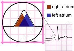
The P wave is the surface record of atrial depolarization. Think of it as two activations stacked into one wave. The impulse fires in the sinoatrial (SA) node, a group of pacemaker cells where the superior vena cava meets the high right atrium. It sweeps across the right atrium, then crosses to the left through interatrial pathways. Its shape shows the size and timing of both atria.
Next, the impulse hits the AV node and slows down. Why? The delay gives the atria time to finish filling the ventricles before the ventricles squeeze. Then it runs the His–Purkinje system and produces the QRS. P wave, PR interval, QRS. Those are three stops on one conduction path.
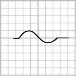
Lead choice changes what you see. Lead II sits close to the normal atrial axis, so both activations add up into one upright deflection. V1 sits over the right atrium and near the left. There, the right activation shows as a small positive deflection and the left as a negative one. Want each atrium on its own? Look at V1.
Normal Values & How to Measure
| Parameter | Adult Normal | Pediatric Note | Where / How to Measure |
|---|---|---|---|
| P duration | <120 ms (0.12 s; under 3 small boxes) | Age-specific; see Pediatrics & Special Populations | Lead II; from P onset to return to baseline |
| P height | <2.5 mm (0.25 mV) in limb leads | Age-specific; see Pediatrics & Special Populations | Lead II; from baseline to peak |
| P axis | 0° to +75° | Age-specific; see Pediatrics & Special Populations | Upright in I and II; inverted in aVR |
| V1 initial positive part | ≤1.5 mm (right atrial component) | Age-specific; see Pediatrics & Special Populations | Lead V1; height of the first positive deflection |
| V1 terminal negative part | <1 mm deep or <40 ms wide | Age-specific; see Pediatrics & Special Populations | Lead V1; depth (mm) and width (ms) of the negative part |
- Pick the lead: use lead II on a rhythm strip of at least 6 seconds for duration and height, and lead V1 for the right and left atrial parts.
- Find onset and end: the P wave starts where the isoelectric baseline (the TP segment) leaves the line and ends where it returns.
- Measure duration: confirm standard calibration first (paper speed 25 mm/s, 10 mm/mV). At 25 mm/s one small box is 40 ms. Count boxes or use calipers, and compare with the 120 ms limit.
- Measure height: count from the baseline to the peak in mm. At 10 mm/mV, ten mm equals 1 mV.
- Read V1 separately: measure the first positive deflection (height) and the terminal negative part (depth in mm and width in ms).
- Check several beats: in an irregular rhythm, measure the widest and tallest P waves, not a single beat.
- Look again before calling it absent: check V1 and the longest strip. A P wave can hide in the T wave or the QRS.
Right Atrial Enlargement (P Pulmonale)
Right atrial enlargement (RAE) shows up as a P-wave pattern, usually after a long pressure or volume load on the right heart. The ECG name is P pulmonale. Why does it look like this? The right atrial part gets taller, so the P wave climbs tall in the inferior leads and the initial positive part in V1 grows. The P duration usually stays normal.
ECG Criteria
- Limb leads: P ≥2.5 mm in II, III or aVF (tall, peaked P)
- Lead V1: initial positive part of the P wave >1.5 mm
- Duration: normal (<120 ms). A wide P wave points to left atrial enlargement or another cause.
Causes
- Pulmonary hypertension of any cause
- Chronic obstructive pulmonary disease (COPD)
- Pulmonary embolism, acute or chronic
- Tricuspid valve disease or pulmonic stenosis
- Congenital heart disease with a right-sided volume or pressure load
Clinical Significance
- The P pattern marks right atrial load. It is not a diagnosis by itself. Pull up a prior ECG if you have one and compare.
- New dyspnea, tachycardia or low SpO₂? Read the P pattern against the whole picture, and keep pulmonary embolism on the list.
Left Atrial Enlargement (P Mitrale)
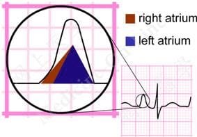
Left atrial enlargement (LAE) is a P-wave pattern from a left atrium under raised pressure or volume. Mitral valve disease, hypertension with left ventricular disease and heart failure are the usual suspects. The ECG name is P mitrale. The left atrium fires last, so a big one shows up late in the P. Expect a wide, notched P in the limb leads and a deep, wide negative part in V1.
ECG Criteria
- Lead II: P wave ≥120 ms and notched (bifid)
- Lead V1: terminal negative part ≥1 mm deep and ≥40 ms wide (P terminal force ≥0.04 mm·s)
Causes
- Mitral stenosis or mitral regurgitation
- Systemic hypertension with left ventricular disease
- Heart failure or cardiomyopathy
- Other conditions that raise left atrial pressure or volume
Clinical Significance
- LAE reflects chronic left atrial load. It is linked to atrial fibrillation risk.
- The ECG shows the electrical pattern and nothing more. Echo measures the left atrium's size and finds the valve or ventricular cause.
Ectopic and Retrograde P Waves
An ectopic P wave starts somewhere other than the sinus node. So its shape, axis or PR interval looks different from a sinus beat. A retrograde P wave is atrial activation running backward, up from the AV junction or an accessory pathway. Look for it negative in II, III and aVF. It can land before, inside or after the QRS.
| Finding | Likely Rhythm | What to Check |
|---|---|---|
| P inverted in II, III, aVF; PR shorter than sinus | Low atrial rhythm | Confirm the inversion on several beats; compare with a prior ECG |
| Negative P in II, III, aVF before, within or after the QRS | Junctional rhythm (retrograde atrial activation) | A P after the QRS suggests the AV junction is driving the rhythm |
| Negative P in II, III, aVF just after the QRS (or pseudo-r′ in V1) in a regular narrow tachycardia | AVNRT or AVRT (also junctional tachycardia) | The retrograde P may hide in the QRS; check V1 and II for pseudo-r′ or pseudo-S, and confirm the rate is regular |
| Three or more P-wave shapes | Wandering atrial pacemaker (rate below 100/min) or multifocal atrial tachycardia (above 100/min) | Count the shapes across the strip; the rate decides the label |
| Sawtooth flutter waves | Atrial flutter | Regular waves in II, III, aVF or V1; typical 2:1 conduction near 150/min |
| No organized P wave, irregular R–R | Atrial fibrillation | Fibrillatory baseline with irregular ventricular response |
Premature atrial complexes (PACs) are ectopic P waves that arrive early with a different shape. Management, plus the junctional and multifocal rhythms, lives on the Atrial Rhythms page. Watch for this: a very early PAC may not conduct to the ventricles.
Sinus Arrhythmia and Wandering Atrial Pacemaker
Sinus Arrhythmia
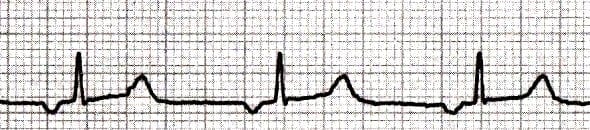
- Rhythm: The R–R intervals move with breathing. They usually shorten on inspiration and lengthen on expiration.
- P waves: sinus in shape, upright in I, II and aVF, inverted in aVR, with a constant PR interval.
- Significance: You see it often in kids, young adults and athletes. With sinus P waves, it is normal and needs no treatment.
Wandering Atrial Pacemaker
- P waves: Three or more shapes and PR intervals. The pacemaker keeps hopping between the sinus node and atrial sites.
- Rate: Usually below 100/min. Cross 100/min and the same pattern becomes multifocal atrial tachycardia. The Atrial Rhythms page covers it.
Absent P Waves (Flutter and Fibrillatory Waves)
No discrete, sinus-type P wave means the atria are not firing that way. The usual culprits are atrial fibrillation, atrial flutter (flutter waves take the P's place) and junctional rhythm. Sinus arrest and sinoatrial exit block also leave no P during the pause. Hyperkalemia can flatten the P until it vanishes. Two things sort these out: how regular the R–R intervals are, and the atrial rate.
- Atrial fibrillation: irregular R–R with fibrillatory waves at 350–600/min and no discrete P waves.
- Atrial flutter: regular sawtooth flutter waves at an atrial rate of 250–350/min. At 2:1 conduction the ventricular rate lands near 150/min. The flutter waves can hide in the T wave or QRS.
- Junctional rhythm: narrow QRS, and no P wave or a retrograde one. Escape rate typically sits at 40–60/min.
- Sinus arrest or sinoatrial exit block: a pause with no P wave. Sinus P waves on both sides of the pause confirm it. In exit block, the pause is a multiple of the usual P–P interval.
- Hyperkalemia: The P flattens, widens and can vanish (sinoventricular rhythm). Peaked T waves, a wide QRS and bradycardia usually come with it. Consider it in any regular wide-complex bradycardia with no P waves, then follow local protocol and medical direction.
Telling AF from flutter changes the anticoagulation and cardioversion decisions (AF guidance: references 5 and 6). The Atrial Rhythms page sets those out.
Differential Diagnosis & Mimics
The table lines up the atrial enlargement patterns so you can compare them at a glance. Each row maps to the criteria in Normal Values & How to Measure and the sections above.
| Feature | Right Atrial Enlargement | Left Atrial Enlargement | Normal Sinus P |
|---|---|---|---|
| Lead II height | ≥2.5 mm, tall and peaked | Usually normal | <2.5 mm |
| Lead II duration and shape | Normal duration | ≥120 ms and notched | <120 ms, smooth |
| Lead V1 initial part | >1.5 mm positive | Normal or small | ≤1.5 mm |
| Lead V1 terminal part | Normal | ≥1 mm deep and ≥40 ms wide | Less than 1 mm deep or 40 ms wide |
| Typical settings | Pulmonary disease, pulmonary hypertension | Mitral valve disease, hypertension, heart failure | Normal findings |
Artifact & Lead Misplacement
- Limb lead reversal: Swap the arm electrodes and the P wave and QRS turn negative in lead I. Check the placement before you read an inverted P in lead I as a non-sinus rhythm.
- Precordial misplacement: V1 sitting high on the chest warps the P-wave shape. Put V1 in the fourth intercostal space at the right sternal border.
- Tremor or motion: Shivering can fake fibrillatory waves. Tremor shifts from beat to beat. Flutter waves stay regular in rate and shape.
- Transport vibration: The vehicle's motion can draw a fibrillatory-looking baseline. Re-check the strip once it stops.
Bedside Management & Escalation
P-wave findings rarely need treatment on their own. The rhythm they belong to, and the patient's status, drive what you do next.
- Stable, sinus P waves: document the finding and continue care. Repeat the ECG if symptoms change.
- Atrial enlargement with symptoms: obtain a 12-lead ECG, check SpO₂ and work of breathing, and report the finding with the history.
- Flutter, AF or junctional rhythm: follow the Atrial Rhythms page for stability assessment, rate or rhythm therapy and notification.
- Flat or absent P waves with a wide QRS and bradycardia: consider hyperkalemia (renal failure, dialysis, crush injury) and follow local hyperkalemia protocol. Obtain a 12-lead ECG and a rhythm strip.
- Hospital work-up: Serial 12-leads, electrolytes (K⁺, Mg²⁺) and an echocardiogram are the usual next steps. They happen in hospital, not on the truck.
Follow local protocols and medical direction.
Pediatric & Special Populations
Pediatrics
- Age-specific limits: P-wave duration and height norms shift with age. Use age-specific limits (see Rijnbeek et al., reference 4). The RAE and LAE thresholds above are adult values. Don't apply them to a child.
- Sinus arrhythmia: Kids have it often, and it is usually normal. No treatment needed.
- Rate context: Read the P wave alongside the child's heart rate. The Pediatric Vital Signs page lists the age ranges.
- Congenital heart disease: A sustained atrial volume load, as with an atrial septal defect, can produce atrial enlargement patterns.
Other Populations
- Athletes: Sinus bradycardia and sinus arrhythmia show up often. Both are usually normal.
- Pregnancy: Sinus tachycardia is common, and the P wave usually keeps its sinus shape. A new atrial pattern or an irregular rhythm still means hunting for causes, pulmonary embolism included.
- Older adults: AF gets more common with age. An irregular rhythm with no P waves should send you looking for it.
- Pacemaker: A paced atrial beat shows a pacing spike, then a P wave. A spike with no P wave behind it points to a capture problem. Follow the device protocol.
- Heart transplant: The sinus node is denervated. The heart rate reacts slowly to demand, and the resting rate often runs faster than normal. You may see two P waves (recipient atrial remnant and donor atrium). Usually only the donor P waves conduct. Atropine does not work here. See ECG Rate.
Monitoring & Transport Considerations
- Monitoring lead: On most monitors, lead II shows the P waves and rhythm best. Switch to V1 when the atrial pattern matters.
- Electrode check: Confirm limb placement before you call an inverted P in lead I or a new axis change. Reversed leads can invert the P in lead I.
- Repeat the 12-lead: Do it when the rhythm changes, when flutter or AF starts or stops, or when the patient changes. Record the time.
- Artifact: Vibration, shivering and movement can fake fibrillatory waves. Stop or re-check the strip before you treat.
- Handoff: Send a 12-lead and a labelled rhythm strip. Include the time, the rate and the P-wave finding.
Step-by-Step Approach & Pitfalls
- Find the P wave: start with lead II, then check V1. Confirm a P wave precedes each QRS.
- Check the shape: a sinus P is smooth, upright in I, II and aVF, and inverted in aVR.
- Measure duration and height: compare with the limits of under 120 ms and under 2.5 mm.
- Read V1: look for a larger initial positive part (right atrial) or a deep, wide negative part (left atrial).
- Check P to QRS: each P should conduct, and the PR interval should be constant unless the rhythm varies.
- Name the rhythm: sinus with variation, ectopic, flutter, AF or junctional, using the Atrial Rhythms page.
Common Pitfalls
- Calling the P wave absent when it hides in the T wave or QRS.
- Missing 2:1 flutter near 150/min and calling it sinus tachycardia.
- Reading an inverted P in lead I before checking limb lead placement.
- Applying adult atrial criteria to children.
- Measuring a single beat in an irregular rhythm.
- Calling RAE or LAE from one ECG, with no clinical picture behind it.
Clinical Pearls
References
- Kligfield P, Gettes LS, Bailey JJ, et al. Recommendations for the standardization and interpretation of the electrocardiogram: part I: the electrocardiogram and its technology. J Am Coll Cardiol. 2007;49:1109–1127. https://doi.org/10.1016/j.jacc.2007.01.024
- Hancock EW, Deal BJ, Mirvis DM, et al. AHA/ACCF/HRS recommendations for the standardization and interpretation of the electrocardiogram: part V: electrocardiogram changes associated with cardiac chamber hypertrophy. J Am Coll Cardiol. 2009;53:992–1002. https://doi.org/10.1016/j.jacc.2008.12.015
- Morris JJ Jr, Estes EH Jr, Whalen RE, et al. P-wave analysis in valvular heart disease. Circulation. 1964;29:242–252 (landmark derivation paper). https://doi.org/10.1161/01.cir.29.2.242
- Rijnbeek PR, Witsenburg M, Schrama E, et al. New normal limits for the paediatric electrocardiogram. Eur Heart J. 2001;22:702–711 (landmark derivation paper). https://doi.org/10.1053/euhj.2000.2399
- Joglar JA, Chung MK, Armbruster AL, et al. 2023 ACC/AHA/ACCP/HRS Guideline for the Diagnosis and Management of Atrial Fibrillation. Circulation. 2024;149:e1–e156. https://doi.org/10.1161/CIR.0000000000001193
- Van Gelder IC, Rienstra M, Bunting KV, et al. 2024 ESC Guidelines for the management of atrial fibrillation developed in collaboration with EACTS. Eur Heart J. 2024;45:3314–3414. https://doi.org/10.1093/eurheartj/ehae176
Medical Disclaimer
- For Educational Purposes Only: This content is intended for educational reference and should not be used for clinical decision-making.
- Not a Substitute for Professional Judgment: Always consult your local protocols, institutional guidelines, and supervising physicians.
- Verify Before Acting: Users are responsible for verifying information through authoritative sources before any clinical application.