

At a Glance
- Represents
- AV-node and His–Purkinje conduction, with almost no surface voltage. A shift from the TP baseline reflects an atrial injury current.
- Normal (Adult)
- Isoelectric, level with the TP baseline. Not measured separately; it is part of the PR interval.
- Measure It
- PR segment (end of P wave to QRS onset) against the TP baseline in lead II and aVR. Use calipers and several beats.
- Abnormal → Think
- PR depression with diffuse ST elevation: pericarditis. PR elevation in aVR with depression elsewhere: pericarditis pattern. Regional change with inferior MI: possible atrial infarction.
- Red Flags
- Hypotension with low QRS voltage or electrical alternans (tamponade), or regional ST elevation (STEMI). Atrial injury raises the risk of atrial arrhythmia, AV block and thromboembolism.
- Bedside Action
- Compare with prior ECGs, repeat the 12-lead and notify the provider. Watch the rhythm for AF or flutter. Follow local protocols and medical direction.
Physiology & What It Represents
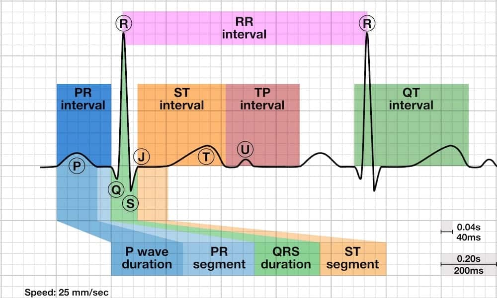
The PR segment is the flat stretch between the end of the P wave and the start of the QRS complex. The P wave is the atria firing. The impulse then slows in the AV node and runs down the bundle of His and the bundle branches. Those tissues carry very little muscle, so the surface ECG records almost nothing there. The trace stays quiet until the ventricles start to depolarize. Atrial repolarization is normally buried inside the QRS.
- Conduction path: SA node, then atria (P wave), then AV node and His–Purkinje system (PR segment), then ventricles (QRS).
- Why it can shift: pericardial inflammation or atrial infarction injures the atrial surface. That injury current moves the baseline during this interval. It is the atrial counterpart of ST-segment deviation.
- Why lead choice matters: aVR looks at the heart from the right shoulder. Its deflections often run opposite to the limb and precordial leads.
Normal Values & How to Measure
| Parameter | Adult Normal | Pediatric Note | Where / How to Measure |
|---|---|---|---|
| PR segment level | Isoelectric, level with the TP baseline | No age-specific PR-segment norms found in the sources reviewed; compare with the child’s own TP baseline | End of P wave to QRS onset; lead II and aVR first |
| TP baseline | End of T wave to start of next P wave | At higher heart rates the TP segment can disappear | Same lead as the PR segment |
| PR interval (context) | 120–200 ms (0.12–0.20 s; 3–5 small boxes) | See the PR Interval page | P wave onset to QRS onset; includes the P wave |
| Deviation | Absent on a normal ECG; reported as present or absent | Same description | Confirm in several leads and beats |
- Choose the leads: start with lead II and the other limb leads, where atrial activity is most visible. Check aVR for PR elevation.
- Find the baseline: use the TP segment (end of the T wave to the start of the next P wave) in the same lead.
- Mark the segment: measure from the end of the P wave to the QRS onset. Do not include the P wave.
- Compare levels: decide whether the PR segment sits above, below or level with the TP baseline.
- Check several leads and beats: a shift in one lead or one beat is weak evidence. Look for a consistent pattern across leads II, III, aVF and V4–V6.
- Measure small deviations: use calipers or a straight edge against the TP level. One millimeter equals 0.1 mV.
PR Segment Depression
Definition
PR depression means the PR segment drops below the TP baseline. It's the atrial version of ST depression.
ECG Criteria
- Level: the PR segment sits under the TP baseline in the same lead. Usually it shows in several leads at once.
- Distribution: in one myopericarditis series, lead II showed the most PR depression (55.9%). Lead aVL showed the least (2.9%).
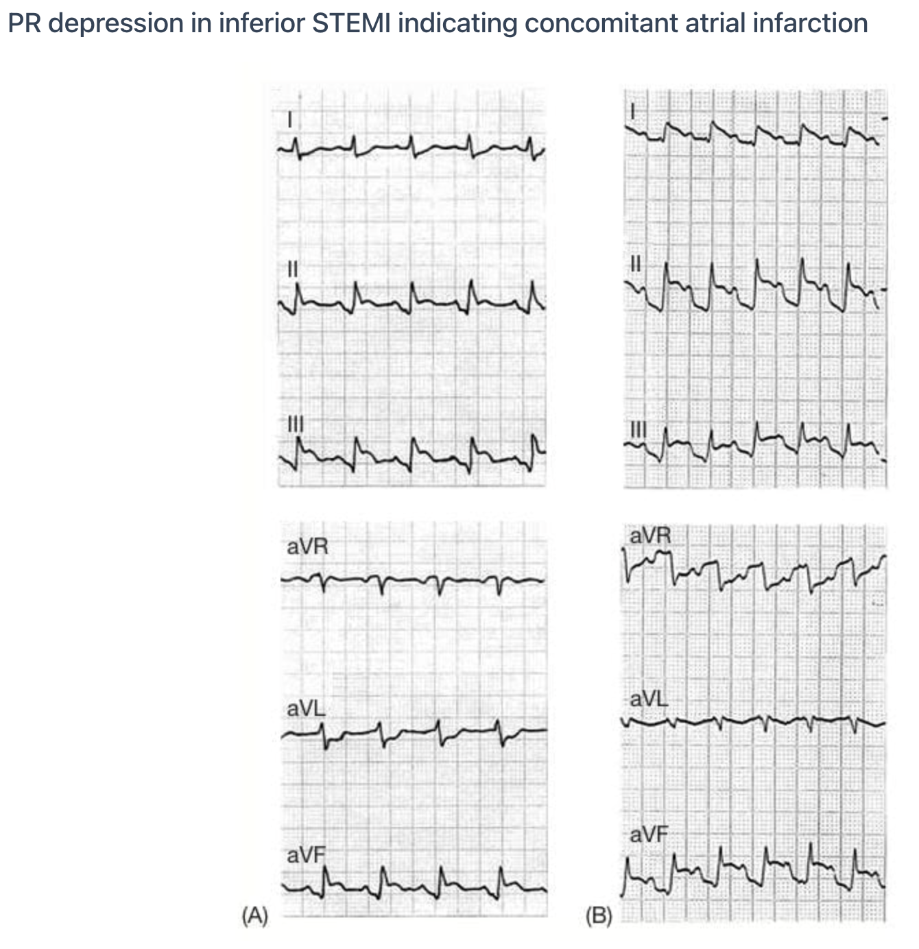
- Pattern: depression across many limb and precordial leads points to pericarditis. A regional version next to an inferior or posterior STEMI can mean atrial infarction.
Causes
| Category | Examples | Notes |
|---|---|---|
| Acute pericarditis | Viral or idiopathic; after myocardial infarction; after cardiac surgery; kidney failure | Diffuse depression across leads |
| Atrial infarction | Ischemia of atrial muscle, usually with an inferior, posterior or right ventricular STEMI | Often regional; see Atrial Infarction below |
| Other pericardial disease | Effusion, constriction, tamponade | Associated with PR depression in one cardiac-surgery series |
| Direct atrial injury | Trauma or surgery involving the atria | Pattern varies by cause |
Acute Pericarditis
PR depression turns up often in the acute phase of pericarditis. Not every patient has it. And a normal PR segment does not rule pericarditis out.
The 2025 ESC guideline groups PR depression with widespread ST elevation as one ECG criterion. It sits among the additional criteria, alongside pericardial rub, raised C-reactive protein and imaging findings. Calling it definite takes a typical clinical presentation plus more than one additional criterion.
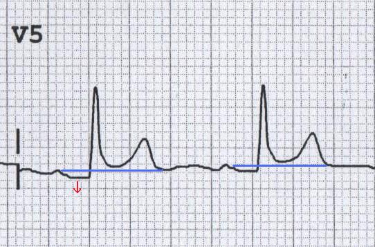
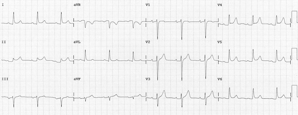
One emergency series enrolled 34 patients with myopericarditis and 46 with STEMI. PR depression in any lead caught myopericarditis with 88.2% sensitivity and 78.3% specificity. Depression in both limb and precordial leads carried a positive predictive value of 96.7% and a negative predictive value of 90%.
Clinical Significance
- Diagnostic: Diffuse ST elevation with PR depression leans toward pericarditis over STEMI. Regional ST elevation keeps STEMI on the list, whatever the PR segment shows.
- Prognostic: in one cardiac-surgery series of pericardial disease, PR depression went with arrhythmias. It also went with a poor composite outcome (death, rehospitalization or heart failure) in 30.6% of patients. Without it, that figure was 7%.
PR Segment Elevation
Definition
PR elevation means the PR segment rides above the TP baseline. You see it less often than depression. Its real teaching value is the reciprocal finding in pericarditis. aVR shows elevation while most other leads show depression.
ECG Criteria
- Lead aVR: Elevation in aVR with depression in most other leads is the pericarditis pattern.
- Other leads: PR elevation alone outside aVR has no defined meaning in the sources reviewed. Read it against the full 12-lead and the clinical picture.
After STEMI
One cohort of 224 STEMI patients had PR displacement in any lead in 31% of cases, and that displacement independently predicted 1-year mortality. Displacement in aVR or aVL was among the findings tied to 1-year mortality.
Atrial Infarction
Atrial infarction is ischemic injury to the atrial muscle. You rarely get it alone. It almost always comes with a ventricular infarction. There are no universally accepted diagnostic criteria.
ECG Criteria
- PR displacement: showed up in 20.5% of atrial infarctions in a 39-patient inferior STEMI series. A 2025 series of 270 patients with atrial-branch involvement found it in about 65%. Its absence does not rule atrial infarction out.
- P-wave duration: a P-wave duration of 95.5 ms or more in lead II separated atrial infarction in the 39-patient series, with 90% sensitivity and 94% specificity. One small study. Not a validated rule.
- Atrial arrhythmias: atrial tachyarrhythmia hit 55–67% of patients with atrial infarction in the 2025 series. The figure depended on the infarct artery.
Causes
- Atherosclerotic occlusion of atrial branches, most often of the right coronary artery. In the 2025 series, the culprit was the right coronary artery in 69% of atrial infarctions. The left circumflex was the culprit in 31%.
- Less often, cor pulmonale and pulmonary hypertension have also been named as causes.
Clinical Significance
- Arrhythmia and clot risk: atrial tachyarrhythmias, thromboembolism, hemodynamic instability and, rarely, atrial rupture all tie back to atrial infarction.
- Mortality: among 224 STEMI patients, PR displacement in any lead tracked with 1-year mortality. That held whether or not atrial infarction was diagnosed.
Look-Alikes
- Pericarditis also produces PR depression, though with diffuse ST elevation. See Differential Diagnosis & Mimics.
Differential Diagnosis & Mimics
In young adults, early repolarization is a common ST-elevation mimic. The ST Segment & Ischemia page sets it beside STEMI and pericarditis.
| Feature | Acute Pericarditis | Regional STEMI | Atrial Infarction |
|---|---|---|---|
| ST elevation | Diffuse, across several territories | Regional, one coronary territory | Occurs with an inferior, posterior or right ventricular STEMI |
| PR segment | Depressed in most leads; aVR may be elevated | Less often depressed than in pericarditis; PR displacement was seen in roughly 20–30% of STEMI patients in the cited series, so it does not exclude STEMI | Displaced in about 20% to 65% of cases in published series |
| Atrial arrhythmia | Possible | Possible | Common in published series (more than half) |
Artifact & Lead Misplacement
- Tachycardia: The P waves run into the T wave, and the PR segment becomes unreadable.
- Baseline wander and tremor: can make the PR level look wrong. Confirm across several beats.
- Limb-lead reversal: flips the polarity of P waves and QRS in the affected leads. Look at lead I and aVR morphology before you read any PR change there.
- Vibration in transport: ambulance motion can imitate a PR or ST change. Reassess once the patient is still.
Bedside Management & Escalation
A PR-segment change is not a diagnosis. Report it, trend it, and treat the cause. Read the pattern against symptoms, the rest of the 12-lead and the patient’s hemodynamics.
- Diffuse ST elevation with PR depression and no regional territory: that is the pericarditis pattern. Tell the treating provider. Shoot another 12-lead and draw troponin as ordered.
- Regional ST elevation that meets STEMI criteria: activate the STEMI pathway per local protocol. The PR finding tells you the patient is higher risk. The pathway stays the same.
- PR change with inferior or posterior MI: the atria may be injured too. Watch for AF or flutter, AV block and hemodynamic change. Tell the receiving team.
- Hypotension with low QRS voltage or electrical alternans: think tamponade and escalate without delay. PR depression won't show you tamponade.
What comes next: serial 12-leads, plus right-sided or posterior leads when the territory is unclear. Electrolytes and echocardiography follow as the provider directs. Follow local protocols and medical direction.
Pediatric & Special Populations
Pediatrics
- Heart rate: a child's normal rate runs faster, so P waves may overlap the T wave. Run a longer rhythm strip. Age-based vital signs are on Vital Signs by Age.
- Norms: the sources reviewed had no age-specific PR-segment reference values. Judge any deviation against the child’s own TP baseline and heart rate.
- Symptoms: a child with chest pain and a new PR or ST change gets the same ECG workup as an adult. Bring the pediatric team in early.
Other Populations
- Early and late post-MI pericarditis: early pericarditis shows up within days of infarction. Late (Dressler) pericarditis shows up weeks later. PR changes can come with either one.
- After cardiac surgery: post-pericardiotomy pericarditis is an inflammatory pericardial syndrome. One cardiac-surgery series of pericardial disease reported PR depression.
- Kidney failure: uremic and dialysis-related pericarditis are both known causes of pericardial inflammation. Ask when dialysis happened and whether any sessions were missed.
Monitoring & Transport Considerations
| Issue | What to Do |
|---|---|
| Best monitoring lead | Lead II for PR and P-wave review, where PR depression was most often seen in one series. Use aVR for PR elevation and V4–V6 when a diffuse precordial pattern is suspected. |
| Repeat 12-leads | Repeat with each change in chest pain, rhythm or hemodynamics. Use the interval set by local protocol. |
| Atrial rhythm | Keep a rhythm strip running. Atrial injury is linked to AF, flutter and AV block. |
| Transmission | Send the 12-lead where local protocol allows so the receiving team can compare it with prior tracings. |
| Hypotension | If hypotension appears with low QRS voltage or electrical alternans, consider tamponade and escalate. |
| Motion artifact | Ambulance motion, shivering or tremor can mimic PR or ST change. Confirm the finding when the patient is still. |
Step-by-Step Approach & Pitfalls
- Find the TP baseline in the lead you are reading: end of the T wave to the next P wave.
- Mark the PR segment from the end of the P wave to the QRS onset.
- Compare its level with the TP baseline in leads II, aVR and V4–V6.
- Check several leads and beats to confirm the deviation is consistent.
- Decide diffuse or regional: diffuse depression with PR elevation in aVR favors pericarditis. Regional change with inferior or posterior ST elevation raises atrial infarction.
- Read the ST segments and T waves against STEMI criteria. Activate the STEMI pathway when criteria are met.
- Review P-wave duration, morphology and the rhythm strip for evidence of atrial injury.
- Correlate with symptoms, troponin, prior ECGs and hemodynamics before deciding what the PR finding means.
Common Pitfalls
- Confusing PR interval with PR segment: the interval takes in the P wave. The segment doesn't.
- Using the wrong baseline: anchor to the TP segment, not the ST segment or T wave.
- Missing PR change at fast rates: P waves run into T waves, and the segment gets hard to read.
- Calling pericarditis from PR depression alone: regional ST elevation still needs the STEMI pathway, whatever the PR segment shows.
- Treating one lead or one beat as proof: check that it holds across leads and beats.
- Forgetting atrial infarction: it can sit alongside a STEMI, and it brings arrhythmia and clot risk with it.
Clinical Pearls
References
- Schulz-Menger J, et al. 2025 ESC Guidelines for the management of myocarditis and pericarditis. Eur Heart J. 2025;46:3952–4041. https://doi.org/10.1093/eurheartj/ehaf192
- Rao SV, et al. 2025 ACC/AHA/ACEP/NAEMSP/SCAI Guideline for the Management of Patients With Acute Coronary Syndromes. Circulation. 2025;151:e771–e862. https://doi.org/10.1161/CIR.0000000000001309
- Di C, Wang H, Wang M, Wang Q, Wu Y, Li L, et al. Acute atrial infarction: a relatively neglected and under-recognized entity in clinical practice. Herz. 2025;50(2):122–134. https://doi.org/10.1007/s00059-024-05272-z
- Lu ML, De Venecia T, Patnaik S, Figueredo VM. Atrial myocardial infarction: A tale of the forgotten chamber. Int J Cardiol. 2016;202:904–909. https://doi.org/10.1016/j.ijcard.2015.10.070
- Lu ML, Nwakile C, Bhalla V, De Venecia T, Shah M, Figueredo VM. Prognostic significance of abnormal P wave morphology and PR-segment displacement after ST-elevation myocardial infarction. Int J Cardiol. 2015;197:216–221. https://doi.org/10.1016/j.ijcard.2015.06.055
- Porela P, Kytö V, Nikus K, Eskola M, Airaksinen KE. PR depression is useful in the differential diagnosis of myopericarditis and ST elevation myocardial infarction. Ann Noninvasive Electrocardiol. 2012;17(2):141–145. https://doi.org/10.1111/j.1542-474X.2012.00489.x
- Yıldız SS, Keskin K, Avsar M, Cetinkal G, Sigirci S, Aksan G, et al. Electrocardiographic diagnosis of atrial infarction in patients with acute inferior ST-segment elevation myocardial infarction. Clin Cardiol. 2018;41(7):972–977. https://doi.org/10.1002/clc.22987
- Kudaiberdiev T, Dzhumagulova A, Joshibayev S, Tilemanbetova K, Imanalieva G. Electrocardiographic abnormalities in patients with pericardial disease: association of PR segment depression with arrhythmias and clinical signs. J Electrocardiol. 2016;49(1):29–36. https://doi.org/10.1016/j.jelectrocard.2015.10.007
Medical Disclaimer
- For Educational Purposes Only: This content is intended for educational reference and should not be used for clinical decision-making.
- Not a Substitute for Professional Judgment: Always consult your local protocols, institutional guidelines, and supervising physicians.
- Verify Before Acting: Users are responsible for verifying information through authoritative sources before any clinical application.