

At a Glance
- Covers
- AVNRT; orthodromic and antidromic AVRT with the accessory pathway; regular narrow-complex tachycardia; the RP interval and adenosine as separators
- Recognize It
- Regular, narrow, abrupt start: AVNRT or AVRT
- P wave well after the QRS (long RP): not typical AVNRT
- Regular and wide: VT until proven otherwise
- Most Dangerous
- Pre-excited AF: irregular, very fast, wide QRS that varies in shape. AV nodal blockers can precipitate VF.
- Unstable Patient
- Synchronized cardioversion now at the device's recommended energy (maximum setting if unknown). Sedate only if it will not delay the shock. Pediatric: 0.5–1 J/kg, then 2 J/kg.
- Stable Patient
- Modified Valsalva, then adenosine 6 mg, then 12 mg (not in asthma or bronchospasm). If it fails: diltiazem, verapamil or metoprolol for regular narrow SVT. Pre-excited AF: procainamide.
- Do Not
- No AV nodal blocker (adenosine, verapamil, diltiazem, metoprolol, digoxin, amiodarone) in an irregular wide rhythm or known pre-excited AF. Regular wide: VT until proven otherwise. No adenosine in asthma or bronchospasm (contraindicated).
Physiology & What It Represents
The AV node is the only normal route from the atria to the ventricles. Narrow-complex tachycardia needs one of two things: a re-entrant circuit or an atrial focus. The ventricles still fire through the normal His–Purkinje system, which is why the QRS stays narrow. Re-entry is a loop with two limbs, one slow and one fast. Add a timing condition that lets the impulse keep circling, and the loop runs.
Three mechanisms explain most regular narrow-complex tachycardias on this page:
- AV nodal re-entry (AVNRT): the circuit sits inside the AV node, usually between a slow and a fast pathway. Atria and ventricles both activate through the same node. That is why the retrograde P wave often hides in the QRS.
- AV re-entry (AVRT): the circuit runs through the AV node and an accessory pathway. That pathway is a strand of conducting tissue outside the node, and it can conduct in one direction or both.
- Atrial tachycardia: a focus in the atria fires fast and needs no AV circuit. The Focal Atrial Tachycardia section below covers it. Flutter and atrial fibrillation run on their own mechanisms, and Atrial Rhythms teaches those.
AVNRT and AVRT are paroxysmal. They start and stop all at once.
Recognition Approach
Every narrow or wide tachycardia gets the same six questions. The table on this page sorts each rhythm with its first-line action. Stability sets the first move. No table replaces the bedside check.
| Rhythm | Rate (bpm) | Regularity | P Waves | PR | QRS | First-Line Action |
|---|---|---|---|---|---|---|
| Sinus tachycardia | Gradual change | Regular | Normal, before each QRS | Normal | Narrow | Treat the cause; rate control rarely needed |
| AVNRT | 150–250 | Very regular | Usually hidden in the QRS; pseudo-r′ in V1 in some | Not measurable | Narrow | Vagal maneuver, then adenosine |
| Orthodromic AVRT | 150–250 | Very regular | Retrograde P after the QRS | Not measurable | Narrow | Vagal maneuver, then adenosine |
| Antidromic AVRT | 150–250 | Regular | Variable; may be hidden | Not measurable | Wide; mimics VT | Treat as VT; no AV nodal blockers |
| Focal atrial tachycardia | Often above 100 | Regular | Abnormal shape; often long RP | Not applicable | Narrow | Treat the cause; stable: IV diltiazem or verapamil (COR 2a) or a beta-blocker; unstable: synchronized cardioversion; see Focal Atrial Tachycardia |
| Pre-excited AF | Often very fast | Irregular | Fibrillatory; no organized P waves | Not applicable | Wide, shape varies | Cardiovert if unstable; procainamide if stable |
| Atrial flutter | About 150 at 2:1 (atrial 250–350) | Regular or variable | Sawtooth flutter waves | Not applicable | Narrow | Rate control if stable; cardioversion if unstable |
- Find the rate: Count the R waves over 6 seconds and multiply by 10. The usual range for these re-entrant tachycardias in adults and older children is 150–250 bpm; infants run faster (see Pediatric & Special Populations).
- Check regularity: Very regular points to AVNRT, AVRT, focal atrial tachycardia or flutter with fixed block. Irregularly irregular means atrial fibrillation, multifocal atrial tachycardia or pre-excited AF.
- Look for P waves: Look in lead II and V1. Where a Lewis lead is available, it shows retrograde P waves more often than standard leads.
- Measure the RP interval: Time it from the R wave to the next P wave. A short RP is the usual pattern for typical AVNRT. A long RP, with the P wave well after the QRS, is not typical AVNRT.
- Measure the QRS: Narrow is supraventricular unless aberrancy is proven. Wide is VT, antidromic AVRT or pre-excited AF until proven otherwise.
- Assess stability first: Hypotension, altered mentation, ischemic chest pain or acute heart failure makes the patient unstable. That patient needs cardioversion now.
Normal Conduction Values
| Parameter | Adult Normal | Note |
|---|---|---|
| PR interval | 120–200 ms (0.12–0.20 s; 3–5 small boxes) | Manifest pre-excitation: PR ≤120 ms, a delta wave and QRS over 120 ms |
| QRS duration | ≤110 ms (≤2.75 small boxes) | 120 ms or more is wide |
| SVT usual rate | 150–250 bpm (adult and older child) | Infants: SVT is usually at or above 220 bpm; sinus tachycardia is usually below 220. A fixed, regular rate near 150 bpm: consider 2:1 flutter. |
AV Nodal Re-entrant Tachycardia (AVNRT)
AVNRT is a paroxysmal re-entrant tachycardia that stays inside the AV node. Along with orthodromic AVRT, it is one of the most frequent kinds of paroxysmal SVT. Expect it in adolescents and adults. It is less common in young children.
ECG Criteria
- QRS: Narrow, under 120 ms, unless there is a bundle branch block or rate-related aberrancy.
- Rhythm: Fast and very regular.
- P waves: The retrograde P often hides inside the QRS. A pseudo-r′ in V1 or a pseudo-S in the inferior leads supports AVNRT when you see it. Don't rule AVNRT out because they are missing. In one study they were present in only 37% and 20% of AVNRT patients.
- Onset and offset: Abrupt. The rhythm starts and stops suddenly.
Causes
- Substrate: Dual AV nodal physiology is the setup. A slow limb and a fast limb sit inside the node, and together they allow the re-entrant loop.
- Age: Less common in young children; more frequent after age 10.
Clinical Significance
- Symptoms: Expect palpitations, lightheadedness and chest discomfort. A rate this fast can drop the blood pressure and bring on angina.
- Treatment: Vagal maneuvers and adenosine can end it. AV nodal blockers can too, because the node is part of the circuit. For recurrent episodes, catheter ablation is the treatment. The 2025 pediatric review lists radiofrequency or cryoablation as first-line.
Look-Alikes
- Orthodromic AVRT: the RP interval separates the two (see the RP section below).
- Sinus tachycardia: see Differential Diagnosis & Mimics.
Atrioventricular Re-entrant Tachycardia (AVRT)
AVRT is a re-entrant circuit built from the AV node and an accessory pathway, a strand of conducting tissue outside the node. Orthodromic AVRT has a narrow QRS, and it is one of the most common paroxysmal SVTs. Antidromic AVRT has a wide QRS and can pass for VT.
ECG Criteria
- Orthodromic (narrow): Anterograde conduction runs down the AV node and retrograde conduction climbs the accessory pathway. The QRS is narrow. The retrograde P follows the QRS, and the RP interval is usually longer than in typical AVNRT.
- Antidromic (wide): Conduction runs down the accessory pathway and back up the AV node. The QRS is wide, so it mimics VT.
- Rate: Usually 150–250 bpm.
- Sinus rhythm clue: In sinus rhythm, a manifest pathway shows PR ≤120 ms, a delta wave and a QRS over 120 ms. The full WPW pattern is taught on PR Interval.
Causes
- Accessory pathway: The pathway is the substrate. A concealed pathway conducts only backward, so it shows no pre-excitation in sinus rhythm.
- Wolff-Parkinson-White (WPW): In fetal life and the first year of life, about 80% of SVTs are linked to WPW. In children older than 10, that falls to about 65%.
Clinical Significance
- The dangerous forms: Antidromic AVRT gets mistaken for VT. An AV nodal blocker given in pre-excited AF can drive the patient into VF.
- Definitive treatment: Ablation of the accessory pathway is the definitive treatment. The 2023 AF guideline recommends it for pre-excited AF. The 2025 pediatric review lists radiofrequency or cryoablation as first-line for AVRT.
Focal Atrial Tachycardia
Focal atrial tachycardia is a fast-firing focus in the atria that uses no AV circuit. It is usually regular. When the P waves are visible, it is a common look-alike for AVNRT. The 2015 ACC/AHA/HRS SVT guideline (References) covers the adult approach.
ECG Criteria
- P-wave morphology: The P wave looks different from a sinus P wave. Its shape and axis reflect where the focus sits. Compare it with a sinus P wave on an earlier strip.
- RP interval: Often long, with the P wave falling well after the QRS. That is the main reason to suspect atrial tachycardia over typical AVNRT.
- Onset: The rate often ramps up gradually and winds down at the end. AVNRT and AVRT start abruptly.
- AV block: 2:1 or other AV block can appear while the atrial rate keeps going.
Adenosine Response
- Adenosine can cause a transient AV block that unmasks the atrial rate. The atrial tachycardia usually keeps going, so an unchanged rhythm after adenosine does not rule out a focus.
- Most focal atrial tachycardias continue, with transient AV block. A minority are adenosine-sensitive and terminate. Termination alone does not fully rule out an atrial focus.
Regular and Irregular Atrial Rhythms
- Regular: focal atrial tachycardia, AVNRT, AVRT and atrial flutter.
- Irregular: multifocal atrial tachycardia (MAT), with at least three P-wave morphologies and a rate usually of 100 bpm or more; wandering atrial pacemaker (WAP), slower than 100 bpm with varying P-wave morphology.
Treat the cause. In stable, regular narrow-complex tachycardia, IV diltiazem or verapamil (COR 2a, LOE B-R) can be effective. IV beta-blockers have also been shown to terminate it. If the patient is unstable, use synchronized cardioversion (2025 AHA Part 9). See Bedside Management & Escalation.
Regular Narrow-Complex Tachycardia and the RP Interval
Most regular narrow-complex tachycardias sort into three groups: typical AVNRT, AVRT and an atrial source. At the bedside, the RP interval and the response to adenosine help separate them. Pinning down the final mechanism may need an electrophysiology study.
RP Interval
- Typical AVNRT: Its RP interval is generally shorter than in AVRT.
- AVRT or focal atrial tachycardia (see its section): A P wave well after the QRS, a long RP, is not typical AVNRT.
- Limit: P waves are hard to see inside a narrow QRS. In one study, a Lewis lead, a bipolar chest lead, showed retrograde P waves in 70% (21 of 30) of AVNRT patients.
Adenosine as a Diagnostic Tool
- Mechanism: Adenosine slows conduction through the AV node. It can interrupt AV-node re-entry, so it treats the circuit and tests it at the same time.
- Termination: An abrupt stop points to an AV-node-dependent circuit. Adenosine does not convert atrial flutter or atrial fibrillation.
- Slowing: In flutter or fibrillation, adenosine can slow the ventricular response a little, and only for a moment. That slowing can unmask flutter waves or AF. Use it to reveal the rhythm, not to treat it.
Differential Diagnosis & Mimics
Compare three things: the rhythm before the tachycardia, the QRS width and the response to a vagal maneuver or adenosine.
| Feature | Typical AVNRT | Orthodromic AVRT | Sinus Tachycardia |
|---|---|---|---|
| Onset and offset | Abrupt | Abrupt | Gradual change |
| QRS | Narrow | Narrow | Narrow |
| P wave | Usually hidden in the QRS | Retrograde P after the QRS | Normal P before each QRS |
| RP interval | Short (typical) | Longer than typical AVNRT | Not applicable |
Artifact & Lead Misplacement
- Motion and shivering can mimic a fast regular rhythm or flutter waves on the monitor. Confirm it on a 12-lead.
- Limb lead reversal can invert the P wave in lead I and shift the apparent P axis. A sinus rhythm can then look atrial.
- Transport vibration adds noise that can look like a regular tachycardia. Pause or steady the patient before you judge the rhythm.
Wide and regular is VT until proven otherwise. For the full picture, see Ventricular Rhythms.
Bedside Management & Escalation
Stability comes first. An unstable patient gets synchronized cardioversion first, and drugs do not delay the shock. The 2025 AHA tachyarrhythmia algorithm (Box 6) also lists adenosine for a regular narrow-complex rhythm. A stable patient with a narrow, regular tachycardia gets a vagal maneuver, then adenosine. Where 2025 AHA states a dose, the doses below follow it. Verapamil, diltiazem and metoprolol doses come from the E3 medication pages.
Stability Approach
| Situation | BLS (EMT) | ALS (AEMT / Paramedic / RN) | Critical Care / Transport |
|---|---|---|---|
| Unstable, narrow or wide with a pulse | Oxygen if SpO₂ <94%; monitor; call ALS and do not delay transport. Pads and CPR only if pulseless. | Synchronized cardioversion now (2025 AHA Part 9, COR 1, LOE B-NR); sedate only if it will not delay the shock. Send a 12-lead ECG if available. | Synchronized cardioversion; anterior-posterior pads; continuous monitoring; notify the receiving team (Paramedic/RN/CCT). |
| Stable, regular, narrow (AVNRT, AVRT) | Notify ALS; help with a vagal maneuver if trained. | Modified Valsalva, then adenosine. If it fails, diltiazem, verapamil or metoprolol (no pre-excitation; no decompensated heart failure). Diltiazem or verapamil: 2025 AHA Part 9, COR 2a, LOE B-R. | Same drug choices; repeat the 12-lead; watch for hypotension after diltiazem or verapamil. |
| Stable, irregular, wide (possible pre-excited AF) | Notify ALS. Give no drugs. | Procainamide if stable. Cardiovert if unstable or if drugs fail. | Defibrillator at bedside; avoid AV nodal blockers; expert consultation. |
| Stable, regular, wide (treat as VT) | Notify ALS. Give no AV nodal blockers. | Treat as VT (VT drugs such as procainamide or amiodarone; synchronized cardioversion if unstable). See Ventricular Rhythms. | Defibrillator at bedside; expert consultation; avoid verapamil and diltiazem. |
Therapy Table
| Therapy | Adult | Pediatric | Key Notes |
|---|---|---|---|
| Modified Valsalva | Strain 15 s at about 40 mm Hg, then lie back with the legs raised (passive leg raise) | Age-appropriate technique; vagal stimulation first (2025 AHA Part 8, COR 1, LOE C-LD). In infants, cold-water face immersion is one option; AHA notes success rates for each maneuver are unknown | In a randomized trial, 43% converted with the modified maneuver vs 17% with the standard one. Use before adenosine when stable. |
| Adenosine (adenosine) | Adenosine is Class 1, LOE B-R (2025 AHA Part 9, rec 2). Doses (2025 AHA ACLS tachyarrhythmia algorithm sidebar): 6 mg rapid IV/IO push, then a rapid saline flush; 12 mg if no conversion in 1–2 min. The label allows one more 12 mg. Max 12 mg per dose. | 0.1 mg/kg rapid IV/IO (max 6 mg), then 0.2 mg/kg (max 12 mg) if needed (2025 AHA PALS tachyarrhythmia algorithm; Part 8 SVT rec 2, COR 1, LOE C-LD). Differs from FDA label: the label starts at 0.05–0.1 mg/kg for children under 50 kg. | High-alert Contraindicated in second- or third-degree AV block and in sick sinus syndrome without a pacemaker. Contraindicated in asthma or active bronchospasm: adenosine can cause severe bronchospasm (2025 AHA Part 9). Differs from FDA label: the PSVT label places asthma under Warnings, not Contraindications. Can provoke atrial fibrillation or flutter; AF can conduct rapidly if an accessory pathway is present. Carbamazepine can increase the degree of AV block. Caffeine and theophylline blunt it; dipyridamole potentiates it. Central line or post-transplant: adenosine may have profound effects, and 1 mg IV may be adequate to terminate the rhythm (2025 AHA Part 9). Defibrillator ready. 2025 AHA Part 9 (COR 2b, LOE B-NR) allows adenosine for a stable, regular, monomorphic wide-complex tachycardia when the cause cannot be determined; it is COR 3: Harm (LOE C-LD) for unstable, irregular or polymorphic wide-complex tachycardia. This page does not teach it for stable wide rhythms. |
| Verapamil (verapamil) | 5–10 mg IV over at least 2 min (at least 3 min in older patients); repeat 10 mg after 30 min if needed. Max 10 mg per dose. | Avoid in infants: severe, sometimes fatal, hemodynamic reactions reported after IV doses in neonates and infants. Older children only with pediatric cardiology guidance. | High-alert Avoid with hypotension, moderate or severe LV systolic dysfunction (2023 AF guideline: Class 3, Harm), sick sinus syndrome, AV block without a pacemaker, or a recent IV beta-blocker. Never for wide-complex tachycardia (2025 AHA Part 9: COR 3: Harm, B-NR) or pre-excited AF. |
| Diltiazem (diltiazem) | 0.25 mg/kg IV over 2 min (about 20 mg for an average adult); at 15 min if needed, 0.35 mg/kg (about 25 mg) over 2 min | Not established in children; specialist use only | High-alert Same cautions as verapamil. The bolus is mg/kg and the infusion is mg/hr; never mix the two. Do not repeat into hypotension. |
| Metoprolol (metoprolol) | 2.5–5 mg IV over 2 min, up to 3 doses (max 15 mg) | Not established for IV use in children | High-alert Avoid with SBP <100 mm Hg, decompensated heart failure or sick sinus syndrome without a pacemaker. Not for pre-excited AF. |
| Procainamide (procainamide) | Stable pre-excited AF: 20–50 mg/min IV until the arrhythmia stops, BP falls, QRS widens by more than 50%, or 17 mg/kg is given (2025 AHA tachyarrhythmia algorithm sidebar) | Specialist use only; see the procainamide page | High-alert Boxed warning: positive ANA and lupus-like syndrome, blood dyscrasias, and CAST mortality in structural heart disease or after MI. Avoid with prolonged QT, heart failure or complete heart block. The 2023 AF guideline recommends IV procainamide or ibutilide as an alternative to elective cardioversion in stable pre-excited AF. |
| Synchronized cardioversion (cardioversion) | Narrow regular: the device's recommended synchronized energy; if unknown, the maximum setting (2025 AHA tachyarrhythmia algorithm). The 2025 electrical cardioversion algorithm lists 100 J for narrow-complex tachycardia. Pre-excited AF or unstable wide rhythm: synchronized shock (see the cardioversion page). | 0.5–1 J/kg, then 2 J/kg if unsuccessful (2025 AHA Part 8, SVT rec 4, COR 2a, LOE C-LD) | Turn SYNC on and confirm a marker on each R wave. Sedate only if it will not delay an unstable shock. |
Pediatric & Special Populations
Pediatrics
- Most common SVT: In infants and young children, AVRT is the most common SVT. In fetal life and the first year, about 80% of SVTs are linked to WPW.
- Heart rate: Infant SVT usually runs at or above 220 bpm. Sinus tachycardia usually stays below 220. Children's SVT is usually 180 bpm or more (2025 AHA PALS tachyarrhythmia algorithm). Check the age-based values on pediatric vital signs.
- Stable: Start with vagal stimulation (2025 AHA Part 8, COR 1, LOE C-LD). In infants, face immersion in cold water (the diving reflex) is one option. AHA notes the success rate of each maneuver is unknown. If vagal maneuvers fail and you have IV or IO access, adenosine is the next step (COR 1, LOE C-LD).
- Unstable: Synchronized cardioversion at 0.5–1 J/kg. If it fails, go to 2 J/kg (COR 2a, LOE C-LD).
- Drugs: Avoid verapamil in infants. Severe, sometimes fatal, hemodynamic reactions have followed IV doses in neonates and infants. Older children get it only with pediatric cardiology guidance. Diltiazem and metoprolol are not established in children. Dose by weight, using the weight-based dosing reference, and cap every drug at its maximum.
Other Populations
- Older adults: Give verapamil over at least 3 minutes. Expect lower doses in older patients, and expect more hypotension and bradycardia.
- Heart transplant: A denervated heart can react strongly to adenosine. 1 mg IV may be enough to terminate the rhythm (2025 AHA Part 9). Have the defibrillator ready.
- Pacemaker or ICD: Check the device before you cardiovert. The device therapy may already have ended the rhythm.
- Pregnancy: Vagal maneuvers and synchronized cardioversion are first-line, and neither depends on drugs. The adenosine label says studies have not been conducted in pregnant women. Use it when the benefit outweighs the risk, with obstetric and cardiology input. Avoid amiodarone and other drugs without specialist review.
- Known WPW: Treat any irregular wide rhythm as possible pre-excited AF. Adenosine can precipitate AF. If that AF turns wide and irregular, treat it as pre-excited AF. The WPW pattern in sinus rhythm is taught on PR Interval.
Monitoring & Transport Considerations
| Issue | What to Do |
|---|---|
| Best lead for P waves | Lead II. Use V1 for pseudo-r′ and retrograde P waves, and a Lewis lead if the monitor can show it. |
| Wide-complex tachycardia | Use V1 or MCL1 to judge morphology, and confirm the rhythm before choosing drugs. |
| Vagal maneuver | Keep the monitor on through the maneuver and the 1-minute check. Record the time. |
| Adenosine | Run a rhythm strip during every push. Expect a brief pause or AV block; the label warns of first-, second- or third-degree block. Defibrillator ready. |
| Diltiazem and verapamil | Watch for hypotension after the bolus and during any infusion. Confirm pump rate and units. |
| Artifact | Shivering, vibration and movement can mimic flutter or a regular tachycardia. Steady the patient before judging the rhythm. |
| Pads | Place anterior-posterior pads before moving an unstable tachycardia patient. |
| After conversion | Repeat the rhythm strip and a 12-lead ECG. Watch for recurrence and post-shock hypotension. |
| Transmission | Send the 12-lead ECG to the receiving team, and note the time of each drug and each shock. |
Step-by-Step Approach & Pitfalls
- Check the patient first: Note blood pressure, mental status, chest pain and heart failure signs. Unstable means cardiovert now.
- Measure the QRS in the widest lead: Narrow or wide sets the drug pathway.
- Check regularity: Very regular points to AVNRT, AVRT or flutter. Irregular points to AF or pre-excited AF.
- Find the P waves and the RP interval: Use lead II and V1, and a Lewis lead if available.
- Choose the pathway: Stable narrow regular goes to a modified Valsalva, then adenosine. Wide is VT until proven otherwise. Irregular wide gets no AV nodal blocker and is cardioverted if unstable.
- Reassess after each step: Check rate, blood pressure and rhythm after every drug or shock.
- Document: Record the time of each drug and shock, and the rhythm after conversion.
Common Pitfalls
- Calling an irregular wide tachycardia SVT before you check for pre-excitation.
- Giving diltiazem or verapamil before you confirm the rhythm is narrow and regular.
- Ruling out AVNRT because pseudo-r′ or pseudo-S waves are missing. They are often missing.
- Giving another adenosine dose after high-level AV block on the first dose. The label says not to give more doses in that case.
- Giving adenosine to a patient with asthma or active bronchospasm. It is contraindicated (2025 AHA Part 9). Use synchronized cardioversion if unstable, or IV diltiazem or verapamil (Class 2a) if stable and regular.
- Treating sinus tachycardia as SVT. Find a normal P wave before each QRS.
ECG Examples
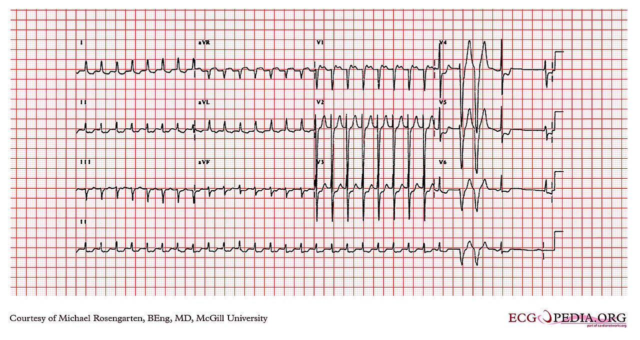
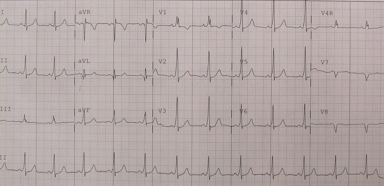
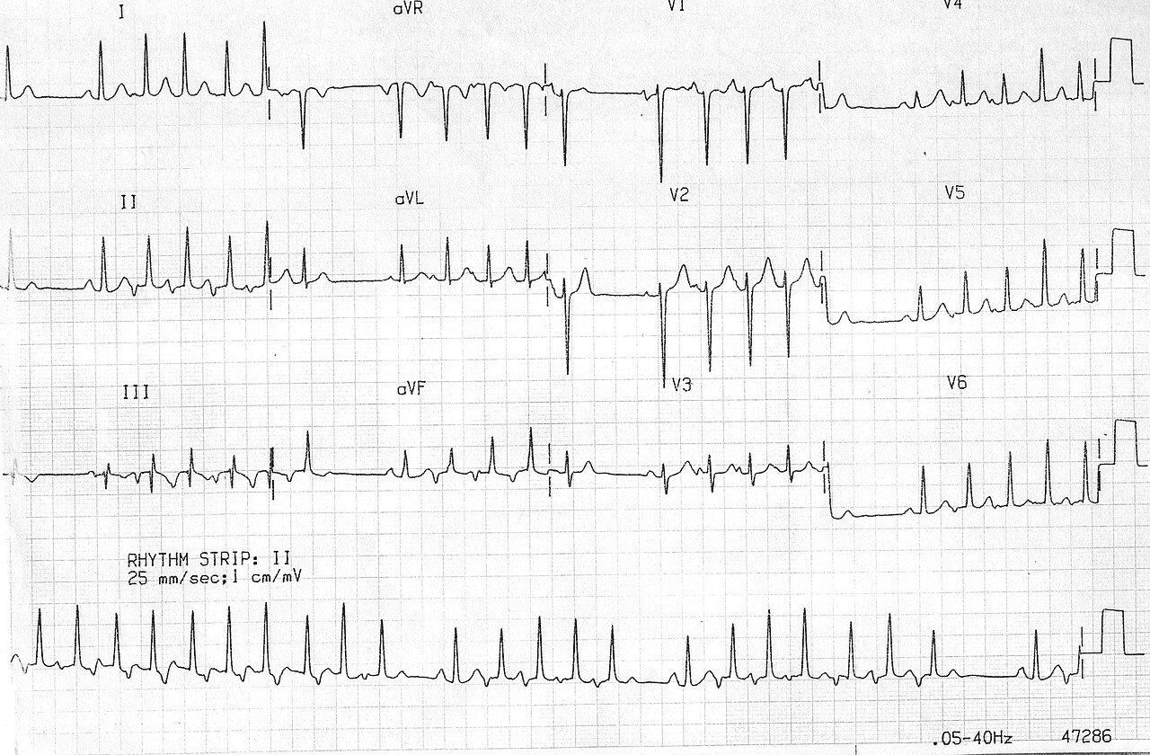
Clinical Pearls
References
- Joglar JA, Chung MK, Armbruster AL, et al. 2023 ACC/AHA/ACCP/HRS Guideline for the Diagnosis and Management of Atrial Fibrillation. Circulation. 2024;149(1):e1–e156. https://doi.org/10.1161/CIR.0000000000001193
- American Heart Association. Part 9: Adult Advanced Life Support: 2025 American Heart Association Guidelines for Cardiopulmonary Resuscitation and Emergency Cardiovascular Care. Circulation. 2025;152(16 suppl 2):S538–S577. https://doi.org/10.1161/CIR.0000000000001376
- Page RL, Joglar JA, Caldwell MA, et al. 2015 ACC/AHA/HRS Guideline for the Management of Adult Patients With Supraventricular Tachycardia. Circulation. 2016;133:e506–e574. https://doi.org/10.1161/CIR.0000000000000311
- Appelboam A, Reuben A, Mann C, et al. Postural modification to the standard Valsalva manoeuvre for emergency treatment of supraventricular tachycardias (REVERT): a randomised controlled trial. Lancet. 2015;386(10005):1747–1753. https://doi.org/10.1016/S0140-6736(15)61485-4
- Yazaki Y, Satomi K, Chikamori T. The Utility of a Lewis Lead for Distinguishing Atrioventricular Reentrant Tachycardia from Typical Atrioventricular Nodal Reentrant Tachycardia. Intern Med. 2022;61(11):1645–1651. https://doi.org/10.2169/internalmedicine.8470-21
- Strangio A, Ielapi J, Sabatino J, et al. Arrhythmias in Pediatric Age: A Narrative Review. Children (Basel). 2025;12(12):1580. https://pmc.ncbi.nlm.nih.gov/articles/PMC12731394/
- American Heart Association. Part 8: Pediatric Advanced Life Support: 2025 American Heart Association Guidelines for Cardiopulmonary Resuscitation and Emergency Cardiovascular Care. Circulation. 2025;152(suppl 2). https://cpr.heart.org/en/resuscitation-science/cpr-and-ecc-guidelines/pediatric-advanced-life-support
- HF Acquisition Co LLC, DBA Health First. Adenosine Injection, Solution [prescribing information]; repackaged label, manufactured by Akorn, Inc. DailyMed; updated October 10, 2023. https://dailymed.nlm.nih.gov/dailymed/lookup.cfm?setid=68f36fc8-8f10-593f-e053-2991aa0a48de
Medical Disclaimer
- For Educational Purposes Only: This content is intended for educational reference and should not be used for clinical decision-making.
- Not a Substitute for Professional Judgment: Always consult your local protocols, institutional guidelines, and supervising physicians.
- Verify Before Acting: Users are responsible for verifying information through authoritative sources before any clinical application.