

Bedside Snapshot
- Indications
- Failed intubation with ventilation still possible (Plan B)
- Primary airway in arrest where protocol and training allow
- Bag-mask ventilation failing, per protocol
- Contraindications / Abort
- Intact gag in an awake patient: poorly tolerated
- Abort if SpO₂ <90% after insertion
- No EtCO₂ waveform after placement: remove and reassess
- High leak or pressure: change size or move to the next plan
- Critical Steps
- Size from the weight-band chart
- Lubricate; insert to the IFU depth
- Inflate the cuff only to the label volume (cuffed devices)
- Confirm with waveform EtCO₂ before moving
- Confirm & Monitor
- Sustained waveform EtCO₂ over 5–6 breaths
- Chest rise and bilateral breath sounds
- Continuous EtCO₂; recheck depth after every move
- Cuff pressure per protocol, and on altitude change
Key Numbers
An SGA is the rescue or primary airway that ventilates without passing a tube through the cords. Confirm it with waveform EtCO₂ before you move the patient or give drugs. Pediatric sizes are in Special Populations.
| Parameter | Value |
|---|---|
| Device choice | Use the device your protocol and training cover. Uncuffed gel (i-gel) or cuffed (classic LMA) per local protocol |
| Size | Adult: by weight band (i-gel 3 = 30–60 kg, 4 = 50–90 kg, 5 = over 90 kg; bands overlap, see Sizing) Peds: i-gel 1 = 2–5 kg, 1.5 = 5–12 kg (see Special Populations) |
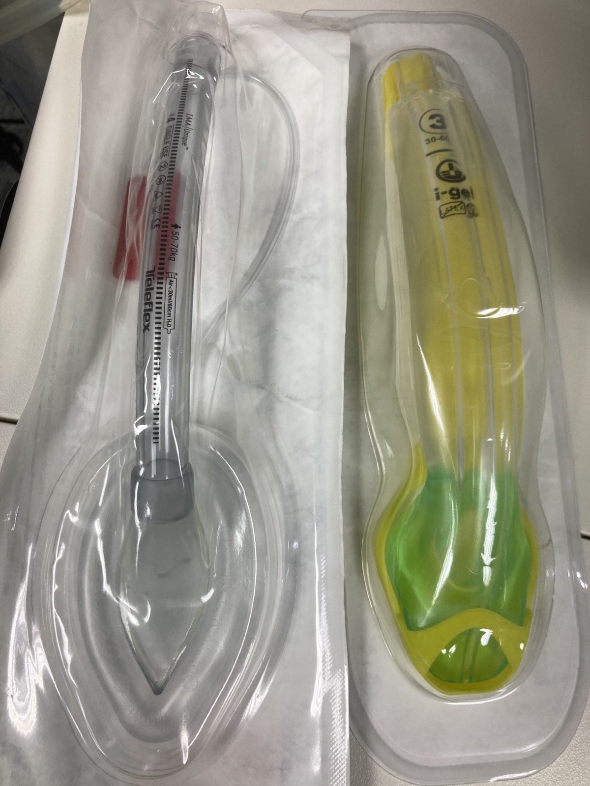
| Cuff (cuffed devices) | Cuff volume is size-specific: read the maximum on the device or pilot balloon label (size 4 label shown: 30 mL max). Intracuff pressure limit 60 cm H₂O at any size. Use a pressure gauge where the protocol requires one |
| Insertion attempts | Limit per local protocol. Reassess ventilation after every attempt |
| Confirm | Sustained waveform EtCO₂ over 5–6 breaths, with chest rise and bilateral breath sounds |
| Abort / reassess | SpO₂ below 90% or no EtCO₂ waveform after placement: stop, ventilate with BVM and PEEP, reassess |
Overview & Principles
A supraglottic airway (SGA) is a tube-like device that sits above the glottis. A bowl or cuff seals around the laryngeal inlet. Air goes into the lungs without a laryngoscope and without a tube passing through the cords. This page covers the i-gel and the classic laryngeal mask airway (LMA). King LT and LMA Supreme are not covered here. Use their IFU for sizing and cuff rules.
How It Works
- Seal: the i-gel has an uncuffed gel bowl. The classic LMA has an inflatable cuff. Either one sits around the laryngeal inlet.
- Ventilation: the bag or ventilator hooks up to a standard 15 mm port. Air moves through the device. It does not go through the glottis.
- Aspiration: the seal gives some protection against regurgitated stomach contents. It does not replace a cuffed endotracheal tube. The NAEMSP statement does not claim full protection.
- Gastric access: some devices have a separate drain channel for gastric decompression. Check the channel and the tube size in the IFU.
Where It Fits
Most failed-airway pathways make the SGA Plan B. Intubation fails, ventilation still works, and you place an SGA to oxygenate. Plan C is the surgical airway, for when neither one works. In cardiac arrest, bag-mask ventilation and advanced airways are both options. The 2025 AHA Guidelines for CPR and ECC, Part 9 (adult advanced life support), count the SGA as an advanced airway. Out of hospital, it is acceptable where intubation success is low or training is minimal (Class 2a, B-R). It can also be an alternative to intubation where intubation success is high or training is optimal (Class 2a, B-R). In in-hospital arrest, expert teams may use either device (Class 2a, B-NR). Bag-mask ventilation or an advanced airway may be considered in any setting (Class 2b, B-R). The two large OHCA trials matter here. The i-gel strategy did not improve 30-day functional outcome versus intubation (AIRWAYS-2: 6.4% vs 6.8% good outcome). Initial ventilation succeeded more often, though. PART tested a different device. Initial laryngeal tube insertion improved 72-hour survival (18.3% vs 15.4%). That is not evidence for the i-gel.
| Device | Seal | Notes |
|---|---|---|
| i-gel | Uncuffed gel bowl | No cuff to inflate. Sized by weight band |
| Classic LMA | Inflatable cuff | Maximum cuff volume is size-specific and printed on the device (size 4 label shown: 30 mL max). Intracuff pressure limit 60 cm H₂O at any size |
Indications & Contraindications
Indications
- Rescue ventilation: failed endotracheal intubation where bag-mask ventilation is inadequate and an SGA can ventilate (Plan B)
- Primary airway in cardiac arrest: where the protocol and the team's training allow it, as an alternative to endotracheal intubation
- Failed bag-mask ventilation with no tube or delay to intubation, per protocol
Absolute Contraindications
Nothing rules it out when the patient needs an airway and cannot be ventilated any other way.
Relative Contraindications & Cautions
- Intact gag or airway reflexes in an awake patient: the patient may not tolerate it well, and it may set off laryngospasm
- High airway pressure or poor compliance: the seal may leak. Consider the next plan early
- Restricted mouth opening: a small opening, see the 3-3-2 assessment, can stop insertion or a seal
- Subglottic or lower-airway obstruction: the SGA cannot get around it. Ventilation fails or pressures climb, so plan the next airway early
- Pharyngeal or laryngeal pathology (caustic injury, foreign body, angioedema, epiglottitis): the surgical airway may be needed sooner, and ventilation may fail
- High aspiration risk (full stomach, bowel obstruction, gastroparesis): the SGA is no substitute for a cuffed tube
Consider an Alternative
When intubation is feasible and the team is trained, intubation is the better choice for aspiration risk. Place the SGA when the tube fails. If the SGA fails too, head to the cricothyrotomy page.
Scope & Team Roles
| Provider Level | Typical Role | Notes |
|---|---|---|
| EMT | Assists | Bag-mask and oxygen. SGA insertion varies by state; some states allow EMT insertion. Confirm your state rules |
| AEMT | Performs (protocol/credentialing required) | Scope varies by state and agency; verify that SGA placement is in your protocol |
| Paramedic | Performs (protocol/credentialing required) | Core rescue device for most EMS systems; train on every device your service carries |
| RN (ED/ICU) | Assists | Maintains the device, cuff and EtCO₂; physician or APP usually inserts |
| Critical Care Transport (CFRN / FP-C / CCP-C) | Performs (protocol/credentialing required) | Often the only advanced airway during transport; re-verify placement after every move |
| Physician / APP | Performs | Plans the airway and makes the decision to convert or to change device |
Team Roles During the Procedure
- Operator: chooses the size, inserts the device, and calls the attempt and the next step.
- Airway/oxygen manager: runs BVM and oxygen, and checks the seal and the bag.
- Monitor and recorder: watches SpO₂, ECG and EtCO₂, times the attempt and documents.
Pre-Procedure Assessment
Anatomic Difficulty
| Component | Assess | Concerning Findings |
|---|---|---|
| L – Look externally | Facial trauma, burns, beard, neck mass | Distorted or obstructed airway landmarks |
| E – Evaluate 3-3-2 | Mouth opening, hyomental distance, thyromental distance | Mouth opening under 3 finger-breadths, hyomental distance under 3 finger-breadths, or thyromental distance under 2 finger-breadths |
| M – Mallampati | Tongue and pharynx view when seated | Class III or IV |
| O – Obstruction | Epiglottitis, abscess, bleeding, angioedema | Any airway swelling or bleeding |
| N – Neck mobility | Range of motion, with C-spine precautions as needed | Fixed or painful neck |
Physiologic Risk
- Hemodynamic risk with positive pressure: SBP under 90 mmHg when positive-pressure ventilation starts. This threshold is borrowed from the RSI and intubation pages
- Shock index: over 0.9 (threshold borrowed from the RSI and intubation pages)
- Hypoxemia: SpO₂ under 93% on the best oxygen available
Site / Device Check
Before you start, make sure SpO₂, ECG, blood pressure and EtCO₂ are all running. Set out at least two SGA sizes, a backup device and the surgical kit. If the device is cuffed, check the cuff and the connector before insertion.
Equipment & Preparation
Equipment
| Item | Size / Spec | Notes |
|---|---|---|
| SGA device | Size by weight band (see Sizing) | Check the expiry and the package integrity before opening |
| Backup SGA | One size smaller and one size larger | Use if the first size does not seal |
| BVM with PEEP valve | Adult and pediatric masks | Oxygen at high flow connected |
| Waveform capnography | Adult and pediatric | Connect before insertion; required for confirmation |
| Lubricant | Water-based | Apply to the back of the device as the IFU directs |
| Suction | Rigid and flexible catheters | Clear the mouth before insertion |
| Surgical airway kit | Per protocol | Open and at the bedside for the CICO plan |
Sizing
| Device / Size | Weight Band | Notes |
|---|---|---|
| i-gel 1 | 2–5 kg | Neonate size; sources agree |
| i-gel 1.5 | 5–12 kg | Infant size; sources agree |
| i-gel 2 | 10–25 kg | Small pediatric; one secondary source gives 12–25 kg. Confirm in the IFU |
| i-gel 2.5 | 25–35 kg | Large pediatric |
| i-gel 3 | 30–60 kg | Shown on packaging. Sources differ (30–60 kg on one chart; 35–50 kg in a letter). Confirm in the IFU or the device label |
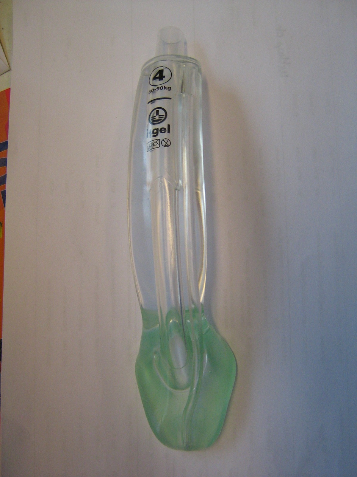
| i-gel 4 | 50–90 kg | Shown on the device |
| i-gel 5 | Over 90 kg | Confirm in the IFU |
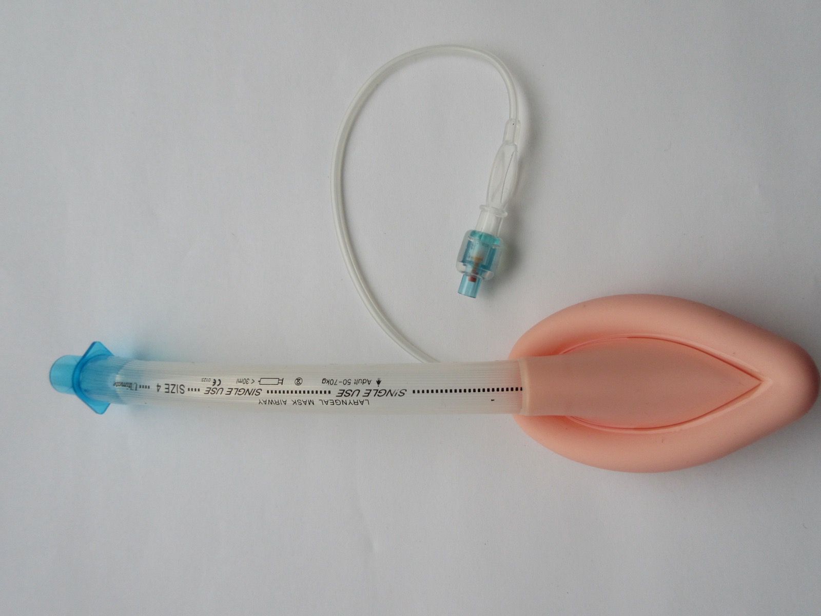
| Classic LMA size 4 | 50–70 kg | Shown on the device; other classic sizes per the manufacturer chart |
Weight bands overlap. Examples: 1.5 and 2 at 10–12 kg, and 3 and 4 at 50–60 kg. When two sizes fit, go with the size the IFU and the device label give for the patient. Check the seal with that size. If it leaks, move to the adjacent size.
Patient Positioning & Preparation
Preoxygenate with a tight-fitting mask and PEEP. If the patient is not apneic, ventilate with BVM before insertion. Put the head in the sniffing position if the neck is not injured. Use a jaw thrust for an unresponsive patient. If the patient may be aspirating, suction first and expect regurgitation on insertion.
Pre-Procedure Checklist
- Weight known or estimated, and the size picked from the chart
- Backup SGA opened, one size up and one size down
- Capnography hooked up and zeroed
- BVM with a PEEP valve, oxygen running at high flow
- Suction tested and within reach
- Lubricant, plus a syringe for any cuffed device
- Surgical airway kit on hand and still sealed
- Plan A, B and C said out loud to the team before the first attempt
Step-by-Step Procedure
Phase 1: Prepare
- Choose the size: match the weight band on the chart. If the weight is unknown, use the protocol's estimation method.
- Preoxygenate: give high-flow oxygen with a tight mask and PEEP for several breaths.
- Position: sniffing position if the neck is not injured; jaw thrust if the patient is unresponsive.
Phase 2: Perform
- Lubricate the device: apply lubricant to the back of the device as the IFU directs.
- Insert: slide the device along the hard palate and into the pharynx, following the IFU's insertion technique, until it stops.
- Inflate the cuff (cuffed devices only): use the volume the device label gives, and keep the pressure within the device limit.
- Ventilate: give a breath and watch for chest rise and EtCO₂.
Phase 3: Confirm & Secure
- Confirm placement: sustained waveform EtCO₂ over 5–6 breaths, with chest rise and bilateral breath sounds (see Confirmation & Monitoring).
- Secure: strap or tape the device; mark the depth on the device and the teeth.
- Gastric decompression: pass a gastric tube through the drain channel if the device has one and the protocol calls for it. Use the tube size and lubricant the IFU gives; this page does not list tube sizes. Passing the tube does not secure the airway or fully empty a full stomach.
- Document and hand off: time, size, depth, attempts and confirmation method.
During CPR
- Compressions continue: with an advanced airway in place, compressions never stop. Ventilation is asynchronous, so you do not pause compressions for each breath.
- Adult rate: 1 breath every 6 seconds (10 per minute) with continuous compressions. Part 9 of the 2025 AHA Guidelines for CPR and ECC says this may be reasonable (Class 2b, C-LD). Don't hyperventilate.
- Pediatric airway choice: in infants and children with out-of-hospital cardiac arrest, the 2025 AHA Guidelines for CPR and ECC, Part 8, say bag-mask ventilation is reasonable rather than an advanced airway such as an SGA or tracheal tube (Class 2a, C-LD). In-hospital, with no advanced airway in place, bag-mask ventilation or an advanced airway may be reasonable (Class 2b, C-LD).
- Pediatric rate: with an advanced airway in place, aim for 20–30 breaths per minute (1 breath every 2–3 seconds), adjusted for age and clinical state. Part 8 of the 2025 AHA Guidelines for CPR and ECC says this may be reasonable (Class 2b, C-LD). Hyperventilation may compromise hemodynamics.
- Leak or no chest rise: a persistent large leak, or no chest rise on compressions? Reseat or reassess the device before the next step.
Alternative Technique: Intubation Through an SGA
Some SGAs let a tube pass through them. The intubating LMA is the usual example, and EMS rarely carries it. This is a protocol and training decision. This page gives no tube-size limit for any i-gel size, no guided-technique outline and no attempt limit. Do not use this technique from this page alone. Every attempt must keep oxygenation going. Ventilate between attempts and do not let SpO₂ fall. Abort and go back to ventilating through the device when SpO₂ is below 90% or the attempt fails. Confirm every tube with EtCO₂. For device limits, read the IFU and the DAS 2025 guideline.
Alternative Technique: Exchange to an Endotracheal Tube
The NAEMSP position is to avoid converting a working SGA to a tube. Convert only when the SGA is failing, and when the patient's condition, the SGA ventilation and the clinical course all support it. For the tube technique, follow the intubation page.
Confirmation & Monitoring
Primary Confirmation
The waveform is the check you trust. Look for sustained EtCO₂ over 5–6 breaths, on every ventilation. It is the standard objective check that air is reaching the lungs. Here's the gap: the 2025 AHA Guidelines for CPR and ECC, Part 9, note that waveform capnography for other advanced airways, such as the laryngeal mask airway, has not been studied. Waveform capnography is recommended (Class 1, C-LD) for confirming endotracheal tube placement.
Secondary Confirmation
- Chest rise with each breath, and bilateral breath sounds on the chest (not the stomach)
- A low leak on the cuff or the seal, with no audible leak at the mouth
- SpO₂ rising or stable on oxygen
- No gastric distension on auscultation over the epigastrium
| Finding | Success | Failure / Malposition |
|---|---|---|
| EtCO₂ waveform | Sustained waveform over 5–6 breaths | No waveform, or a flat trace: remove and reassess. In arrest a low value is expected, but an absent waveform still means malposition |
| Chest rise | Symmetric with each breath | Rise only over the epigastrium, or none |
| Breath sounds | Equal bilaterally | Gurgling or silence over the chest |
| Leak | Low; ventilation at the set pressure | High leak, or high pressure needed to ventilate |
| SpO₂ | Rising or stable | Falling after insertion |
Ongoing Monitoring
- Continuous waveform EtCO₂ (every minute during transport)
- SpO₂ and BP every 3–5 min
- Recheck depth and seal after every move, and on any change in the patient
- Cuff pressure (cuffed devices) per protocol, and on altitude change
- In cardiac arrest: compressions continuous, ventilation 1 breath every 6 seconds (adult), see During CPR in Step-by-Step
Troubleshooting
| Problem | Likely Cause | Fix |
|---|---|---|
| No EtCO₂ waveform | Device not in the airway, or wrong size | Remove, check size, reinsert once, then move to the next plan |
| High leak or high pressure | Too small, mis-seated, or a cuff problem | Check size and cuff; try the next size; reposition the head |
| Gastric insufflation | Ventilating too hard, or esophageal placement | Lower the pressure; decompress with the gastric tube if fitted; confirm position |
| Regurgitation | Full stomach, or insufflation | Suction the mouth; turn the patient; decompress the stomach |
| Desaturation after insertion | Leak, displacement, or laryngospasm | Reposition; ventilate by BVM with PEEP and reassess the seal and leak. Sedation or analgesia only in a non-arrest context and per protocol |
| Device dislodges en route | Movement, poor securing | Re-check depth and waveform after every move; re-secure |
If It Still Fails
- Ventilate: BVM with two-hand seal and PEEP, and reoxygenate.
- Try one more device or size: per protocol, once.
- Intubate if feasible: per protocol, with the attempt limit (see the intubation page).
- Declare CICO: when no device ventilates and no tube can be placed, go to the cricothyrotomy page without delay.
Complications
| Complication | Recognition | Prevention | Management |
|---|---|---|---|
| Immediate: malposition | No EtCO₂ waveform, gurgling, one-sided breath sounds | Correct size; full insertion; waveform check every time | Remove, reassess, reinsert once or move to the next plan |
| Immediate: laryngospasm | Stridor, high pressure, desaturation | Adequate depth of anesthesia where the protocol allows; avoid stimulation during insertion | BVM with PEEP; treat per protocol; reposition |
| Immediate: gastric insufflation | Epigastric distension, high pressure, falling EtCO₂ | Lower pressure; use the gastric tube if fitted | Decompress; reassess placement |
| Immediate: dental or mucosal injury | Loose tooth, bleeding | Gentle insertion; check the teeth first | Suction, manage the bleeding, document |
| Delayed: aspiration | Fever, new infiltrate, hypoxemia after the event | Decompress the stomach; secure the device; suction | Treat per protocol; the SGA does not prevent aspiration fully |
| Rare: cuff-related mucosal or nerve injury | Hoarseness or sore throat after the event | Use the cuff volume the label gives; do not over-inflate | Document; reassess at handoff |
Special Populations
Pediatrics
Pediatric SGA sizes come from the manufacturer's weight bands. Size by weight, and check the chart before each use. On whether SGAs are used in children, follow local protocol. This page does not cover cuffed pediatric LMA sizes or cuff volumes, so use the device IFU for those. Surgical airway age limits are contested between sources. See the cricothyrotomy page.
| Device / Size | Weight Band | Notes |
|---|---|---|
| i-gel 1 | 2–5 kg | Neonate; confirm the IFU before use |
| i-gel 1.5 | 5–12 kg | Infant |
| i-gel 2 | 10–25 kg | Small pediatric |
| i-gel 2.5 | 25–35 kg | Large pediatric |
| i-gel 3 | 30–60 kg | Adult size; the band overlaps larger children. Confirm in the IFU |
In a child, the dose for any drug-assisted insertion is weight-based. See Weight-Based Dosing, Pediatric Equipment Sizing and Pediatric Airway.
Older Adults
- Teeth and mucosa can be fragile. Insert gently
- Less reserve, so check EtCO₂ and SpO₂ closely after insertion
Obesity
- Weight-band sizing may underestimate the device needed. Use the chart and check the seal
- Reduced functional capacity, so preoxygenate fully before insertion
Pregnancy
- Aspiration risk is higher. Where intubation is feasible, an SGA is no substitute for a cuffed tube
- Follow the local obstetric airway protocol
Trauma and Cervical Spine
- Maintain manual in-line stabilization during insertion
- Facial trauma may stop a seal from forming. Go to the next plan early
Post-Procedure Care & Transport
Immediate Aftercare
- Recheck waveform EtCO₂, SpO₂ and chest rise immediately after securing
- Reassess sedation and analgesia per protocol
Ongoing Maintenance
- Cuff pressure per the device label or protocol, and after any altitude change
- Keep the device strapped. Check depth after every move
- Suction the mouth and pharynx as needed
Transport Considerations
- Reassess the device after every move: stretcher, ambulance, elevator and stairs
- In noise and vibration, trust EtCO₂ and the monitors. A sensor alarm means check the device
- Carry a backup device and the surgical kit
- Recheck cuff pressure on climbs and descents when the device is cuffed
Document & Hand Off
- Time, device, size, depth at the teeth or lip, attempt count
- How you confirmed it, plus the EtCO₂ value and waveform quality
- Cuff volume or pressure, if a cuffed device went in
- Medications, with doses and weights, plus any complications
- How the patient responded to the device, and the plan for the next move
Clinical Pearls & Pitfalls
Pearls
- Size by the chart: Eyeballing the patient is not sizing. Use the weight band on the device chart.
- Waveform, not sight: A bag squeeze and chest rise do not prove placement. The waveform does.
- Plan B early: Place the SGA the moment the tube fails, as long as ventilation is possible.
- Re-check after every move: Depth and waveform. Every time, not just the first time.
Common Pitfalls
- Pitfall: one wrong size, then attempt after attempt → fix: change to the next size once, then go to the next plan.
- Pitfall: swapping a working SGA for a tube → fix: keep it unless there is a specific reason.
- Pitfall: pushing the device through resistance → fix: remove, check size and the head position, and reinsert once.
Education & Skill Maintenance
Skill Maintenance
- Put a manikin on the table every quarter and practice with every device your service carries
- Rehearse choosing a size by weight band. Pediatric bands count too
- Every use gets a team debrief, and the confirmation step is part of it
- Every equipment check should cover the device stock and the size chart
References
- Benger JR, et al. Effect of a strategy of a supraglottic airway device vs tracheal intubation during out-of-hospital cardiac arrest on functional outcome: the AIRWAYS-2 randomized clinical trial. JAMA. 2018;320(8):779. https://doi.org/10.1001/jama.2018.11597
- Costa BS, Colla MCP, et al. Supraglottic airway versus endotracheal intubation in out-of-hospital cardiac arrest: a systematic review and meta-analysis. Resusc Plus. 2026. https://doi.org/10.1016/j.resplu.2026.101491
- National Association of EMS Physicians. Prehospital Supraglottic Airways: An NAEMSP Position Statement and Resource Document. Prehosp Emerg Care. 2022;26(sup1):32-41. https://doi.org/10.1080/10903127.2021.1983680
- American Heart Association. Part 9: Adult Advanced Life Support: 2025 American Heart Association Guidelines for Cardiopulmonary Resuscitation and Emergency Cardiovascular Care. Circulation. 2025;152(16 suppl 2):S538-S577. https://doi.org/10.1161/CIR.0000000000001376
- Ahmad I, El-Boghdadly K, et al. Difficult Airway Society 2025 guidelines for management of unanticipated difficult tracheal intubation in adults. Br J Anaesth. 2026;136(1):283-307. https://doi.org/10.1016/j.bja.2025.10.006
- Lasa JJ, Dhillon GS, Duff JP, et al. Part 8: Pediatric Advanced Life Support: 2025 American Heart Association Guidelines for Cardiopulmonary Resuscitation and Emergency Cardiovascular Care. Circulation. 2025;152(suppl 2):S479-S537. https://cpr.heart.org/en/resuscitation-science/cpr-and-ecc-guidelines/pediatric-advanced-life-support
- Wang HE, et al. Effect of a strategy of initial laryngeal tube insertion vs endotracheal intubation on 72-hour survival in adults with out-of-hospital cardiac arrest: a randomized clinical trial. JAMA. 2018;320(8):769. https://doi.org/10.1001/jama.2018.7044
- Sachidananda R, et al. Comparison of standard weight-based and thenar eminence dimension-based selection of I-gel in pediatric patients: a randomized controlled study. J Anaesthesiol Clin Pharmacol. 2023;39(4):642-647. https://doi.org/10.4103/joacp.joacp_215_22
Medical Disclaimer
- For Educational Purposes Only: This content is intended for educational reference and should not be used for clinical decision-making.
- Not a Substitute for Professional Judgment: Always consult your local protocols, institutional guidelines, and supervising physicians.
- Verify Before Acting: Users are responsible for verifying information through authoritative sources before any clinical application.