

Bedside Snapshot
- Indications
- CICO: intubation failed, SGA failed, mask ventilation impossible
- Upper-airway obstruction where oral and nasal access fail
- Massive facial trauma, oropharyngeal bleeding, rapidly progressing burn
- Contraindications / Abort
- Children under about 8 years: needle or tracheotomy per protocol
- Laryngotracheal transection: false passage, distal airway lost
- Critical Steps
- Declare CICO; extend the neck; keep oxygen running
- Laryngeal handshake; stab through skin and membrane
- Rotate blade 90°; bougie in; advance 6.0 cuffed ETT
- Hold-up under 5 cm: pre-tracheal bougie, re-check
- Confirm & Monitor
- Waveform EtCO₂ on every ventilation
- Chest rise and bilateral breath sounds
- Bleeding, cuff leak, emphysema; plan definitive airway
Key Numbers
Cricothyrotomy is the rescue step once cannot-intubate, cannot-oxygenate (CICO) is declared. Use the adult values below; pediatric limits are in Special Populations.
| Parameter | Value |
|---|---|
| Tube | Adult: 6.0 mm cuffed ETT (DAS 2015 guideline) Peds: not used under about 8 years (see Special Populations) |
| Blade | Scalpel with #10 blade, broad enough to match the tube width (DAS 2015 guideline) |
| Bougie | Coudé (angled) tip; advance 10–15 cm into the trachea (DAS 2015 guideline) |
| Hold-up sign | Bougie stops at under 5 cm: likely pre-tracheal, re-check (DAS 2015 guideline) |
| Impalpable membrane | Midline vertical skin incision 8–10 cm, then blunt dissection (DAS 2015 guideline) |
| Needle cricothyrotomy | Temporizing only. Jet ventilation with a pressure limit: start at a low driving pressure (about 1 bar) and raise it only until chest rise is seen (BJA Education jet ventilation review). Needs an open exhalation path. Follow device IFU and local protocol |
| Confirm | Waveform EtCO₂ on every ventilation, plus chest rise and auscultation |
Overview & Principles
A cricothyrotomy is a surgical airway. You cut through the cricothyroid membrane, between the thyroid and cricoid cartilages, and into the trachea. It is Plan D of the difficult airway algorithm. You reach it only after intubation, supraglottic ventilation and face-mask ventilation have all failed.
How It Works
- Bypasses the upper airway: the cuffed tube sits below the cords. Normal tidal ventilation works again on a standard breathing system.
- Scalpel is the preferred rescue route: DAS 2015 and NAEMSP 2022 favour the scalpel over needle-jet techniques for success.
- Needle cricothyrotomy is temporizing: jet ventilation through a narrow-bore cannula takes high pressure, and that pressure carries barotrauma risk.
- Waveform capnography confirms placement: chest rise and breath sounds alone are not enough. Trust the waveform.
Where It Fits
Plan A is intubation. Plan B is a supraglottic airway. Plan C is face-mask ventilation. Plan D is emergency front-of-neck access (DAS 2025 guideline). Declare CICO early, once no non-surgical means can oxygenate the patient. Wait for arrest and you have no margin left.
Indications & Contraindications
Indications
- CICO: failed intubation, failed supraglottic airway, and face-mask ventilation impossible or inadequate
- Upper-airway obstruction where oral and nasal access is impossible: massive facial trauma, oropharyngeal bleeding, rapidly progressing airway burn or angioedema
Absolute Contraindications
In true CICO there are none. Losing oxygenation outweighs the usual cautions.
Relative Contraindications & Cautions
- Children under about 8 years: the cricothyroid membrane is small and hard to palpate. Use needle cricothyrotomy or surgical tracheotomy per protocol
- Laryngotracheal transection or disruption: the tube may pass into a false passage, and distal airway control may be lost. Consider a surgical tracheotomy
- Distorted anatomy (tumor, prior neck surgery, obesity, edema). You need a vertical skin incision and blunt dissection
Consider an Alternative
Can the patient be oxygenated by mask or supraglottic airway? Then work Plan A to C first, and reattempt intubation as described on the intubation and RSI pages.
Scope & Team Roles
| Provider Level | Typical Role | Notes |
|---|---|---|
| EMT | Not in scope | Recognize CICO, call for help, bring equipment |
| AEMT | Not in scope | Support oxygenation and ventilation until the paramedic arrives |
| Paramedic | Performs (protocol/credentialing required) | Not in national model: state/agency credentialing |
| RN (ED/ICU) | Assists | Equipment, suction, medication and oxygen support |
| Critical Care Transport (CFRN / CCT / FP-C / CCP-C) | Performs (protocol/credentialing required) | Agency-specific; verify before procedure is listed on a protocol |
| Physician / APP | Performs | Operator for most ED and hospital cases |
Team Roles During the Procedure
- Operator: lands landmarks, makes incision, places tube
- Airway/oxygen manager: keeps upper-airway oxygen going until the surgical airway is ventilating
- Monitor/recorder: reads EtCO₂ and SpO₂, times each step and calls out changes
- Equipment support: hands over scalpel, bougie and tube in order
Pre-Procedure Assessment
At this stage, assessment means recognition and landmarks. It is not a full airway workup.
Anatomic Difficulty
| Finding | Assess | Concerning Finding |
|---|---|---|
| Neck anatomy | Landmarks by feel | Membrane not palpable; thick neck or obesity |
| Upper airway | Swelling, bleeding, burns | Rapid progression despite oxygen |
| Neck mobility | Trauma, C-spine precautions | Limited extension; use the best position possible |
Physiologic Risk
- Hypoxemia: SpO₂ is falling despite BVM with an SGA. Start the procedure. Do not wait for a trend
- Hemodynamics: hypotension or shock pushes the procedure up the list, not down it
Site / Device Check
- Equipment: have the scalpel, bougie, 6.0 cuffed ETT and suction on hand before you decide
- Capnography: have it ready to confirm placement
Anatomy & Landmarks
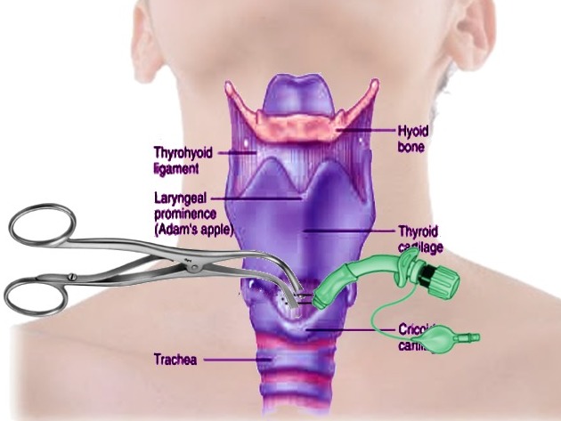
Your target is the cricothyroid membrane. It is the soft space between the thyroid cartilage above and the cricoid cartilage below.
Key Structures
- Thyroid cartilage: the largest laryngeal prominence. The membrane sits just below it
- Cricothyroid membrane: a soft, slightly indented space. This is your target
- Cricoid cartilage: a firm, complete ring below the membrane. It sets the lower limit
Landmarking: Laryngeal Handshake (DAS 2015 guideline)
- Grasp the larynx: use the non-dominant hand; thumb and index on the hyoid and thyroid laminae
- Stabilize: hold the thyroid cartilage between thumb and middle finger
- Find the membrane: move the index finger down to the soft space
- Confirm: the firm cricoid ring should be just below it
Pediatric Anatomy
Children aged 1–8 years are the hard case. In one cited series (2026 pediatric review), even pediatric anesthesiologists got the membrane right by palpation only 29% of the time. The membrane is smaller. Hitting the ideal needle angle of 45° or less without neck hyperextension is hard.
Equipment & Preparation
Wherever advanced airway management happens, have a complete cricothyrotomy kit or a standardized scalpel-bougie-tube setup.
Equipment
| Item | Size / Spec | Notes |
|---|---|---|
| Scalpel | #10 blade | Broad enough to match the tube width (DAS 2015 guideline) |
| Bougie | Coudé (angled) tip | Advance into trachea; clicks may be felt over rings |
| Cuffed ETT | 6.0 mm | Lubricate before railroading |
| Syringe | 10 mL | Cuff inflation |
| Hook or dilator | Optional | Keeps the incision open; not required (DAS 2015 guideline) |
| Suction | Rigid tip | Clear blood and secretions early |
| Capnography | Waveform | Confirms placement |
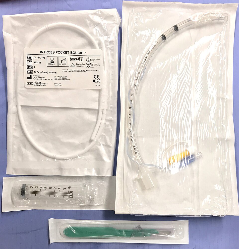
Many services use commercial percutaneous or Seldinger kits, such as the Melker. Follow the device IFU and local protocol. DAS 2015 notes that a smaller cuffed tube can be used if it fits over the bougie, and that includes a Melker.
Sizing
- Adult tube: 6.0 mm cuffed ETT (DAS 2015 guideline)
- Bougie: adult coudé bougie. It must reach at least 10–15 cm past the skin
- Pediatric: see Special Populations. Needle sizes differ, and local protocol sets them
Patient Positioning & Preparation
- Neck extension: a pillow under the shoulders, or the head dropped over the end of the stretcher
- Oxygen: keep upper-airway oxygen running until the surgical airway is ventilating
- Paralysis: make sure the complete neuromuscular block is in place before rescue (DAS 2015 guideline). Local protocols may differ for a patient who is already deeply unresponsive or in arrest. Sugammadex given? Then use a neuromuscular blocker other than rocuronium or vecuronium (DAS 2015 guideline)
Pre-Procedure Checklist
- CICO declared, team called
- Neck in extension
- Scalpel, bougie, 6.0 cuffed ETT and syringe open
- Suction running and tested
- Capnography connected
- Upper-airway oxygen flowing
- Needle cricothyrotomy kit and jet source ready as backup
Step-by-Step Procedure
Phase 1: Prepare
- Declare CICO: state it aloud so the team moves to Plan D.
- Position the neck: extend it fully and keep upper-airway oxygen running.
Phase 2: Perform (Scalpel-Bougie-Tube)
- Stabilize the larynx: use the laryngeal handshake and keep the non-dominant hand on the landmarks.
- Make a transverse stab incision: through skin and membrane with the blade edge facing you, for a palpable membrane.
- Rotate the blade 90°: turn the sharp edge toward the feet while keeping the handle vertical to the skin.
- Place the bougie: slide the coudé tip down the side of the blade into the trachea, then advance up to 10–15 cm.
Phase 3: Confirm & Secure
- Railroad the tube: thread the 6.0 mm cuffed ETT over the bougie, rotating as you advance, and avoid excessive advancement.
- Remove the bougie: inflate the cuff and ventilate.
- Confirm placement: waveform EtCO₂, chest rise and auscultation (see Confirmation & Monitoring).
- Secure the tube: with tape or a tube holder; manage bleeding with pressure around the tube, not on it.
Alternative Technique: Impalpable Membrane (Scalpel-Finger-Bougie, DAS 2015)
- Make a midline vertical skin incision: 8–10 cm (DAS 2015 guideline) if the membrane cannot be felt or a single stab may fail.
- Blunt dissect: separate tissue with fingers of both hands; stabilize the larynx with the non-dominant hand.
- Find the membrane: repeat the scalpel-bougie-tube steps above.
- Watch for a hold-up under 5 cm: that suggests the bougie is pre-tracheal. Re-check the anatomy.
Alternative Technique: Needle Cricothyrotomy (Temporizing)
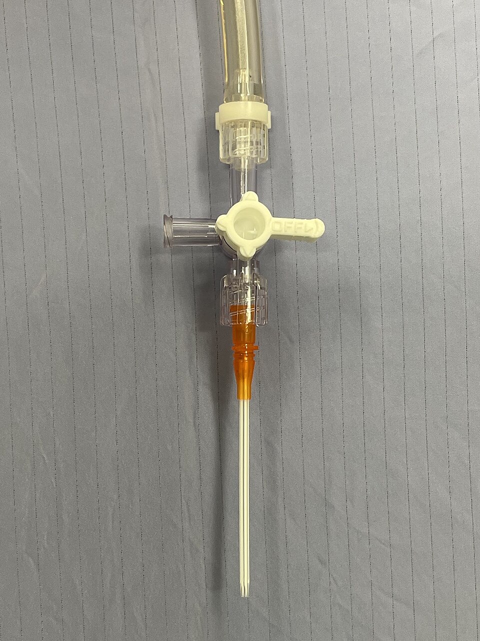
Needle cricothyrotomy buys time, and only time. It does not give reliable CO₂ elimination, and a definitive airway must follow. In the sources reviewed here, a narrow-bore cannula needs high-pressure jet ventilation, with a serious barotrauma risk (DAS 2015 guideline). DAS 2015 limits the technique to clinicians who use high-pressure devices in routine practice.
- Identify and stabilize the membrane: as above.
- Insert the catheter: caudally at 45° or less, aspirating with a saline-filled syringe.
- Confirm: stop when air appears in the syringe, then advance the catheter and withdraw the needle.
- Connect jet ventilation: use a pressure-limited source. Start at a low driving pressure (about 1 bar, BJA Education jet ventilation review) and raise it only until chest rise is seen; the device IFU and local protocol set the exact values.
Confirmation & Monitoring
Primary Confirmation
How do you know the tube is in the trachea? Sustained waveform capnography (EtCO₂) on every ventilation, plus visible chest rise and bilateral breath sounds. DAS 2015 calls capnography the gold standard for confirming ventilation.
Secondary Confirmation
- Bilateral chest expansion with each ventilation
- Auscultation over both lung fields and the stomach
- Improvement in SpO₂ and heart rate
| Finding | Success | Failure / Malposition |
|---|---|---|
| EtCO₂ waveform | Present and sustained | Absent or flat: stop, re-check |
| Chest rise | Bilateral with ventilation | Unilateral or none |
| Bougie | Clicks over rings; no hold-up | Hold-up under 5 cm: pre-tracheal |
| Cuff | Inflated; leak-free | Air leak or the tube too deep |
| Subcutaneous tissue | No swelling | Spreading emphysema |
Ongoing Monitoring
- Continuous: waveform EtCO₂, SpO₂ and chest rise
- After each move: re-check the tube position and seal
- Frequently: bleeding around the tube and tube security
Troubleshooting
| Problem | Likely Cause | Fix |
|---|---|---|
| No EtCO₂ | False passage or wrong space | Stop, re-palpate, consider repeating the procedure while CICO persists |
| Bougie hold-up under 5 cm | Pre-tracheal bougie | Withdraw and re-check landmarks; redo the incision |
| Cannot palpate membrane | Obesity, edema, anatomy | Vertical incision and blunt dissection |
| Bleeding obscures view | Vessels or thyroid isthmus | Suction; pressure around the tube; proceed |
| Tube resists | Tube too large or false passage | Confirm bougie is in trachea; try a smaller tube only if the protocol allows |
| Cuff leak or low ventilation | Tube too deep or too shallow | Adjust depth while maintaining seal; check the cuff |
If It Still Fails
- Re-declare CICO: keep oxygen and ventilation running.
- Switch to the second approach: scalpel-finger-bougie for an impalpable membrane, with an 8–10 cm vertical incision and blunt dissection, per DAS 2015.
- Escalate: a trained clinician performs surgical tracheotomy, or you use needle cricothyrotomy per protocol.
Complications
These complication rates come from a 2019 systematic review of emergency surgical airways: 13 cricothyrotomy studies, 1,219 patients, mean age 39.8 years. That review excluded pediatric data.
| Complication | Recognition | Prevention | Management |
|---|---|---|---|
| Immediate: failure to obtain airway (1.6%) | No EtCO₂, no chest rise | Practice landmarks; declare early | Re-declare CICO; try the alternative approach |
| Immediate: injury to cartilage (1.6%) | Crepitus, hoarseness | Careful blade and bougie control | Secure airway; plan definitive airway |
| Immediate: hemorrhage (0.7%) | Bleeding at incision or thyroid isthmus | Vertical incision in difficult anatomy | Pressure around the tube; suction; proceed |
| Immediate: pneumothorax (0.2%) | Reduced breath sounds, hypoxia | Passive exhalation with any jet | Decompression per protocol |
| Delayed: airway stenosis (1.1%) | Stridor or dyspnea after recovery | Early conversion to a definitive airway | ENT or surgery |
Special Populations
Pediatrics
- Under about 8 years: this is contested. The DAS 2015 paediatric guidance (ages 1–8), as the 2026 pediatric review summarizes it, says this: if ENT is unavailable, start with needle cricothyrotomy and pressure-limited jet ventilation. Move to surgical cricothyrotomy or tracheotomy if that fails. The 2026 pediatric review argues for surgical tracheotomy rather than needle cricothyrotomy. AIDAA 2025 prefers tracheotomy up to age 12 when trained surgical help is available. Without that help, needle cricothyrotomy at ages 5–12. Defer to local protocol and training.
- Age 8 and over: use scalpel-bougie-tube as for adults. The DAS adult eFONA guidance is defined for patients over 8 years, per the 2026 pediatric review. Local protocol sets the age cutoff. The fixed 6.0 mm cuffed tube is an adult size and is not appropriate for a small child. Choose the tube size for the child.
- Needle set: catheter gauge, angle (45° or less) and jet pressure must be set for the child. Ideally, a definitive airway replaces it within about 40 minutes. Catheter gauge is device- and protocol-specific. The DAS 2015 text cites a 14 gauge cannula in one wet-lab technique.
Need the pediatric numbers? Start with these pages: pediatric airway, equipment sizing, weight-based dosing, Broselow, vascular access.
Obesity
- Membrane: often deeper. Use a vertical incision and blunt dissection
- Tube: the bougie may need more force and length
Trauma and C-Spine
- Neck: extension may be limited. Keep manual in-line stabilization
- Bleeding: expect more blood. The incision is often larger
Physiologically Difficult
- Shock: bleeding and reduced cardiac output make the procedure more urgent
- Hypoxemia: continue oxygen. Do not wait for a perfect position
Post-Procedure Care & Transport
Immediate Aftercare
- Confirm tube position with waveform EtCO₂ and chest rise, and document it
- Control bleeding around the tube with pressure, not pressure on the tube
- Start sedation and analgesia per protocol if the patient is awake or paralyzed
Ongoing Maintenance
- Security: tape or a tube holder. Check the seal after every move
- Cuff: keep it inflated and check the pressure per protocol
- Incision site: check for bleeding, swelling or infection
Transport Considerations
- Reassess after each move with EtCO₂, SpO₂ and chest rise
- Vibration and noise can hide a dislodged tube. Rely on waveform EtCO₂
- Plan the definitive airway: the cricothyrotomy is usually temporary
Handoff & Documentation
Document & Hand Off
- When you declared CICO, and when the tube went in
- Tube size, depth and the technique used
- Attempts made, and any complications
- Confirmation method and EtCO₂ readings
- Medications given and oxygen settings
- Patient response and transport plan
Clinical Pearls & Pitfalls
Pearls
- Declare early: CICO is a decision. It is not a last resort after arrest.
- Stabilize and keep your hand on the landmarks: lose the landmarks and you lose time.
- Hold-up under 5 cm: that means a pre-tracheal bougie. Re-check.
- Scalpel over needle: the scalpel gives higher success in the emergency setting (NAEMSP 2022 statement).
- Needle cricothyrotomy is temporary: plan a definitive airway.
Common Pitfalls
- Delay until arrest: start Plan D while the patient still has a pulse.
- Blind bougie without landmarks: palpate again, and redo the incision.
- Tube too deep: excessive advancement causes endobronchial intubation. Stop when the cuff passes the membrane.
- Relying on chest rise: chest rise does not confirm it. Use waveform EtCO₂.
Education & Skill Maintenance
Skill Maintenance
- Practice landmarks and the scalpel-bougie-tube sequence on a task trainer at least every 6 months
- Run CICO simulations where cricothyrotomy is the correct answer
- Check kits for completeness at every shift
- Debrief each case: was CICO declared early enough
References
- Frerk C, Mitchell VS, McNarry AF, et al. Difficult Airway Society 2015 guidelines for management of unanticipated difficult intubation in adults. Br J Anaesth. 2015;115(6):827-848. https://doi.org/10.1093/bja/aev371
- Ahmad I, El-Boghdadly K, Iliff H, et al. Difficult Airway Society 2025 guidelines for management of unanticipated difficult tracheal intubation in adults. Br J Anaesth. 2026;136(1):283-307 (published online 2025). https://doi.org/10.1016/j.bja.2025.10.006
- Apfelbaum JL, Hagberg CA, Connis RT, et al. 2022 American Society of Anesthesiologists practice guidelines for management of the difficult airway. Anesthesiology. 2022;136(1):31-81. https://doi.org/10.1097/ALN.0000000000004002
- Reardon RF, Robinson AE, Kornas R, et al. Prehospital surgical airway management: an NAEMSP position statement and resource document. Prehosp Emerg Care. 2022;26(1):96-101. https://doi.org/10.1080/10903127.2021.1995552
- Lehman AMB, Amstutz P, Moore JE, et al. Training for pediatric cannot intubate cannot oxygenate: surgical airway should replace needle cricothyrotomy. Front Disaster Emerg Med. 2026. https://doi.org/10.3389/femer.2026.1772381
- Lyons C, Badiger S. Jet ventilation. BJA Educ. 2025;25:131-138. https://doi.org/10.1016/j.bjae.2024.11.004
- DeVore EK, Redmann A, Howell R, Khosla S. Best practices for emergency surgical airway: a systematic review. Laryngoscope Investig Otolaryngol. 2019;4(6):602-608. https://doi.org/10.1002/lio2.314
Medical Disclaimer
- For Educational Purposes Only: This content is intended for educational reference and should not be used for clinical decision-making.
- Not a Substitute for Professional Judgment: Always consult your local protocols, institutional guidelines, and supervising physicians.
- Verify Before Acting: Users are responsible for verifying information through authoritative sources before any clinical application.