

Bedside Topline
- What It Does
- Delivers breaths and sets pressure, volume and flow. When a ventilated patient deteriorates, first find whether the patient, the tube, the circuit or the machine is the cause.
- Key Settings & Targets
- Plateau pressure at or below 30 cm H₂O (ARDS)
- Tidal volume 4–8 mL/kg predicted body weight, usually 6 (ARDS)
- Cuff pressure 20–30 cm H₂O
- Check Every Hour
- Chest rise, breath sounds, SpO₂, EtCO₂ waveform, peak and plateau pressure, exhaled volume, and any alarm history.
- Top Alarm → First Move
- High peak pressure: check patient, suction, check tube kinks and bite, then disconnect and bag if the patient worsens.
- Never
- Silence an alarm without finding its cause. Leave a disconnected patient without bagging.
- Escalate Now If
- SpO₂ falls despite more FiO₂; blood pressure falls with rising peak pressure (think tension pneumothorax); or the EtCO₂ waveform disappears.
Overview & How It Works
A mechanical ventilator pushes positive-pressure breaths through an endotracheal tube (ETT) or tracheostomy. On every breath it measures pressure, flow and volume. An alarm means the measured breath is not the set breath, or the patient is breathing in a way the machine does not expect. Most sudden alarms come from the patient, the tube or the circuit. Not the machine.
Key Components
| Component | Where It Sits | What It Does |
|---|---|---|
| Ventilator console | Bedside or transport unit | Mixes gas, sets mode and breath type, and measures pressure and flow |
| Inspiratory and expiratory limbs | Patient circuit | Carry gas to and from the patient; water traps collect condensate |
| Humidifier or heat-moisture exchanger | Inspiratory limb | Conditions gas; a filter adds dead space and resistance |
| ETT or tracheostomy with cuff | Patient airway | Delivers gas; the cuff seals the airway |
| Flow sensor and pressure line | Y-piece | Source of the pressure and flow waveforms and most alarms |
| Exhalation valve and PEEP valve | Expiratory limb | Releases gas and sets positive end-expiratory pressure (PEEP) |
Flow Path
- Gas source (wall oxygen and air, or compressor) enters the ventilator and blender.
- Gas passes through the inspiratory limb and humidifier to the Y-piece with the flow sensor.
- Gas enters the ETT and the lungs.
- Expired gas returns through the expiratory limb, passes the exhalation valve (PEEP is set here) and exits the ventilator.
Physiology & Hemodynamic Effects
What It Does
| Parameter | Effect | Why It Matters |
|---|---|---|
| Intrathoracic pressure | ↑ during each breath | Reduces venous return; blood pressure can fall, especially with high PEEP or auto-PEEP |
| Lung volume at end-expiration | ↑ with PEEP | Recruits collapsed alveoli and improves oxygenation in ARDS, but adds lung stretch |
| Lung stretch | ↑ with tidal volume and driving pressure | Ventilator-induced lung injury; in pooled ARDS trial data, driving pressure was the ventilator variable most associated with survival |
| Air trapped at end-expiration | ↑ with auto-PEEP | Hyperinflation, higher work of breathing, lower blood pressure and missed triggers |
| CO₂ elimination | ↓ when minute ventilation falls | PaCO₂ rises; the EtCO₂ trend shows it before the blood gas does |
| Oxygenation | ↑ with FiO₂ and PEEP | Falls when the tube is displaced, the circuit leaks or the lung becomes stiffer |
What It Depends On
- Preload: low volume status makes positive pressure hit blood pressure harder.
- Compliance and resistance: stiff lungs (ARDS, pneumothorax, abdominal distension) and narrow airways (secretions, bronchospasm) change every pressure you read.
- Patient effort: sedation, pain and paralysis all reshape the waveform.
- Tube and circuit: position, cuff seal, kinks and leaks.
Types, Modes & Configurations
| Mode | What the Machine Targets | What to Watch |
|---|---|---|
| Volume control (assist-control) | Set tidal volume and flow; pressure varies | Peak pressure rises with secretions or stiffer lungs; read the plateau |
| Pressure control | Set inspiratory pressure; volume varies | Exhaled volume falls when compliance drops or the circuit leaks |
| Pressure support | Patient sets rate and timing; machine supports each breath | Patient-ventilator mismatch: check trigger sensitivity and cycling |
| SIMV (mandatory plus spontaneous) | Mandatory breaths at set rate; spontaneous breaths between them | A mandatory breath landing on top of an effort can stack breaths |
Settings That Change the Waveform
- Tidal volume (volume modes): the volume you set is the volume delivered, and that sets peak and plateau pressure.
- Inspiratory flow and time: a short inspiratory time or low flow cuts expiration short and promotes auto-PEEP.
- Respiratory rate: a higher rate leaves less expiratory time. That is the main driver of dynamic hyperinflation, as the 2026 Marini review on airflow obstruction describes.
- PEEP: it sets the end-expiratory baseline.
- Trigger sensitivity: too insensitive and you miss efforts. Too sensitive and the machine auto-triggers.
Indications, Contraindications & Evidence
Indications
- Acute respiratory failure with hypoxemia or hypercapnia that does not respond to noninvasive support or that requires a secure airway.
- Acute respiratory distress syndrome (ARDS): use lung-protective settings (tidal volume 4–8 mL/kg predicted body weight, usually 6, and plateau pressure at or below 30 cm H₂O; ARDS Network 2000, Fan 2017).
- Inability to protect the airway or clear secretions, with or without a rising PaCO₂.
Cautions: Treat or Adjust, Do Not Withhold Ventilation
| Condition | What to Do |
|---|---|
| Pneumothorax, tension or simple (untreated) | Ventilate if the patient needs it, and decompress or drain at the same time per protocol. Positive pressure can enlarge a simple pneumothorax and turn it into a tension pneumothorax (general teaching). The Leigh-Smith and Harris review (2005) covers the recognition and decompression of tension pneumothorax. |
| Severe hypotension | Resuscitate (fluid, vasopressor) in parallel with intubation, and anticipate hypotension after intubation. Use the lowest rate and PEEP the protocol allows while oxygenation is maintained, and recheck blood pressure after each change. |
| Severe bronchospasm with auto-PEEP | Treat the bronchospasm, use a slower rate and longer expiratory time, and disconnect briefly to deflate the lungs if the patient is unstable. |
- Positive pressure cuts venous return. Blood pressure can fall with it.
- A clean waveform does not rule out hypoxemia, a displaced tube or a pneumothorax.
- Waveforms and calculated values are unreliable while the patient coughs, fights the ventilator or has a leaking cuff. Don't trust the numbers then.
Key Evidence
| Trial or Guideline (Year) | Population | Bottom Line |
|---|---|---|
| ARDS Network (2000) | ARDS, randomized | Tidal volume 6 mL/kg predicted body weight with plateau pressure at or below 30 cm H₂O improved survival compared with 12 mL/kg |
| Amato (2015) | ARDS, pooled trial data | Driving pressure (plateau minus PEEP) was most strongly associated with survival; each 1 SD (about 7 cm H₂O) increase raised mortality risk |
| Taran (2026) | Acute brain injury, secondary analysis | Higher driving pressure was associated with ICU mortality; the association was similar for static driving pressure |
| Fan (ATS/ESICM/SCCM, 2017) | ARDS, guideline | Tidal volume 4–8 mL/kg predicted body weight and plateau pressure below 30 cm H₂O |
| ATS/ACCP (2017) | Ventilated adults over 24 hours | Protocolized ventilator liberation; cuff leak test before extubation in selected patients (conditional recommendation) |
Setup, Settings & Targets
| Parameter | Typical Target | If Below | If Above |
|---|---|---|---|
| Plateau pressure | At or below 30 cm H₂O (ARDS) | — | Lower tidal volume in 1 mL/kg steps toward 4 mL/kg predicted body weight (ARDS Network protocol); check compliance and chest |
| Driving pressure (plateau minus PEEP) | As low as feasible (protocol-dependent) | — | Lower tidal volume or adjust PEEP per protocol |
| Tidal volume | ARDS: 4–8 mL/kg predicted body weight, usually 6 (typical; follow institutional protocol). Non-ARDS: 6–8 mL/kg (typical) | Check leak and exhaled volume | Lower tidal volume; check plateau |
| ETT cuff pressure | 20–30 cm H₂O | Add air; check for leak and aspiration | Remove air; check for tracheal injury |
| PEEP | Per protocol or ARDS table | Check oxygenation trend | Check blood pressure and driving pressure |
| FiO₂ | Lowest that meets the ordered SpO₂ goal | Check tube, circuit and lung first | Wean as tolerated |
Initial Settings
- ARDS: volume control at 4–8 mL/kg predicted body weight, usually 6 (ARDS Network 2000; Fan 2017). Keep plateau at or below 30 cm H₂O. Set PEEP and FiO₂ per the ARDS table or protocol.
- Non-ARDS adult (typical starting point): volume control at 6–8 mL/kg predicted body weight, rate about 12–20, PEEP about 5, FiO₂ titrated to the SpO₂ goal; keep plateau at or below 30 cm H₂O (typical; follow institutional protocol).
- Obstructive lung disease (asthma, COPD): low rate (about 8–12/min) and a long expiratory time (I:E 1:4 or longer) so expiratory flow returns to zero. Accept hypercapnia within protocol limits. Check auto-PEEP with an end-expiratory hold (see the 2026 Marini review).
- Acute brain injury: avoid hyperventilation unless it is ordered for herniation; target normocapnia per protocol. Driving pressure is associated with outcome in this group (2026 Taran secondary analysis).
- Set rate for the target minute ventilation and expiratory time that allows flow to return to zero (typical; follow institutional protocol).
- Confirm alarm limits before you leave the bedside.
Sizing & Positioning
Predicted body weight comes from height and sex. Actual weight does not enter into it. The formulas are in the calculations section. After every move, confirm ETT depth at the lip marking and the cuff seal.
Calculations & Derived Values
| Variable | Formula | Normal or Target | Why You Care |
|---|---|---|---|
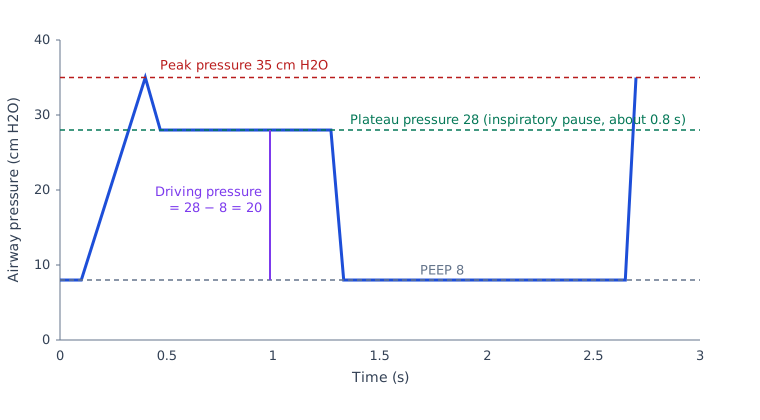
| Driving pressure | Plateau − PEEP | As low as feasible | Tracks lung stretch; trend it, not one value |
| Static compliance | Vt ÷ (plateau − PEEP) | Trend from baseline | Falls with ARDS, pneumothorax, distension or a stiff chest |
| Airway resistance | (Peak − plateau) ÷ flow | Trend from baseline | Rises with secretions, kinks, bite and bronchospasm |
| Predicted body weight | Height and sex formula | Set tidal volume from this | Actual weight overestimates lung size in obesity |
Circuit Pressures & Console
Normal
- Volume control: flow comes in as a square wave. Pressure spikes briefly, then holds flat on the plateau during the inspiratory pause. Expiratory flow is back at zero before the next breath.
- Pressure control: pressure hits target fast and holds. Flow slows to zero before the next breath.
- Pressure support: the patient's effort shows as a small negative deflection just ahead of the machine breath.
Abnormal Patterns
Asynchrony terms come from the two 2026 scoping reviews on patient-ventilator asynchrony, listed in the References.
| Pattern | What You See | Cause | Fix |
|---|---|---|---|
| High peak, normal plateau | Peak rises; plateau unchanged | Airway resistance: secretions, kink, bite, bronchospasm, small tube | Suction, check circuit and bite block, treat bronchospasm per order |
| High peak and high plateau | Both rise together | Lower compliance: pneumothorax, mainstem intubation, distension, edema, ARDS | Check breath sounds, tube depth and chest; treat the cause per protocol |
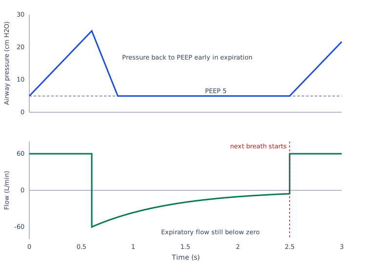
| Expiratory flow not back to zero | Next breath starts before flow ends | Auto-PEEP (dynamic hyperinflation) | Lower rate or tidal volume, lengthen expiration, relieve obstruction; disconnect to deflate the lungs if unstable |
| Ineffective triggering | Patient effort with no machine breath | Trigger too insensitive, auto-PEEP, sedation shift | Adjust trigger, check auto-PEEP, reassess sedation |
| Double triggering | Two machine breaths for one patient effort | Mandatory breath lands during an effort; common in volume control | Review rate and sedation; adjust mode per protocol |
| Auto-triggering | Machine breath with no patient effort | Water or leak in the circuit, cardiac oscillations, trigger too sensitive | Drain water, check for leaks, adjust trigger sensitivity |
| Flow starvation | Patient effort meets a scooped (concave) pressure curve; patient is short of air | Set inspiratory flow too low for the patient's demand in volume modes | Raise inspiratory flow per protocol or change mode; reassess effort and sedation |
| Premature cycling | Machine ends the breath while the patient is still inhaling | Cycle criterion too sensitive, leak, or patient effort larger than the set breath | Adjust cycle criterion per protocol; check for leaks; reassess the patient |
| Delayed cycling | Patient has begun to exhale while the machine keeps delivering inspiratory flow | Cycle criterion too low, or pressure support too high | Adjust cycle criterion or pressure support per protocol; reassess sedation |
| Reverse triggering | A patient effort follows a passive machine breath | Strong respiratory drive or light sedation; the machine breath triggers the patient's own effort | Reassess sedation and drive per protocol; review mode and settings |
Loops
- Flow-volume loop, leak: the expiratory limb should be back at zero flow before the next breath. If the loop does not close, think leak.
- Flow-volume loop, sawtooth: a jagged, sawtooth expiratory limb points to secretions or water in the circuit.
- Flow-volume loop, obstruction: a scooped expiratory limb points to airflow obstruction, as in asthma or COPD.
- Pressure-volume loop, beaking: an upper inflection on the inspiratory limb points to overdistension. Reduce tidal volume and check the plateau.
Monitoring & Ongoing Care
| Frequency | What to Check | Expected or Action Threshold |
|---|---|---|
| Hourly | Chest rise, breath sounds, SpO₂, EtCO₂ waveform, peak and plateau, exhaled volume | Plateau at or below 30 cm H₂O (ARDS); act on any new alarm |
| Every 4–6 h | ETT depth at the lip, cuff pressure, humidifier and water traps | Cuff 20–30 cm H₂O; re-check after any move |
| Every 12–24 h | Blood gas trend (PaO₂/FiO₂, PaCO₂, pH), FiO₂ and PEEP, sedation target | |
| Daily | Sedation interruption, weaning screen, oral care, head-of-bed position | Light sedation target (2018 PADIS guideline); pneumonia-prevention bundle |
Positioning & Mobility
- Keep the head of the bed raised as the bundle requires; log-roll and secure the tube when turning.
- Check the circuit and tube after every position change.
Anticoagulation & Hematology
The ventilator does not deliver anticoagulants. Bleeding hits ventilation three ways. Suctioning and tube changes can bleed from the airway. A pneumothorax in an anticoagulated patient can bleed into the pleural space. Hemoptysis can occlude the tube. Before chest decompression or a tube change, confirm the patient's anticoagulant status.
- Anticoagulants such as heparin are High-alert. Confirm the agent and dose on the order.
- Plan suction and tube changes with the team when a patient is heavily anticoagulated.
Key Medications & Drug Considerations
| Medication | Role Here | Typical Adult Dose / Target | Ventilator-Specific Notes |
|---|---|---|---|
| Propofol | Sedation of ventilated patients | Titrate to the sedation target; see medication page | High-alert Can drop blood pressure after a change in positive pressure; check the circuit before blaming the drug |
| Dexmedetomidine | Light sedation with less respiratory depression | Label (adult ICU sedation): 1 mcg/kg over 10 minutes, then 0.2–0.7 mcg/kg/hr IV infusion; many protocols omit the load | High-alert Bradycardia and hypotension; do not confuse concentrations |
| Rocuronium | Neuromuscular blockade for intubation or ventilator synchrony | RSI: 1 mg/kg is a common dose within the label range of 0.6–1.2 mg/kg IV. Ventilator synchrony: per ICU protocol; the label starts an infusion at 10–12 mcg/kg/min only after early recovery from the intubating dose (0.6 mg/kg). IO use per protocol (see rocuronium page) | High-alert Paralysis hides effort and dyssynchrony; the patient must be sedated first |
| Albuterol | Bronchospasm and obstruction-related auto-PEEP | Per order; see medication page | Treats resistance, not compliance; reassess the peak and expiratory flow after each dose |
| Norepinephrine | Hypotension from positive pressure, auto-PEEP or disconnect | Titrate to the MAP target; see medication page | High-alert Treat the ventilator cause at the same time |
Doses vary by protocol; follow local protocols and medical direction.
Alarms & Troubleshooting
| Alarm or Problem | What You See | Likely Causes | First Actions |
|---|---|---|---|
| High pressure (peak) | Peak alarm; breath stops or cycles early | Secretions, kink, bite, bronchospasm, cough, mainstem intubation, pneumothorax | 1) Look and listen. 2) Suction. 3) Check the tube for kinks and bite. 4) If still high, disconnect and bag, then reassess. |
| Low pressure or disconnect | Low pressure, low exhaled volume | Circuit disconnect, cuff leak, tube out of position | 1) Check connections and cuff. 2) If the tube is out, bag and re-intubate per airway protocol. |
| Low exhaled volume (leak) | Exhaled volume below set volume | Cuff underinflated, circuit or humidifier leak | Check cuff pressure (20–30 cm H₂O), circuit joins and humidifier seal |
| Apnea | No breath within the set time | Patient apnea, deep sedation, disconnect | Stimulate the patient, bag with oxygen, check connection |
| Desaturation or EtCO₂ drops | SpO₂ falls; EtCO₂ waveform flat or lower | Tube displaced, disconnect, obstruction, pneumothorax | Bag with oxygen, check tube, then work through DOPES below |
| High respiratory rate | Patient breathing faster than set rate | Pain, anxiety, hypoxemia, sepsis, auto-PEEP, trigger too sensitive | Assess the patient first; then check trigger, sedation and auto-PEEP |
| Low PEEP | Low PEEP alarm; set PEEP not reached | Leak at the cuff or circuit, disconnect, PEEP valve fault | 1) Check connections and cuff. 2) Check the PEEP valve and water traps. 3) Bag if oxygenation falls. |
| Low minute ventilation | Low minute-volume alarm; EtCO₂ rising | Apnea, deep sedation, leak, disconnect, tube out | 1) Look at the patient and the chest. 2) Check connections and the EtCO₂ waveform. 3) Bag if the patient is not breathing adequately. |
| Power or console failure | Alarm, blank screen | Mains loss, depleted battery, console fault | Switch to backup power; bag with oxygen; use a spare ventilator if needed |
DOPES: Sudden Deterioration on a Ventilator
- D, Displacement or disconnection: tube out, cuff deflated, or circuit apart. Check the EtCO₂ waveform and tube depth.
- O, Obstruction: kinked, plugged or bitten tube. Secretions. A mucus plug.
- P, Pneumothorax: rising pressure, falling SpO₂ and one-sided breath sounds. If blood pressure falls too, think tension (see Emergencies).
- E, Equipment failure: a ventilator, humidifier or valve fault. Switch to a bag.
- S, Stacked breaths: auto-PEEP is trapping air. Disconnect to let the lungs empty.
Complications & Red Flags
| Complication | How You Recognize It | Immediate Action | Prevention |
|---|---|---|---|
| Barotrauma and pneumothorax | Sudden peak rise, falling SpO₂, one-sided breath sounds, hypotension if tension | Disconnect, bag, decompress per protocol | Lung-protective tidal volume and low driving pressure (ARDS Network trial; pooled ARDS driving-pressure analysis) |
| Hypotension from positive pressure or auto-PEEP | Blood pressure falls when PEEP rises or after disconnect | Check for auto-PEEP; disconnect briefly; vasopressor per order | Lower rate, adequate sedation, volume status per order |
| Ventilator-induced lung injury | Rising driving pressure or falling compliance over hours to days | Lower tidal volume and driving pressure per protocol | Lung-protective settings from the first breath |
| Aspiration around the cuff | Secretions above the cuff; cough; new infiltrate | Suction; check cuff pressure | Cuff 20–30 cm H₂O (usual target); oral care |
| Tube displacement or extubation | EtCO₂ waveform lost; sudden desaturation | Bag with oxygen; re-intubate per airway protocol | Secure the tube; check depth at the lip after every move |
| Ventilator-associated pneumonia | Fever, new infiltrate, purulent secretions | Cultures and antibiotics per protocol | Head-of-bed position, oral care, cuff pressure per bundle |
- Blood pressure falls as peak pressure rises, with falling SpO₂ or one-sided absent breath sounds
- SpO₂ keeps falling despite more FiO₂
- No EtCO₂ waveform on an intubated patient
- Air leak or bubbling in a chest drain after a ventilator change
Emergencies: Arrest, Power Loss & Device Failure
Sudden Hypoxemia or Hypotension on the Ventilator (DOPES)
- Disconnect and bag with 100% oxygen to rescue the patient first (Paramedic/RN/CCT, per protocol). If the patient needs PEEP, use a PEEP valve on the bag.
- Check the tube: depth, cuff and EtCO₂ waveform.
- Listen to both chests. One-sided absent breath sounds with rising pressure and hypotension point to tension pneumothorax: decompress per protocol.
- Suction the tube if it is obstructed.
- If the tube is out and the patient is unstable, re-intubate per the airway protocol.
- Call for help and check the console only after the patient is oxygenating.
Cardiac Arrest on the Ventilator
- Start CPR per AHA adult and ACLS protocol.
- With an advanced airway in place, give 1 breath every 6 seconds (10 breaths/min) with continuous chest compressions (2025 AHA Guidelines for CPR and ECC, Part 9; Class 2b, LOE C-LD).
- Confirm the tube with waveform capnography (2025 AHA Guidelines for CPR and ECC, Part 9; Class 1, LOE C-LD).
- If the ventilator is not delivering reliable breaths, or auto-PEEP is suspected (breath stacking), disconnect and bag with 100% oxygen at 1 breath every 6 seconds without pausing compressions.
- Follow reversible causes, including the DOPES list above.
Power or Ventilator Failure
- Switch to battery or backup power on the console.
- If there is no backup, bag with oxygen and watch chest rise.
- Check the tube and circuit for disconnect or kink.
- Use a spare ventilator with the same tube if one is available.
- Keep EtCO₂ monitoring connected throughout.
Circuit Separation or Tube Out
- Bag immediately with oxygen.
- Do not reconnect until the tube position is confirmed by EtCO₂ and breath sounds.
- Suction and check the cuff.
- Re-intubate per protocol if the tube is out; see the intubation guide.
Weaning & Discontinuation
Readiness Criteria
- The cause of respiratory failure is treated or improving.
- Oxygenation adequate on low FiO₂ and PEEP per protocol.
- Stable on no or low-dose vasopressor support.
- Able to protect the airway and clear secretions.
Weaning Steps
- Run a ventilator liberation protocol, with daily sedation interruption and a spontaneous breathing trial (ATS/ACCP 2017 guideline).
- Reduce pressure support or move to a trial per protocol; watch for patient-ventilator mismatch.
- Before extubation in patients at higher risk of post-extubation stridor, consider a cuff leak test (conditional recommendation, ATS/ACCP 2017 guideline).
Failure Criteria: Step Back Up
- Rising work of breathing, tachypnea, accessory muscle use or paradoxical breathing.
- Falling SpO₂ or rising PaCO₂ on the trial.
- Agitation or new dyssynchrony that sedation does not fix.
Removal & Post-Removal Care
- Suction before extubation; watch for stridor and airway edema after removal.
- Keep humidified oxygen available and the patient upright unless the order says otherwise.
Transport Considerations
Before You Move
- Check battery run time and the backup power source.
- Check oxygen cylinder contents and duration at the planned flow.
- Secure the tube, circuit and all lines; carry a spare bag-valve.
- Confirm alarm limits and EtCO₂ monitoring.
- Brief the receiving team on settings, sedation and recent alarms.
If You Are Not the Device Specialist
- Watch SpO₂, EtCO₂, blood pressure and chest rise.
- Do not change the mode or settings without an order.
- If the screen goes blank, bag the patient with oxygen.
- Do not silence alarms you do not understand; call for help.
During Transport
- Recheck tube depth and cuff pressure after every move and after any change in altitude.
- Keep connections short and secured; avoid tugging the circuit.
Air Medical & Altitude
Gas in the cuff and the circuit expands as ambient pressure falls with altitude (Boyle's law). That is why cuff pressure rises on ascent and falls on descent. Every altitude change means a fresh manometer check of the cuff. Whether the transport ventilator compensates for altitude depends on the device. Some services inflate the cuff with saline instead. Follow local protocol.
Handoff
- Mode and working settings, including PEEP and FiO₂.
- Last plateau pressure, last blood gas and recent alarms with the fix.
- Sedation, paralysis and anticoagulation status.
- Tube size, depth, cuff pressure and any leak.
Special Populations
Pediatric & Neonatal
The adult 6 mL/kg target and the adult predicted-body-weight formulas do not apply to children. For pediatric ARDS, the PALICC-2 executive summary suggests tidal volume 6–8 mL/kg predicted body weight. Go below 6 mL/kg only as needed to keep plateau pressure at or below 28 cm H₂O and driving pressure at or below 15 cm H₂O. These are conditional recommendations. Below 4 mL/kg, go only with caution. Tube choice and the rest of pediatric ventilation run on the institutional pediatric protocol. Pediatric weight-based dosing is on the weight-based dosing page. See also pediatric airway.
Other
- Obesity: calculate tidal volume from predicted body weight, not actual weight.
- Pregnancy: follow the obstetric critical care protocol. Don't apply adult targets without it. Left uterine displacement relieves aortocaval compression from about 20 weeks. Normal pregnancy PaCO₂ runs about 30 mmHg. Avoid hypercapnia. Avoid over-correcting it too, because a low PaCO₂ can reduce uterine blood flow. The obstetric team sets maternal oxygenation targets. Avoid maternal hypoxemia, which harms the fetus.
Bedside Pearls & Teaching Script
Systematic Walk-Through
- Is the patient breathing and oxygenating? Look and listen first.
- Check the tube position and the EtCO₂ waveform.
- Read the pressure waveform: peak against plateau.
- Read the expiratory flow for auto-PEEP.
- Check the circuit, humidifier and water traps.
- Check the console settings and alarm limits.
- Decide: fix the cause, bag, or call for help.
Pearls
- A rising peak with a normal plateau points to the airway. A rising plateau points to the chest and the lung.
- Bag first when the patient gets worse, not when the alarm gets loud.
- Auto-PEEP hides on most screens. Look at expiratory flow. Measure it with an end-expiratory hold.
- Paralysis hides the patient's effort. Re-check the waveform after rocuronium.
References
- Acute Respiratory Distress Syndrome Network; Brower, R. G., Matthay, M. A., Morris, A., Schoenfeld, D., Thompson, B. T., & Wheeler, A. (2000). Ventilation with lower tidal volumes as compared with traditional tidal volumes for acute lung injury and the acute respiratory distress syndrome (landmark trial). New England Journal of Medicine, 342(18), 1301–1308. https://doi.org/10.1056/NEJM200005043421801
- Amato, M. B., Meade, M. O., Slutsky, A. S., Brochard, L., Costa, E. L., Schoenfeld, D. A., Stewart, T. E., Briel, M., Talmor, D., Mercat, A., Richard, J.-C., Carvalho, C. R., & Brower, R. G. (2015). Driving pressure and survival in the acute respiratory distress syndrome. New England Journal of Medicine, 372(8), 747–755. https://doi.org/10.1056/NEJMsa1410639
- Fan, E., Del Sorbo, L., Goligher, E. C., et al. (2017). An official American Thoracic Society/European Society of Intensive Care Medicine/Society of Critical Care Medicine clinical practice guideline: Mechanical ventilation in adult patients with acute respiratory distress syndrome. American Journal of Respiratory and Critical Care Medicine, 195(9), 1253–1263. https://doi.org/10.1164/rccm.201703-0548ST
- Leigh-Smith, S., & Harris, T. (2005). Tension pneumothorax: time for a re-think? Emergency Medicine Journal, 22(1), 8–16. https://pubmed.ncbi.nlm.nih.gov/15611534/
- Girard, T. D., Alhazzani, W., Kress, J. P., et al. (2017). An official American Thoracic Society/American College of Chest Physicians clinical practice guideline: Liberation from mechanical ventilation in critically ill adults. Rehabilitation protocols, ventilator liberation protocols, and cuff leak tests. American Journal of Respiratory and Critical Care Medicine, 195(1), 120–133. https://doi.org/10.1164/rccm.201610-2075ST
- Devlin, J. W., Skrobik, Y., Gélinas, C., et al. (2018). Clinical practice guidelines for the prevention and management of pain, agitation/sedation, delirium, immobility, and sleep disruption in adult patients in the ICU. Critical Care Medicine, 46(9), e825–e873. https://doi.org/10.1097/CCM.0000000000003299
- Marini, J. J. (2026). Airflow obstruction and dynamic hyperinflation. Clinics in Chest Medicine, 47, 499–510. https://doi.org/10.1016/j.ccm.2026.04.005
- Taran, S., Citerio, G., Taccone, F. S., et al. (2026). Dynamic driving pressure and clinical outcomes in mechanically ventilated patients with acute brain injury: a secondary analysis of the VENTIBRAIN study. Intensive Care Medicine, 52(1), 31–41. https://doi.org/10.1007/s00134-025-08241-0
- Popayán, A. M. E., Vaporidi, K., Parada-Gereda, H. M., et al. (2026). Challenges and strategies for the identification and correction of patient-ventilator asynchronies in daily practice: a scoping review. Acute and Critical Care, 41, 493–505. https://doi.org/10.4266/acc.006050
- Perez-Garzon, M., Robayo-Amortegui, H., Fernandez-Sarmiento, J., et al. (2026). Classification of patient-ventilator asynchronies, development of an updated conceptual framework: scoping review. Respiratory Medicine, 262, 109081. https://doi.org/10.1016/j.rmed.2026.109081
- Emeriaud, G., López-Fernández, Y. M., Iyer, N. P., et al. (2023). Executive summary of the second international guidelines for the diagnosis and management of pediatric acute respiratory distress syndrome (PALICC-2). Pediatric Critical Care Medicine, 24(2), 143–168. https://doi.org/10.1097/PCC.0000000000003147
- American Heart Association. (2025). Part 9: Adult advanced life support: 2025 American Heart Association guidelines for cardiopulmonary resuscitation and emergency cardiovascular care. Circulation, 152. https://doi.org/10.1161/cir.0000000000001376
Medical Disclaimer
- For Educational Purposes Only: This content is intended for educational reference and should not be used for clinical decision-making.
- Not a Substitute for Professional Judgment: Always consult your local protocols, institutional guidelines, and supervising physicians.
- Verify Before Acting: Users are responsible for verifying information through authoritative sources before any clinical application.