

Bedside Snapshot
- Recognize
- Vaginal bleeding late in pregnancy. Painless bright-red bleeding suggests placenta previa; painful bleeding with a tender or firm uterus suggests abruption. Visible loss can under-count.
- Red Flags
- Shock index 0.9 or higher (over 1 is critical), syncope or hypotension; firm, tender or tetanic uterus; fetal heart-rate abnormality; sudden pain with loss of contractions after prior uterine surgery
- First Actions
- Call for obstetric help early
- No digital vaginal exam if previa is possible
- IV access and monitoring; fluids per protocol
- Measure and record blood loss
- Transport to obstetric care
- Key Doses
- Rho(D) immune globulin 300 mcg IM or IV within 72 hours for non-sensitized Rh(D)-negative or unknown Rh status; more if fetal RBCs exceed 15 mL
- Don't Miss
- A digital exam that triggers previa bleeding; abruption with a normal ultrasound; uterine rupture mistaken for abdominal pain; Rh(D)-negative status not checked
- Escalate & Transport
- Obstetric facility with cesarean capability, blood bank and neonatal team; higher level of maternal care for refractory bleeding or fetal compromise
Definition & Pathophysiology
Antepartum hemorrhage (APH) is bleeding from the genital tract at or after 24+0 weeks of gestation (the RCOG definition) and before birth. Here's the catch: US practice and local protocols may set a different gestational cutoff, so check the protocol. Bleeding before that point goes down the early-pregnancy or miscarriage pathway. APH is a sign. It is not a diagnosis. Three causes matter most: placenta previa, placental abruption and uterine rupture. Their presentations overlap. Treat the bleeding and the fetal status first, and sort out the cause in the hospital.
Placenta previa means the placenta lies over or near the internal cervical os. As the lower uterine segment stretches, the placenta pulls away from the placental bed. That is where the blood comes from.
Placental abruption is the placenta separating from the decidua before birth. About 0.4% to 1.0% of all pregnancies are reported with abruption. The mechanism is still not fully understood. Blood can leave through the cervix, stay hidden behind the placenta, or do both.
- Placenta previa: the classic pattern is painless, bright-red bleeding. It can recur, and it can start with no warning
- Placental abruption: bleeding with abdominal pain, a tender or tetanic (firm, contracting) uterus, or fetal heart-rate abnormalities. Don't trust the visible loss. It may under-estimate the blood lost
- Uterine rupture: a tear in the uterine wall. Most often it happens in a patient with a prior cesarean birth who is in labor. Watch for sudden pain, loss of fetal station and fetal distress. It can also cause massive bleeding
- Vasa previa: fetal vessels cross the membranes over the internal os with no cord or placenta to protect them. When the membranes rupture, that can cause fetal hemorrhage. It is on this list because it can look like painless bleeding at the moment the membranes rupture
Gestational age, fetal status and maternal hemodynamic stability set the pathway. Mother and fetus are treated together. That means the mother's hemodynamics come first.
Recognition & Red Flags
Presentations
- Painless bleeding: bright-red vaginal bleeding with no pain and no uterine tenderness. Typical of previa
- Painful bleeding with a firm or tender uterus: typical of abruption. It can stay concealed, with little blood showing
- Painful bleeding with a change in fetal heart rate or sudden loss of contractions: consider uterine rupture or severe abruption
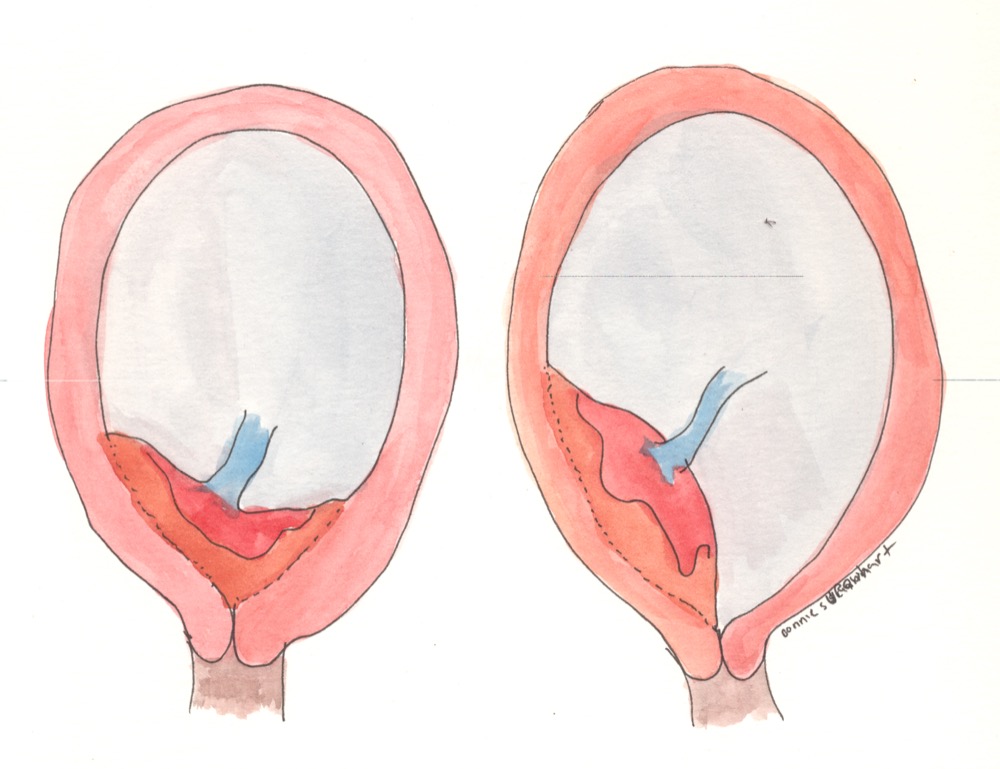
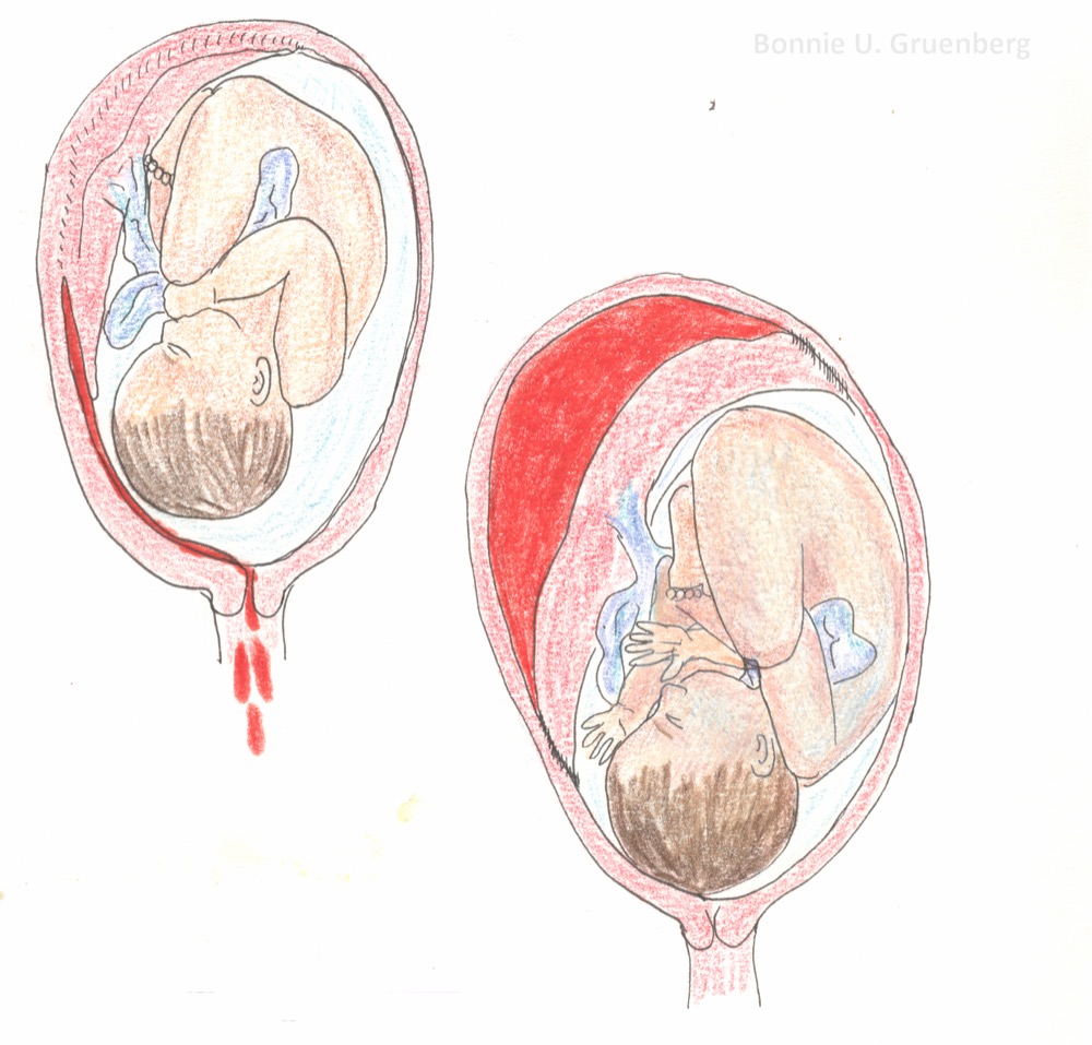
Red Flags
- Any vaginal bleeding in late pregnancy (see the gestational cutoff in Definition), with any sign of hypovolemia: syncope, pallor, cool skin, tachycardia, or a shock index of 0.9 or higher (over 1 is critical)
- A firm, tender or tetanic uterus with bleeding, or abdominal pain out of proportion to the visible blood
- Fetal heart-rate abnormality, or fetal movement that has stopped, with bleeding
- Sudden severe abdominal pain with loss of contractions, or a palpable fetal part in an abnormal place, in a patient with prior uterine surgery
- Heart rate and shock index can climb before the pressure drops. A normal blood pressure does not rule out major loss in pregnancy
Assessment & Diagnostics
In the field, you are assessing hemodynamics and fetal status. Imaging and labs come after handoff, so do not delay transport for them.
| Check | Why | Act If |
|---|---|---|
| Vital signs and shock index | Shock index (pulse divided by systolic BP) shows hypovolemia before BP falls. Pregnancy raises heart rate and lowers BP, so normal values run higher than in non-pregnant adults. In a postpartum cohort, mean shock index was 0.68 to 0.74 in the first two hours; in vaginal births needing transfusion for PPH it was about 0.9 at 30 minutes. Judge the trend as well as the value | Shock index 0.9 or higher (over 1 is critical), a rising trend, or new hypotension: escalate and reassess at each intervention |
| Visible blood loss | Visual estimates under-count. Concealed abruption can hide the blood | Weigh pads or estimate per protocol and record time. Treat the hemodynamics, not the estimate |
| Uterine tone and pain | A firm, tender or tetanic uterus suggests abruption. Pain with bleeding is a feature of abruption | Tender or tetanic uterus: transport to obstetric care as an abruption. Do not palpate for previa |
| Fetal status | Fetal heart-rate abnormality changes the urgency of delivery | Abnormal fetal heart rate or no fetal movement: notify the receiving obstetric team now |
| Fibrinogen, platelets, hemoglobin, PT/INR | Abruption and massive loss can cause coagulopathy. Early values guide replacement | |
| Rh type | Non-sensitized Rh(D)-negative patients need immune globulin after bleeding. A positive antibody screen alone does not show active sensitization: it can be passive anti-D from an earlier Rh immune globulin dose, which the receiving team sorts out with titers and history | Rh(D)-negative or unknown: see the Rho(D) step in ED and ICU Management |
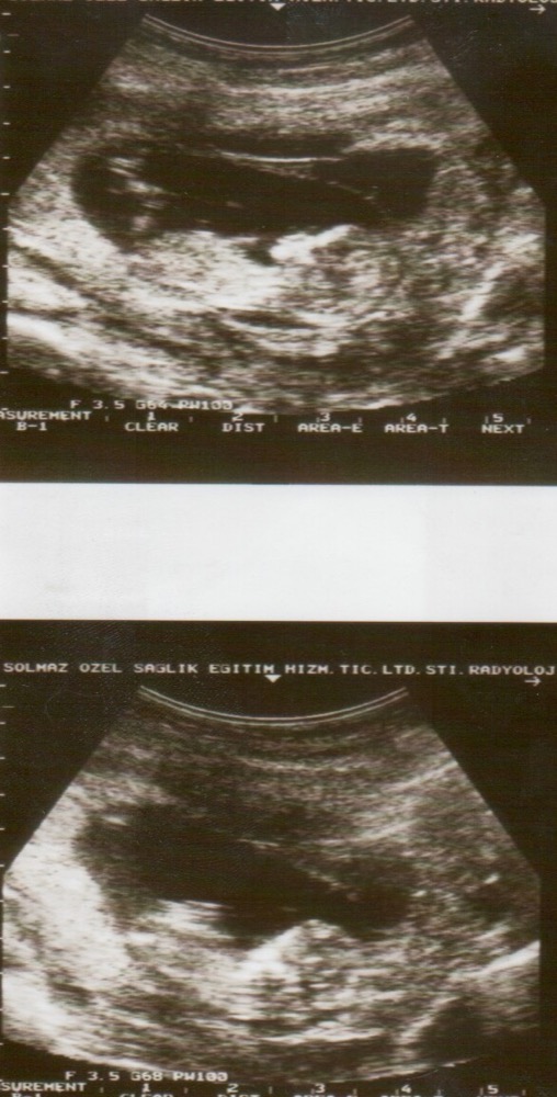
Imaging in the hospital establishes placenta location, by transabdominal or transvaginal ultrasound. A visual check in the field is no substitute. The radiology reviews of the contemporary placenta previa classification are in References. Report the location and the imaging result at handoff.
Differential & Mimics
- Postpartum hemorrhage: bleeding after birth. Go to Postpartum Hemorrhage and Uterine Inversion
- Cervical or vaginal lesion: a bleeding source low in the genital tract can cause painless bleeding. A firm uterus with no pain or contractions points here. Even so, skip the digital exam if previa is possible
- Bloody show of labor: small-volume, blood-tinged mucus that comes with contractions. Heavy bleeding is not bloody show
- Trauma: abdominal or pelvic trauma causes abruption or uterine injury. Treat the trauma per protocol, on the trauma pathway
BLS Management
- Call for ALS and obstetric help early. Give the receiving facility the bleeding, gestational age and fetal status.
- Do not perform a digital vaginal exam when placenta previa is possible. Do not put anything into the vagina.
- Position for hemodynamics. If the uterus is at or above the umbilicus (about 20 weeks or later), tilt the patient to the left (about 15 to 30 degrees) or displace the uterus manually to the left. Avoid the supine position. Lying flat compresses the aorta and vena cava. If spinal injury is suspected, keep spinal precautions and use manual displacement. Keep the patient warm.
- Oxygen: give oxygen for SpO₂ below 94% or hypoxia, and keep SpO₂ at 94% or higher per local protocol. Oxygen cannot stand in for restoring perfusion with fluids, blood and hemorrhage control.
- Measure and record: weigh pads, record each measurement with the time, and note any clots or tissue.
- Vital signs: check pulse, BP, mental status and skin perfusion at each intervention and frequently while bleeding.
- Do not delay transport for drugs or IV access the crew cannot complete quickly. Continue monitoring in transit.
ALS Management
Doses vary by protocol; follow local protocols and medical direction. Bleeding and hemodynamics come first. Drugs and fluids run alongside them.
- IV access (Paramedic/RN/CCT): two large-bore IV lines, or IO access per protocol. See IO Access and IV Insertion
- Fluids (Paramedic/RN/CCT): crystalloid per protocol, with reassessment after each bolus. Watch for overload. In shock, get blood products going early. See Blood Products
- Rh(D)-negative or unknown Rh status (ED/ICU): give Rho(D) immune globulin 300 mcg IV or IM within 72 hours of the bleeding event, for a non-sensitized Rh(D)-negative patient (or unknown Rh status, pending typing). The label excludes patients with evidence of active anti-D sensitization. The receiving team tells that apart from passive anti-D from an earlier Rh immune globulin dose. So a positive antibody screen alone does not exclude the dose. The Rhophylac label lists obstetric complications as indications, transplacental hemorrhage from antepartum hemorrhage among them. See Rho(D) Immune Globulin
- Coagulopathy (Paramedic/RN/CCT, per protocol): replace fibrinogen and blood products per protocol when labs or ongoing bleeding show a deficit. See Fibrinogen
- Positioning (Paramedic/RN/CCT): for late-pregnancy patients, keep left lateral tilt or manual left uterine displacement as in BLS step 3 and avoid the supine position
- Cardiac arrest in pregnancy: follow the pregnancy-specific guidance in AHA 2025 Part 10 (pregnancy is listed among its special circumstances) and local protocol. Note the gestational age and call obstetric help early
- Drugs not routinely used prehospital: uterotonics such as oxytocin belong to postpartum hemorrhage. They are not the first step for antepartum bleeding. Don't give them to stop previa or abruption bleeding unless the obstetric team directs it. Tranexamic acid here depends on protocol and gets decided with obstetrics
ED & ICU Management
- Diagnosis: use transabdominal or transvaginal ultrasound to confirm placenta location before any vaginal exam. When ultrasound is normal, abruption stays a clinical diagnosis
- Delivery decision: gestational age, fetal status and maternal hemodynamic stability set the plan. Obstetrics makes the delivery call. When previa or abruption threatens the mother or fetus, cesarean delivery is the usual route
- Massive hemorrhage: activate the hospital's massive-hemorrhage protocol early. Check fibrinogen, platelets, hemoglobin and PT/INR and repeat per protocol. See Hemorrhagic Shock for the shock approach
- Rh(D) immune globulin: for non-sensitized Rh(D)-negative patients with transplacental hemorrhage, 300 mcg IV or IM within 72 hours. Send fetal-cell quantification (for example Kleihauer-Betke or flow cytometry). If it shows more than 15 mL of fetal RBCs, the Rhophylac label adds 20 mcg per mL above 15 mL; if the excess cannot be quantified, it adds an additional 300 mcg. See Rho(D) Immune Globulin
- Preterm delivery: antenatal corticosteroids and magnesium sulfate are obstetric calls, made with the receiving team per protocol
- Consults: obstetrics, anesthesia, neonatal team, blood bank and interventional radiology when available for refractory bleeding
- Disposition: admit to obstetric care with monitoring and blood available until bleeding is controlled and the fetus is stable
Transport Considerations
- Destination: pick the nearest facility that can give obstetric care, cesarean delivery, blood bank support and a neonatal team. Use regional maternal levels of care (Levels of Maternal Care, ACOG 2019). A patient who may need a higher level goes to a level III or IV center once stable enough.
- Do not: perform a vaginal exam, and do not delay transport for imaging or labs
- Continue in transit: monitoring, IV access, fluids, and blood product or replacement per protocol
- Position in transit: for late-pregnancy patients, keep left lateral tilt or manual left uterine displacement for the whole transport and avoid the supine position
- Measured losses: carry the pads and record every measurement with the time
- Handoff essentials: gestational age, time of bleeding onset, measured loss and trend, pain and tone, fetal status, Rh type, fluids and products given, and vital-sign trend
- Equipment: pads or a calibrated drape, IV and IO supplies, monitor with fetal-status awareness per local protocol, and blood product transfer per protocol
Special Populations
- Pediatrics: an adolescent who is pregnant is treated as an adult with an adult pregnancy, per protocol. Pediatric weight-based dosing does not apply to the mother. The newborn is covered on the Neonatal Resuscitation page
- Pregnancy: the fetus is part of every decision. Gestational age and fetal status drive delivery planning, and obstetrics owns that call. Mother's hemodynamics come first, because maternal shock threatens the fetus. From about 20 weeks, keep the patient off the supine position (left lateral tilt or manual left uterine displacement). This section sets no drug-specific adjustment beyond the dose and route on this page. Check the medication page before giving any drug
- Rh(D)-negative patients: give Rho(D) immune globulin 300 mcg IV or IM within 72 hours after bleeding if the patient is non-sensitized, per the dose rules above
- Hypertensive disorders: late-pregnancy bleeding with hypertension calls for the Preeclampsia and Eclampsia pathway as well. Keep the two problems separate on the handoff
- Patients on Anticoagulants: anticoagulant exposure raises bleeding risk. Reversal decisions follow the Anticoagulant Reversal reference and local protocol
Pitfalls & Safety Checks
- Error: doing a digital vaginal exam when placenta previa is possible. Don't. Lean on the abdominal exam, vital signs, fetal monitoring and imaging.
- Error: calling it a "minor" bleed because the pads look light. Concealed abruption and previa can cause large losses. Treat the shock index, not the pad count.
- Error: missing an abruption because the ultrasound looks normal. Part of the abruption call is clinical. A firm, tender uterus with bleeding is enough to transport as abruption.
- Error: missing Rh(D)-negative status. Give Rho(D) immune globulin 300 mcg IV or IM within 72 hours to a non-sensitized Rh(D)-negative patient. Check the Rh type and record it on the handoff.
- Error: leaving a late-pregnancy patient supine with hypotension. Use left lateral tilt or manual left uterine displacement.
- Error: giving uterotonics for antepartum bleeding as routine. Oxytocin and related drugs belong to postpartum hemorrhage. Ask obstetrics first.
- Error: holding transport for imaging, labs or IV access the crew cannot finish quickly. Transport to obstetric care is the treatment.
- Error: writing uterine rupture off as ordinary abdominal pain. Sudden pain with loss of contractions or fetal distress, in a patient with prior uterine surgery, needs immediate obstetric transport.
Clinical Pearls
References
- Jauniaux E, Bhide A, Hussein AM, Lees C, D'Antonio F, Robson SC, Kaelin Agten A, Jurkovic D; Royal College of Obstetricians and Gynaecologists. Placenta Praevia and Placenta Accreta Spectrum: Diagnosis and Management: Green-Top Guideline No. 27a. BJOG. 2026;133. https://doi.org/10.1111/1471-0528.70239
- Schneider E, Kinzler WL. Placental Abruption: Pathophysiology, Diagnosis, and Management. Clin Obstet Gynecol. 2025;68:98–104. https://doi.org/10.1097/GRF.0000000000000903
- Oyelese Y. Imaging of Antepartum and Postpartum Hemorrhage: Contemporary Classification of Placenta Previa. RadioGraphics. 2024;44. https://doi.org/10.1148/rg.240127
- American College of Obstetricians and Gynecologists. Obstetric Care Consensus No. 9: Levels of Maternal Care. Obstet Gynecol. 2019;134:e41–e55. https://doi.org/10.1097/AOG.0000000000003383
- Part 10: Adult and Pediatric Special Circumstances of Resuscitation: 2025 American Heart Association Guidelines for Cardiopulmonary Resuscitation and Emergency Cardiovascular Care. Circulation. 2025;152. https://doi.org/10.1161/CIR.0000000000001380
- RHOPHYLAC (Rho(D) immune globulin) prescribing information. CSL Behring. DailyMed, revised July 2025. https://dailymed.nlm.nih.gov/dailymed/drugInfo.cfm?setid=c1b5d801-336f-4541-a7d9-a8d4a110a4cc
- Royal College of Obstetricians and Gynaecologists. Antepartum Haemorrhage: Green-top Guideline No. 63 (2011). https://www.rcog.org.uk/guidance/browse-all-guidance/green-top-guidelines/antepartum-haemorrhage-green-top-guideline-no-63/
- Borovac-Pinheiro A, Ribeiro FM, Morais SS, Pacagnella RC. Shock index and heart rate standard reference values in the immediate postpartum period: a cohort study. PLoS One. 2019;14(6):e0217907. https://doi.org/10.1371/journal.pone.0217907
- Borovac-Pinheiro A, Pacagnella RC, Puzzi-Fernandes C, Cecatti JG. Case-control study of shock index among women who did and did not receive blood transfusions due to postpartum hemorrhage. Int J Gynaecol Obstet. 2018;140(1):93–97. https://doi.org/10.1002/ijgo.12343
Medical Disclaimer
- For Educational Purposes Only: This content is intended for educational reference and should not be used for clinical decision-making.
- Not a Substitute for Professional Judgment: Always consult your local protocols, institutional guidelines, and supervising physicians.
- Verify Before Acting: Users are responsible for verifying information through authoritative sources before any clinical application.