

Bedside Snapshot
- Recognize
- Sudden painless one-eye vision loss (CRAO); eye pain after chemical exposure or trauma; painful red eye with headache, nausea and blur (angle closure); new flashes or floaters (retinal detachment)
- Red Flags
- Reduced acuity, RAPD, peaked pupil or prolapsed iris or lens (open globe); proptosis and tense globe (orbital compartment syndrome); cherry-red spot (CRAO); hard eye on palpation
- First Actions
- Irrigate chemical exposure until pH is normal; rigid shield for suspected open globe, no pressure and no irrigation; lateral canthotomy for orbital compartment syndrome; time last known well for CRAO; ophthalmology the same day
- Key Doses
- Acetazolamide 500 mg IV once if canthotomy is not possible (OCS); angle closure: 250 mg every 4 h (label)
- Mannitol 1.5–2 g/kg IV over at least 30 min, once (IOP)
- Tetracaine 0.5%, 1 drop per eye, before irrigation
- Don't Miss
- Globe rupture with no visible wound; CRAO with a normal-looking early fundus; irrigating an open globe; delaying canthotomy, since sight can be lost within 90 minutes
- Escalate & Transport
- Ophthalmology the same day for all; stroke pathway for CRAO (4.5 h); open globe repair ideally within 24 h; orbital compartment syndrome to an ED with OR access; keep irrigating in transit until pH is normal
Definition & Pathophysiology
Six emergencies can take a patient's sight. EMS, ED and transport teams all meet them: chemical burns, open globe injury, orbital compartment syndrome from retrobulbar hemorrhage, central retinal artery occlusion (CRAO), acute angle-closure glaucoma and rhegmatogenous retinal detachment. Wait on any of them and the vision loss is permanent. The first move is different every time.
- Chemical burns: an acid or alkali on the ocular surface. Two things set the severity: the pH of the agent and how long it stays in contact. It is an ocular emergency and accounts for 11.5–22.1% of ocular injuries. Irrigate immediately and copiously. That is the first step every time.
- Open globe injury: a full-thickness hole in the eye wall. A sharp penetrating object can cause it, and so can blunt rupture. Blunt rupture most often happens behind the rectus muscle insertions, so the front of the eye may show no wound at all.
- Orbital compartment syndrome: bleeding behind the globe (retrobulbar hematoma) raises orbital pressure. That squeezes the retinal vessels, and the retina goes ischemic. Permanent sight loss can follow within 90 minutes of injury.
- CRAO: a sudden blockage of the central retinal artery. Think of it as a stroke of the eye. Over 90% are nonarteritic and thromboembolic. The risk factors match stroke: diabetes, hypertension, coronary and carotid disease, TIA and smoking.
- Acute angle-closure glaucoma: intraocular pressure (IOP) rises fast when the anterior chamber angle closes. Incidence is 2.2–4.1 per 100,000 persons per year in Europe. Markedly elevated IOP can damage vision permanently.
- Rhegmatogenous retinal detachment: a retinal tear lets fluid get under the neurosensory retina and lift it off the pigment epithelium. Macula-on and macula-off detachments carry very different visual prognoses.
Recognition & Red Flags
Chemical Exposure
- Pain, tearing, light sensitivity and reduced vision after exposure to an acid or alkali, whether liquid, powder or gas. Find out the agent and how much of it reached the eye.
- Severity: lid swelling, corneal clouding and a white eye (limbal ischemia) point to a deeper injury. The Roper-Hall grade, built from limbal ischemia and corneal haze, sets the prognosis.
Globe Rupture or Laceration
- Mechanism: blunt trauma, a sharp or impaled object, grinding, metal cutting or explosion. Think open globe with any eyelid laceration, any eye with reduced acuity or an impaired red reflex.
- Signs: a relative afferent pupillary defect (RAPD) is common, usually with hyphema. A peaked pupil, lost iris or lens, a "step" in the conjunctiva or extruded intraocular tissue is diagnostic. IOP may be low (collapsed globe) or raised if the patient presents late.
No visible wound does not exclude rupture: the break is most often at the back, behind the rectus insertions. Blunt orbital trauma with reduced vision is the cue. Suspect it.
Closed-Globe Injury and Hyphema
- Signs: blunt trauma with acute vision loss and no wound through the eye wall. Hyphema, blood in the anterior chamber, is common. Judge how much of the pupil it covers, and check the red reflex.
- Risk: about half of total hyphemas (52%) push IOP high enough to need surgery. Losing the red reflex may mean a severe hyphema, vitreous hemorrhage or retinal detachment.
Orbital Compartment Syndrome
- Signs: eye pain, reduced acuity or lost color sensitivity after blunt orbital injury. The eye is proptosed and tense. A RAPD is often present.
Central Retinal Artery Occlusion
- Presentation: sudden, painless, monocular vision loss. Acuity is usually under 20/200, and a RAPD is present in the affected eye.
- Classic findings: retinal whitening from edema, a cherry-red spot at the macula and a boxcar look to the retinal vessels.
- Early exam: a very early fundus exam may not show ischemia yet. A normal-looking fundus does not exclude CRAO.
- Arteritic cause: over 50 with jaw claudication, temporal headache or scalp tenderness? Suspect giant cell arteritis and treat it as urgent (see ED and ICU Management).
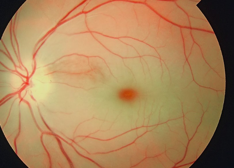
Painless vision loss is a stroke equivalent: sudden painless one-eye loss gets handled as CRAO. Stay on that path until ophthalmology or the stroke team tells you otherwise.
Acute Angle-Closure Glaucoma
- Symptoms: eye redness, worsening vision, other visual disturbances, headache and nausea. The symptoms do not always point to the eye, so patients often show up first to a non-ophthalmologist.
- Exam: a hard, tense globe on palpation points to markedly elevated IOP. You don't need to be an ophthalmologist to feel it.
Retinal Detachment
- Symptoms: new floaters and flashes of light. In one 2025 series, floaters were the top presenting complaint (75%), then flashes (38%).
- Field loss: a peripheral visual field defect that can expand over days. Macular involvement causes a large drop in central vision.
Red Flags
- Sudden painless monocular vision loss (CRAO) or painful vision loss with a proptosed, tense eye (orbital compartment syndrome)
- Eye trauma with reduced vision, a RAPD or an irregular pupil (open globe)
- Chemical exposure with pain and reduced vision
- Painful red eye with blurred vision, headache and vomiting (angle closure)
- New flashes, floaters or a visual field defect (retinal detachment)
Assessment & Diagnostics
Ocular emergencies are diagnosed at the bedside, not on a scan. Don't hold treatment for a test. Irrigation, shielding and the first drug doses don't depend on imaging.
| Check | Why | Act If |
|---|---|---|
| Visual acuity, pupils and color vision | A RAPD signals retinal or optic-nerve damage; common in open globe and orbital compartment syndrome | Reduced acuity or RAPD: treat as sight-threatening and escalate per this pathway |
| Ocular surface pH | Sets the end point for chemical irrigation | Test with litmus or universal indicator paper in the conjunctival fornix. Not neutral: keep irrigating and retest |
| Eyelid and globe inspection | Lacerations, proptosis, peaked pupil, hyphema, prolapse | Suspected open globe: rigid shield, no pressure, no irrigation, no removal of objects |
| Fluorescein with cobalt-blue light (Seidel test) | A dark stream diluting the fluorescein shows an aqueous leak | Positive: open globe. Not used when an open globe is obvious |
| Intraocular pressure | Raised in angle closure and orbital compartment syndrome | Contraindicated in suspected open globe. Otherwise measure if available; a hard globe on palpation needs ophthalmology the same day |
| CT of the orbit (noncontrast) | Orbital fractures, intraocular foreign bodies, retrobulbar hemorrhage | Use CT when the exam is limited. MRI is contraindicated if a metallic foreign body is suspected |
| Fundus exam and OCT | Cherry-red spot and retinal whitening; OCT detects inner retinal hyper-reflectivity | Suspected CRAO: rapid ophthalmic opinion, telemedicine if it speeds consultation |
| Time last known well, stroke screen and glucose | CRAO is treated within the stroke window | Within 4.5 h of onset with no contraindication: stroke team for thrombolysis per protocol |
Differential & Mimics
- Corneal abrasion: fluorescein stains the abrasion. A dark stream that dilutes the dye is an aqueous leak. That means open globe, not abrasion.
- Orbital swelling without vision loss: swelling alone does not fit the orbital compartment syndrome picture. Look for vision loss, RAPD, proptosis and a tense globe.
- CRAO with cilioretinal sparing: the cilioretinal artery can spare central vision. That leaves peripheral field loss only, with acuity of 20/50 or better. Peripheral-only loss still needs ophthalmic review.
- Angle closure versus headache or migraine: redness, blurred vision and a hard globe on palpation point to angle closure. Headache alone does not.
- Hyphema or vitreous hemorrhage after blunt trauma: this is a closed-globe injury, not a simple bruise. With a visual field defect, suspect retinal detachment.
BLS Management
- Chemical exposure: get the agent off the face and out of the eye, and start irrigation at once with drinking water, 0.9% sodium chloride or Ringer's solution. Pull contact lenses only if it is quick and doesn't delay irrigation. Don't wait for pH testing, a neutralizing agent or the ED. Irrigation is the treatment.
- Suspected open globe: don't press on the eye, evert the lids, pull out an impaled object or irrigate. Put a rigid eye shield on, over both eyes if bilateral injury is suspected.
- Blunt eye injury without an open globe: don't press on the eye. Keep the patient still and calm, and raise the head of the bed 45 degrees when spinal precautions allow. Report hyphema or vision loss, and any NSAID or aspirin use.
- Limit vomiting and straining: vomiting raises IOP and can worsen an open globe. Antiemetic per protocol (see ALS).
- Sudden painless vision loss in one eye: record the time last known well and run the stroke screen per local protocol. Report it as possible CRAO.
- Painful red eye with headache, nausea or blurred vision: report it early as possible angle closure. Don't treat it as a routine headache.
- Orbital injury with a bulging or tense eye and vision loss: avoid pressure on the orbit, keep the patient calm and call early for orbital compartment syndrome.
- Flashes, floaters or new visual field loss: arrange urgent ophthalmology referral.
- Report: the agent and concentration, time of exposure or injury, pH if known, vision before and after, and any drops given.
ALS Management
Doses vary by protocol; follow local protocols and medical direction. Ophthalmology or ED care is where most ocular emergencies end up. Treat what follows as early work only.
- Pre-dose checks for acetazolamide and mannitol (Paramedic/RN/CCT): acetazolamide: check for sulfonamide hypersensitivity, and don't give it with depressed serum sodium or potassium, adrenal (suprarenal) failure, hyperchloremic acidosis, cirrhosis or marked kidney or liver disease (label). Mannitol: don't give it with anuria, severe hypovolemia, severe pulmonary congestion or edema, or active intracranial bleeding (label). Mannitol can cause or worsen hypovolemia. So avoid or defer it in hypotensive or hypovolemic trauma, and when intracranial bleeding is suspected, unless the treating team directs otherwise. If a contraindication applies, reach for another IOP-lowering approach with medical direction, or definitive decompression.
- Chemical exposure (Paramedic/RN/CCT): tetracaine 0.5%, 1 drop per eye, for analgesia before irrigation (dose from the E3 tetracaine page; EXTRACCT names tetracaine without a dose). See Tetracaine. Keep irrigating until the conjunctival-fornix pH is normal. Use at least 3 L if pH paper is unavailable. Evert the lids and sweep the fornices to clear out particles. If you're using IV tubing, run it onto a nasal cannula placed at the inner canthus. Don't use IV 1% lidocaine as an eye drop, because its pH is about 6.
- Open globe: antiemetic (Paramedic/RN/CCT): ondansetron 4–8 mg IV or PO, repeated per protocol to prevent emesis (EXTRACCT: up to three times daily). Screen for long QT before you give it (see the E3 ondansetron page). Ondansetron is the preferred choice over metoclopramide. See Ondansetron. Analgesia per protocol; see Fentanyl.
- Open globe: antibiotics and tetanus (Paramedic/RN/CCT): IV or oral antibiotics per the local ophthalmology or emergency protocol (for example, a fluoroquinolone when no ophthalmologist is available, per EXTRACCT), plus tetanus prophylaxis for a penetrating wound. Don't hold up transfer for drugs.
- Orbital compartment syndrome: lateral canthotomy (per local protocol and credentialing): a sight-saving bedside procedure that non-ophthalmologists can perform. Infiltrate the lateral skin with 1% lidocaine (with or without epinephrine 1:200,000). Clamp the skin for 1 minute. Make a 1 cm incision toward the orbital rim, and cut the inferior canthal tendon. If the globe does not recess, cut the superior limb. Then check pupils and acuity, start antibiotics and refer urgently. See Lidocaine.
- Orbital compartment syndrome: osmotic and carbonic anhydrase drugs (Paramedic/RN/CCT): if you can't do a canthotomy locally, give acetazolamide 500 mg PO or IV as a single dose (EXTRACCT), after the pre-dose checks above, then transfer. Mannitol 1.5–2 g/kg IV over at least 30 minutes, once, lowers IOP (label dose). EXTRACCT 2026 uses 1 g/kg over 30–60 minutes for raised IOP after closed-globe trauma. Its orbital compartment section does not list mannitol. The dose follows local protocol and medical direction. See Mannitol.
- Acute angle-closure glaucoma (Paramedic/RN/CCT): acetazolamide IV for rapid relief, after the pre-dose checks above. The label prefers 250 mg every 4 hours. An initial 500 mg, then 125–250 mg every 4 hours, is the label alternative for some acute cases. The label prefers direct IV administration. Reconstitute each 500 mg vial with at least 5 mL sterile water. Don't give it intramuscularly. EXTRACCT's oral regimen for raised IOP is 500 mg PO, then 250 mg four times daily. It is contraindicated in sickle cell disease or trait. The label indication is an adjunct where surgery is delayed. Ophthalmology gives the definitive treatment. Mannitol 1.5–2 g/kg IV over at least 30 minutes (20% solution: 7.5–10 mL/kg), after its checks. Avoid pupil-dilating drugs (mydriatics and anticholinergics) in suspected angle closure. Ophthalmology starts the topical miotic and beta-blocker drops.
- CRAO (Paramedic/RN/CCT, per stroke protocol): thrombolysis within 4.5 hours of onset when there are no contraindications. It is a weak recommendation based on very low-quality evidence. Your part is the time last known well, glucose and rapid transfer to the stroke pathway.
- CRAO: IOP-lowering measures (ED): ocular massage (compress the globe for 10–15 seconds, then release, repeated per protocol), acetazolamide 500 mg IV and mannitol 1.5–2 g/kg IV, after the pre-dose checks. None of them has been shown to change final vision.
- Retinal detachment: no ALS drug treatment. Urgent ophthalmology referral.
- Monitoring: BP and pulse after acetazolamide and mannitol. Urine output after mannitol. Check electrolytes (sodium, potassium) and acid-base status when a lab is available. Acetazolamide can cause electrolyte imbalance and metabolic acidosis (label). Check renal, cardiac and pulmonary status before mannitol (label).
ED & ICU Management
- Chemical burn: get ophthalmology to review it once the surface is neutral. Then: topical antibiotic and cycloplegic (cyclopentolate 1% three times a day for 1 week), artificial tears and analgesia. Add a topical steroid (dexamethasone 0.1% at least four times a day) only with ophthalmology input. Severe alkali injury: doxycycline 200 mg PO once daily, sodium citrate or ascorbate 10% drops hourly where available, and oral ascorbic acid 2 g four times a day. Patients must not apply non-prescribed ointments.
- Open globe: noncontrast CT of the orbit when the exam is limited. Keep the patient NPO. General anesthesia is the preferred route for repair. Primary repair within 12 to 24 hours improves visual outcome and lowers endophthalmitis risk. A protruding foreign body comes out only in the operating room. MRI is contraindicated if a metallic foreign body is suspected.
- Orbital compartment syndrome: emergency decompression under general anesthesia, ideally within 6 hours of injury. Recovery is still possible after 6 hours. Check visual acuity every 2 hours for at least 24 hours after orbital fracture surgery. It is both an ophthalmic and a maxillofacial emergency.
- CRAO: thrombolysis within 4.5 hours through the stroke pathway. Past the window, no guideline-endorsed treatment exists. Anterior chamber paracentesis showed little visual benefit in a retrospective series. Nd:YAG embolus lysis is a specialist option, with a risk of vitreous hemorrhage. Suspected arteritic CRAO from giant cell arteritis needs urgent medicine and specialist management.
- Angle closure: ophthalmology the same day, ideally the day symptoms start. Keep IOP-lowering therapy going until the ophthalmologist takes over definitive treatment.
- Closed globe and hyphema: cyclopentolate 1% or atropine 1% three times a day, head of bed 45 degrees, no NSAIDs or aspirin, and daily visual acuity and IOP checks for rebleeding, especially days 3–5 (EXTRACCT). Prednisolone 1% four times a day only when there is no corneal injury. For raised IOP, EXTRACCT lists timolol 0.5% twice a day (not in asthma or heart block), acetazolamide (not in sickle cell disease or trait), 3% hypertonic saline or mannitol. A hyphema that covers the pupil with raised IOP needs early evacuation by an ophthalmologist.
- Retinal detachment: surgery is the treatment. In high-income settings, a macula-on detachment is typically operated on within 24 hours. It is considered a surgical emergency.
Transport Considerations
- EMS: transport suspected open globe, orbital compartment syndrome, CRAO, angle closure and any chemical exposure with ongoing symptoms to an ED. Retinal detachment needs urgent ophthalmology referral.
- Chemical exposure in transit: keep irrigating until the pH is neutral. If no other system is available, use IV tubing and a nasal cannula.
- Open globe in transit: keep the rigid shield on. Give antiemetics per protocol. Avoid anything that raises pressure in the eye. Vomiting and straining increase the risk of intraocular expulsion.
- CRAO: the time last known well starts the clock. Route to the stroke center per local protocol. The thrombolysis window is 4.5 hours.
- Orbital compartment syndrome: write down the canthotomy time and the vision before and after. Do the canthotomy locally when trained and credentialed. If not, give acetazolamide and transfer. Decompression is ideally within 6 hours at the receiving center.
- Handoff: agent and concentration, irrigation volume and pH readings, time of exposure or injury, acuity and pupils, canthotomy time, and every drug dose with its time.
Special Populations
- Pediatrics: mannitol labeling includes pediatric use for intracranial and intraocular pressure, but the optimal dose is not defined. Use weight-based dosing. Acetazolamide safety and effectiveness in children are not established. Growth retardation has been reported with long-term use. Tetracaine is 1 drop of 0.5% per eye, the same as adults. If a child can't be examined, examination under sedation may be needed, with the same airway safeguards as any sedation.
- Pregnancy: acetazolamide is teratogenic (limb defects) in animals. No adequate controlled studies exist in pregnant women. Use it only if the benefit justifies the risk. Mannitol crosses the placenta and can shift fluid to the fetus. Case reports, though, show no drug-associated major birth defects. Weigh these label risks against the eye emergency with the treating team. For nursing mothers, follow the label on acetazolamide. It advises deciding whether to continue nursing or the drug.
- Older Adults: CRAO presents on average in the early sixties, with men more often affected. Acetazolamide can cause severe metabolic acidosis in older adults with reduced renal function.
- Renal Impairment: evaluate renal, cardiac and pulmonary status, and correct fluid and electrolyte imbalances, before mannitol (label). Acetazolamide is contraindicated in marked kidney disease or dysfunction (label). It also carries a risk of metabolic acidosis when renal function is reduced.
- Hepatic Impairment: acetazolamide is contraindicated in cirrhosis, because of the risk of hepatic encephalopathy, and in marked liver disease or dysfunction (label). Reach for another IOP-lowering approach with medical direction, such as mannitol if it is otherwise suitable, or definitive decompression. Mannitol's label contraindications don't include liver disease. Its own checks still apply.
- Sulfonamide Hypersensitivity: acetazolamide is contraindicated in hypersensitivity to acetazolamide or its excipients. Cross-sensitivity with sulfonamides is possible (label).
- Patients on Anticoagulants: bleeding risk is part of the thrombolysis decision for CRAO. Check it per stroke protocol.
Pitfalls & Safety Checks
- Error: irrigating a suspected open globe. Irrigation is contraindicated. Shield the eye and refer.
- Error: pressing on the eye, measuring IOP in a suspected open globe, or pulling out an impaled object or prolapsed tissue.
- Error: stopping irrigation after a set volume instead of at a neutral pH. When pH paper is unavailable, use at least 3 L.
- Error: using 1% IV lidocaine as an eye drop. Its pH is about 6. Reach for tetracaine 0.5% for analgesia.
- Error: sending a patient with a suspected metallic foreign body for an MRI.
- Error: waiting for an ophthalmologist before doing a canthotomy in orbital compartment syndrome. Permanent sight loss can follow within 90 minutes.
- Error: brushing off sudden painless vision loss, or taking a normal early fundus as proof against CRAO.
- Error: giving acetazolamide to a patient with sulfonamide hypersensitivity, depressed sodium or potassium, adrenal failure, hyperchloremic acidosis, cirrhosis or marked kidney or liver disease (label).
- Error: giving mannitol 1.5–2 g/kg (about 105–140 g at 70 kg) to a patient who is anuric, severely hypovolemic, in pulmonary edema or has active intracranial bleeding. Osmotic diuresis can drop a hypotensive trauma patient further. Check first.
Clinical Pearls
Discriminator: sudden painless vision loss in one eye? Think CRAO. Painful red eye with headache, nausea and blurred vision? Think angle closure.
Palpation: a hard globe on palpation signals markedly raised IOP. Your hands can find it. No ophthalmic equipment needed.
CRAO evidence: the ESO 2025 guideline suggests thrombolysis within 4.5 hours of onset. The recommendation is weak, built on very low-quality evidence. Eligibility belongs to the stroke team.
Orbital pressure: a canthotomy releases pressure in the orbital compartment, and it is a simple procedure. Its benefit depends on doing it inside the window.
References
- Bourquard F, Foy JP, Hennocq Q, et al. Diagnosis and management of orbital compartment syndrome caused by retrobulbar hematoma following orbital fractures. J Craniomaxillofac Surg. 2026;54(3):104445. https://doi.org/10.1016/j.jcms.2026.104445
- Explosive Weapons Trauma Care Collective (EXTRACCT); Butterfield E, Bolt A, Clare G, et al. Explosive Weapons Trauma Care Collective (EXTRACCT) Blast Injury Clinical Practice Guideline: Ocular Trauma. World J Surg. 2026;50(2):289–297. https://doi.org/10.1002/wjs.70204
- European Stroke Organisation (ESO). ESO guideline on visual impairment in stroke. Eur Stroke J. 2025;10:1087–1159. https://doi.org/10.1177/23969873251314693
- Nüßle S, Reinhard T, Lübke J. Acute Closed-Angle Glaucoma—An Ophthalmological Emergency. Dtsch Arztebl Int. 2021;118(45):771–780. https://doi.org/10.3238/arztebl.m2021.0264
- Madike R, Cugati S, Chen C. A review of the management of central retinal artery occlusion. Taiwan J Ophthalmol. 2022;12(3):273–281. https://doi.org/10.4103/2211-5056.353126
- Zhou Y, DiSclafani M, Jeang L, Shah AA. Open Globe Injuries: Review of Evaluation, Management, and Surgical Pearls. Clin Ophthalmol. 2022;16:2545–2559. https://doi.org/10.2147/OPTH.S372011
- Sharma N, Kaur M, Agarwal T, Sangwan VS, Vajpayee RB. Treatment of acute ocular chemical burns. Surv Ophthalmol. 2018;63(2):214–235. https://doi.org/10.1016/j.survophthal.2017.09.005
- Ahmed A, Appiagyei E, Sedhom J, et al. Clinical characteristics and quality of life for rhegmatogenous retinal detachment in a tertiary centre in Ghana: a case control study. J Patient Rep Outcomes. 2025;9:116. https://doi.org/10.1186/s41687-025-00952-8
- OSMITROL (mannitol) injection, solution: prescribing information. Baxter Healthcare Corporation. DailyMed label, August 2025. https://dailymed.nlm.nih.gov/dailymed/drugInfo.cfm?setid=0d914965-7001-45cb-ba51-d7c5964b05bc
- Acetazolamide for injection (acetazolamide sodium): prescribing information. Mylan Institutional LLC. DailyMed label, revised February 2026. https://dailymed.nlm.nih.gov/dailymed/drugInfo.cfm?setid=c9bd730d-c06e-47ef-b003-4dde6336f8ae
Medical Disclaimer
- For Educational Purposes Only: This content is intended for educational reference and should not be used for clinical decision-making.
- Not a Substitute for Professional Judgment: Always consult your local protocols, institutional guidelines, and supervising physicians.
- Verify Before Acting: Users are responsible for verifying information through authoritative sources before any clinical application.
AI Assistance Notice
AI was used to assist in organizing, paraphrasing, and formatting this information.