

Bedside Snapshot
- Recognize
- Head injury with a GCS of 12 or below, a falling GCS, or a pupil change. Severe TBI is a GCS of 3 to 8 after resuscitation (BTF).
- Red Flags
- Pupil asymmetry over 1 mm or fixed pupil; Cushing triad (hypertension, bradycardia, irregular breathing); posturing; GCS below 9 (airway; not a herniation trigger alone); SpO₂ below 90%; systolic BP below 100 mm Hg or 150 mm Hg or higher
- First Actions
- Oxygen to SpO₂ 90% or higher; airway if GCS below 9 or failing; EtCO₂ 35 to 40 mm Hg (eucapnia); systolic BP 110 mm Hg or higher with isotonic fluid or blood; GCS and pupils every 30 min
- Key Doses
- Mannitol per protocol for active herniation only (label ICP dose 0.25 g/kg IV over at least 30 min); not for prophylaxis
- Hypertonic saline per protocol (prehospital, GCS below 8 with suspected raised ICP)
- Prehospital TXA: not for TBI
- Don't Miss
- Expanding hematoma after a normal first GCS or CT (higher risk on anticoagulants); hypoxia or hypotension from other injuries; sedatives or intoxication masking the GCS; hyperventilation without herniation
- Escalate & Transport
- Moderate to severe TBI directly to a facility with CT, neurosurgery and ICP monitoring; children to a pediatric trauma center where available; ICP above 22 mm Hg is treated; refractory ICP goes to neurosurgery
Definition & Pathophysiology
Traumatic brain injury (TBI) means structural damage or dysfunction of the brain, caused by a force to the head. Grade it with the Glasgow Coma Scale (GCS, scored 3 to 15) once the patient is resuscitated. A score of 3 to 8 is severe TBI (Brain Trauma Foundation, BTF, 4th edition). Your care in the field strongly shapes the outcome. The BTF prehospital guideline aims at moderate to severe injury.
The primary injury happens at impact, and you cannot undo it. Secondary injury is the one you can act on. It unfolds over minutes to days, and that window is where prehospital and early hospital care does its work. The secondary insults to prevent are hypoxia, hypotension, hypertension, hyperventilation and abnormal temperature. Low oxygen in the brain also makes its vessels dilate, and that raises intracranial pressure (ICP).
- Common lesions: contusion, subdural and epidural hematoma, diffuse axonal injury and skull fracture. A growing hematoma or swelling raises ICP. It can push brain tissue across the midline or through the tentorium. That shift is herniation.
- Cerebral perfusion pressure (CPP): mean arterial pressure minus ICP. Think of it as the pressure pushing blood into the brain. The BTF severe TBI guideline targets a CPP of 60 to 70 mm Hg.
- Clinical course: a normal first GCS can still turn bad as a hematoma grows. Reassess serially. That is part of the pathway.
Recognition & Red Flags
Typical presentation
Look for head injury with reduced awareness, a GCS below 15, vomiting, headache, amnesia or a focal deficit. Score the GCS after you address airway, breathing and circulation. Score it before sedatives or paralytics when you can. Those drugs can make the score unreliable (BTF prehospital).
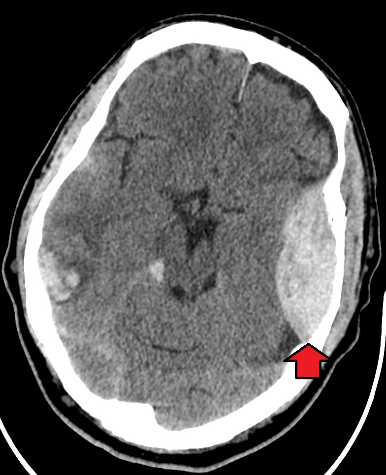
Red Flags
- GCS below 9, or 9 and decompensating: airway indicated (BTF prehospital). BTF also counts GCS below 9 as a herniation sign. On this page, though, it does not trigger hyperosmolar therapy or hyperventilation alone (see ALS Management).
- Pupils: asymmetry greater than 1 mm, or a fixed pupil with less than 1 mm response to bright light (BTF prehospital).
- Cushing triad: hypertension, bradycardia and irregular breathing. BTF calls this a sign of active herniation.
- Posturing, lateralizing findings or progressive decline: signs of active herniation (BTF).
- Hypoxemia or hypotension: SpO₂ below 90% or systolic BP below 100 mm Hg (BTF prehospital monitoring thresholds).
- Hypertension: a systolic BP of 150 mm Hg or higher is a BTF prehospital monitoring threshold. Report it. Do not lower it reflexively in a Cushing response (see Pitfalls).
- Seizure: treat through the Status Epilepticus pathway. Both the seizure and the postictal state confound the GCS.
Assessment & Diagnostics
Stabilize first, and assess at the same time. Imaging is the key diagnostic step. Until the patient gets it, the bedside checks below decide what you do next.
| Check | Why | Act If |
|---|---|---|
| GCS | Level of consciousness and the trend that shows herniation | Score after airway, breathing and circulation are addressed; report every 30 min and at any change |
| Pupils | Asymmetry or fixed dilation signals herniation | Asymmetry over 1 mm or a fixed pupil: treat as active herniation and report it |
| SpO₂ and EtCO₂ | Hypoxia is a secondary insult; over-ventilation lowers EtCO₂ and reduces cerebral blood flow | SpO₂ below 90%: oxygen, reposition, bag-valve mask, advanced airway if needed. EtCO₂ target 35 to 40 mm Hg (eucapnia), monitored within 35 to 45 mm Hg; 30 to 35 mm Hg only for active herniation (capnography) |
| Blood pressure | Hypotension is associated with worse outcome | Adult prehospital target systolic BP 110 mm Hg or higher (BTF, weak); measure every 5 to 10 min or continuously. Report 150 mm Hg or higher and do not lower BP reflexively in a Cushing response |
| Temperature | Euthermia is the target | Target 36 to 37 °C (98 to 99 °F); treat fever or hypothermia per protocol |
| Head CT | Hematoma, contusion, swelling, herniation or compressed basal cisterns | Abnormal CT with GCS 3 to 8 after resuscitation: monitor ICP and call neurosurgery (BTF 4th edition) |
| Anticoagulant or antiplatelet use | Raises the risk of an enlarging bleed | Anticoagulant or antiplatelet other than aspirin monotherapy: NICE NG232 advises considering a CT within 8 hours of injury (within 1 hour if presenting after 8 hours) when there is no other CT indication. A normal first CT or GCS does not exclude delayed bleeding; reversal per protocol (anticoagulant reversal) |
| PT/INR and aPTT | Coagulopathy worsens intracranial bleeding | Abnormal values: correct per protocol (PT/INR) |
| Spine | Cervical injury can coexist | Immobilize per spinal motion restriction protocol when indicated |
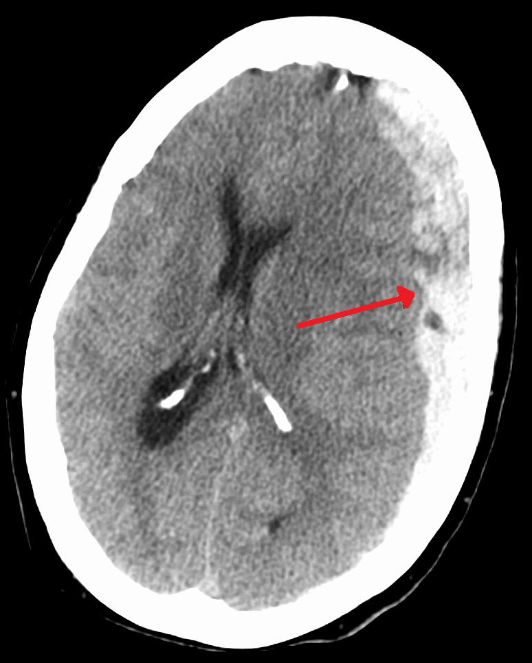
A head CT is indicated within 1 hour for adults 16 and older with any of these: GCS 12 or below on first assessment, GCS below 15 at 2 hours, suspected open or depressed skull fracture, signs of basal skull fracture, post-traumatic seizure, focal neurological deficit, or more than one episode of vomiting (NICE NG232, UK guidance). Confirm the local CT rule. In children, use a validated pediatric head-trauma CT rule such as PECARN, which has separate branches for under 2 years and for 2 years and older, per local protocol.
Differential & Mimics
- Intoxication or drug effect: alcohol, opioids, sedatives and other drugs all pull the GCS down. BTF recognizes ingestion and medications as confounders. See Toxidromes and Opioid Overdose.
- Postictal state: a depressed level of consciousness after a seizure can look like progressive injury. Document it, then reassess (BTF).
- Shock from injury elsewhere: bleeding or low oxygen from a chest or abdominal injury drags the GCS down through secondary injury. See Hemorrhagic Shock.
- Non-traumatic coma: hypoglycemia, stroke or intracranial hemorrhage can cause coma with no trauma at all. See Hemorrhagic Stroke and glucose.
BLS Management
- Scene safety and mechanism: confirm scene safety, identify the mechanism, and apply spinal precautions per protocol.
- Oxygen: give continuous supplemental oxygen to every patient with suspected severe TBI, even when SpO₂ is normal (BTF prehospital 3rd edition, strong).
- Airway: position the airway to correct hypoxemia. If SpO₂ stays below 90% despite oxygen, reposition, then ventilate with a bag-valve mask and an airway adjunct (bag-valve mask).
- Ventilation rate: in adults on positive-pressure ventilation, about 10 breaths per minute with capnography, keeping EtCO₂ at 35 to 40 mm Hg. Do not hyperventilate without signs of herniation. Children need age- and weight-based rates per PALS or local protocol.
- Reassess: GCS and pupils at least every 30 minutes and at any change. Record and report each finding, along with any confounders.
- Glucose: check blood glucose when consciousness is impaired and treat hypoglycemia per protocol (BTF prehospital algorithm).
- Bleeding: control external bleeding (hemorrhage control).
- Pediatric sizing: use appropriately sized oximetry sensors and blood pressure cuffs for children (BTF, strong).
- Transport and notification: notify the receiving facility early and transport to the destination described in the Transport section.
ALS Management
Doses vary by protocol; follow local protocols and medical direction. One pathway covers every level of care. Anything above BLS scope carries a tag.
- Intubation indications (Paramedic/RN/CCT): GCS below 9, or 9 and decompensating; inability to maintain the airway; or hypoxemia not corrected by oxygen and airway positioning (BTF). See RSI and Intubation.
- Confirm the tube (Paramedic/RN/CCT): bilateral breath sounds plus EtCO₂ detection or capnography. Monitor SpO₂, EtCO₂ and frequent blood pressure after intubation (BTF, strong).
- Hypotension (Paramedic/RN/CCT): isotonic IV or IO fluid, or blood products, to restore systolic BP to 110 mm Hg or higher; limit the duration of hypotension (BTF, strong). See IV insertion.
- Herniation or active ICP crisis (Paramedic/RN/CCT): the trigger is clinical herniation: a fixed or dilated pupil, posturing, a Cushing response or progressive neurologic decline (BTF, strong). A GCS below 9 alone is an airway indication. On its own, it does not trigger hyperosmolar therapy or hyperventilation. BTF prehospital gives no mannitol dose and advises against prophylactic hyperosmolar therapy, with or without herniation signs (weak). That leaves the choice and dose of any mannitol or hypertonic saline to protocol and medical direction. For reference, the BTF 4th edition hospital range is 0.25 to 1 g/kg IV. The mannitol label ICP dose is 0.25 g/kg IV over at least 30 minutes, repeatable every 6 to 8 hours. While herniation is active, ventilate to EtCO₂ 30 to 35 mm Hg (BTF, strong). See Mannitol.
- Mannitol safety: correct hypovolemia first, and keep SBP at 90 mm Hg or higher (BTF 4th edition avoids SBP below 90 with mannitol). The label contraindicates anuria, severe hypovolemia, pulmonary edema or pulmonary vascular congestion, and active intracranial bleeding, except during craniotomy. Infuse over at least 30 minutes unless protocol says otherwise. Use an administration set with a final in-line filter, because mannitol crystals can form. Osmotic diuresis can worsen hypovolemia and hypotension in a trauma patient, so reassess volume and blood pressure after each dose. Monitor fluid and electrolytes, serum osmolality, urine output, and renal, cardiac and pulmonary function. Stop if these worsen (label).
- Hypertonic fluid (Paramedic/RN/CCT): may be given to a patient with a GCS below 8 when raised ICP is suspected (BTF, weak). Volume and rate follow protocol. See Hypertonic Saline 3%.
- Prophylactic hyperosmolar therapy: BTF advises against it in the prehospital setting, whether or not herniation signs are present (BTF, weak). Treat clinical herniation only, per protocol.
- Tranexamic acid: prehospital TXA is not generally indicated for TBI, suspected intracranial hemorrhage or raised ICP (BTF, strong). Extracranial hemorrhagic shock has its own protocol (Tranexamic Acid).
- Seizures (Paramedic/RN/CCT): treat through the Status Epilepticus pathway.
- Sedation (Paramedic/RN/CCT): measure the GCS before sedatives when possible, and document each sedative given. Sedation obscures the exam (BTF).
- Glucose (Paramedic/RN/CCT): check blood glucose when consciousness is impaired and treat hypoglycemia per protocol (BTF prehospital algorithm). See Glucose.
ED & ICU Management
ICP treatment comes in tiers (SIBICC consensus, 2019). Treatments within one tier are considered equivalent. Higher tiers carry more risk. Move up only when the tier below has not worked or the patient is getting worse. Thresholds follow the BTF 4th edition.
- Tier zero, every monitored patient: keep normothermia, and avoid fever above 38 °C. Give sedation and analgesia for comfort and ventilator tolerance. They do not lower ICP by themselves. Raise the head of the bed about 30 degrees, and no more than 45 degrees, once the spine is cleared (NCS 2020, conditional recommendation).
- Tier one, ICP above 22 mm Hg: if an external ventricular drain is in place, drain CSF through it. Continuous drainage may lower ICP more effectively than intermittent drainage (BTF Level III). Next, give hypertonic saline or mannitol. SIBICC treats the two as equivalent. For hypertonic saline, an upper serum sodium of about 155 to 160 mEq/L may be reasonable (NCS, conditional). Adjusting sedation or analgesia to lower ICP is also a tier-one measure.
- Tier two, temporizing only: brief hyperventilation for herniation or an acute ICP crisis, with SjO₂ or PbtO₂ monitoring. Think of it as a bridge, not a plan. This is a BTF 3rd-edition Level III statement, restated in the 4th edition without a formal recommendation. The same guidance advises avoiding hyperventilation in the first 24 hours after injury. NCS supports brief episodes for acute ICP elevation (strong). Neuromuscular blockade is also tier two, with a trial period and early discontinuation (SIBICC).
- Tier three, refractory ICP: high-dose barbiturate after a test dose, once the patient is hemodynamically stable, with EEG monitoring (BTF Level IIB; SIBICC tier three). For ICP that does not respond to first-tier therapy, decompressive craniectomy is considered. See below.
- Not to be used (SIBICC): routine steroids; routine therapeutic hypothermia below 35 °C; high-dose propofol to attempt burst suppression; routinely lowering PaCO₂ below 30 mm Hg; lumbar CSF drainage.
- Prophylactic hypothermia: BTF does not recommend it to improve outcome in diffuse injury (BTF 4th edition, Level IIB).
- Prophylactic barbiturates: burst suppression to prevent intracranial hypertension is not recommended, even as prophylaxis (BTF 4th edition, Level IIB).
- Imaging and neurosurgery: get an early head CT. Any hematoma or mass effect needs neurosurgical evaluation. Moderate to severe TBI needs immediate CT, neurosurgery and ICP treatment capability (BTF).
- ICP monitoring: monitor ICP in salvageable patients who have a GCS of 3 to 8 after resuscitation and an abnormal CT. A normal CT still calls for monitoring if 2 or more of these are present at admission: age over 40, unilateral or bilateral posturing, or systolic BP below 90 mm Hg (BTF 4th edition).
- ICP threshold: treat ICP above 22 mm Hg (BTF 4th edition, Level IIB). Values above this are linked to higher mortality.
- CPP target: 60 to 70 mm Hg (BTF 4th edition, Level IIB). Avoid aggressive attempts to keep CPP above 70 mm Hg with fluids and vasopressors. They carry a risk of respiratory failure (Level III).
- Blood pressure: the systolic target depends on age. Aim for 100 mm Hg or higher at ages 50 to 69, and 110 mm Hg or higher at ages 15 to 49 or over 70 (BTF 4th edition, Level III). Avoid systolic BP below 90 mm Hg with mannitol.
- Osmotherapy: mannitol 0.25 to 1 g/kg is effective for raised ICP (BTF 4th edition dose range). The label ICP dose is 0.25 g/kg every 6 to 8 hours. Before ICP monitoring, restrict mannitol to patients with signs of transtentorial herniation, or with progressive decline that extracranial causes do not explain.
- Ventilation: prolonged prophylactic hyperventilation to PaCO₂ 25 mm Hg or lower is not recommended (BTF 4th edition, Level IIB). BTF repeats its 3rd-edition Level III statements without a formal recommendation. Hyperventilation is a temporizing measure for raised ICP. Avoid it in the first 24 hours after injury. If it is used, monitor it with jugular venous oxygen saturation or brain tissue oxygen.
- CSF drainage: through an external ventricular drain, continuous drainage may lower ICP more effectively than intermittent drainage (Level III).
- Decompressive craniectomy: in RESCUEicp, craniectomy for ICP above 25 mm Hg that medical care did not control lowered 6-month mortality (26.9% vs 48.9%). It also raised vegetative state (8.5% vs 2.1%) and severe disability. BTF recommends a large frontotemporoparietal craniectomy (at least 12 × 15 cm) over a small one. Bifrontal craniectomy is not recommended to improve outcome in severe TBI with diffuse injury (no mass lesion) and ICP above 20 mm Hg for more than 15 minutes within 1 hour, refractory to first-tier therapy (BTF 4th edition, Level IIA). The decision belongs to neurosurgery and the family.
- Seizure prophylaxis: phenytoin is recommended to reduce early post-traumatic seizures within 7 days of injury (BTF 4th edition, Level IIA). It is not recommended for late seizures. The evidence is not strong enough to prefer levetiracetam over phenytoin. See Fosphenytoin / Phenytoin.
- DVT prophylaxis: LMWH or low-dose unfractionated heparin may be used with mechanical prophylaxis. Either one raises the risk of an expanding intracranial hemorrhage. Consider pharmacologic prophylaxis only when the brain injury is stable and the benefit is judged to outweigh the risk. BTF finds insufficient evidence on agent, dose or timing (Level III).
- Steroids: high-dose methylprednisolone is contraindicated in severe TBI (BTF 4th edition).
- Nutrition: start feeding to reach basal caloric needs by day 5 and no later than day 7 (BTF 4th edition, Level IIA).
- Anticoagulated patients: reversal per protocol and timing per neurosurgery; see anticoagulant reversal.
Transport Considerations
- Destination: moderate to severe TBI goes straight to a facility with immediate CT, prompt neurosurgery, and the ability to monitor ICP and treat intracranial hypertension (BTF prehospital, strong). When direct transport is not possible, stabilize at the nearest facility and transfer within an established trauma system (weak).
- Pediatric patients: a pediatric trauma center, or an adult trauma center with added pediatric qualifications, is preferred over a Level I or II adult center without them. In a metro area, severe pediatric TBI goes straight to a pediatric trauma center where one is available (BTF, strong).
- Mode: choose the transport mode that minimizes time to definitive care (BTF, strong).
- Monitoring in transit: continuous SpO₂ and EtCO₂, blood pressure every 5 to 10 min or continuously, GCS at least every 30 minutes, and repeat pupil checks. Keep SpO₂ at 90% or higher and systolic BP at 110 mm Hg or higher.
- Handoff: report the GCS and any change at every contact and on arrival. Relay pupil findings and document time, assessment and treatment (BTF, strong).
- Ventilation in transit: aim for EtCO₂ 35 to 40 mm Hg (eucapnia), with 35 to 45 mm Hg as the acceptable monitoring range. Reserve 30 to 35 mm Hg for active herniation (BTF, strong).
Special Populations
- Pediatrics: the same pathway applies. Age-based systolic targets come from the BTF prehospital guideline (weak, 75th percentile or higher). Up to 28 days, aim above 70 mm Hg. From 1 to 12 months, above 84. From 1 to 5 years, above 90. From 6 years up, above 100. Use the pediatric GCS for preverbal children, and the adult GCS after age 2 (BTF, strong). Dose mannitol and hypertonic saline by weight per the weight-based dosing page. Use the pediatric vital signs page for normal ranges. ICP and CPP targets for children depend on the PICU protocol. The BTF pediatric severe TBI guideline (2019) is cited in References, but this page did not re-verify its ICP and CPP targets. For pediatric head CT decisions, use PECARN, which has separate branches for under 2 years and for 2 years and older, per local protocol.
- Pregnancy: this page did not verify a pregnancy-specific TBI target. Keep maternal oxygenation, ventilation and blood pressure at the same targets. From about 20 weeks' gestation, use left lateral uterine displacement and bring in obstetric care early, per the obstetric trauma protocol (general trauma guidance, not TBI-specific).
- Older Adults: adults 16 and older with some loss of consciousness or amnesia, plus a risk factor such as age 65 or older, should get a head CT within 8 hours of injury (NICE NG232, UK guidance). Older adults also carry the higher age-based systolic target above (110 mm Hg or higher over age 70).
- Renal Impairment: the label contraindicates mannitol in anuria. Check renal, cardiac and pulmonary status before mannitol. Correct any fluid or electrolyte imbalance first (label).
- Obesity: this page did not verify a TBI-specific adjustment. For weight-based drugs such as mannitol, use the dosing weight your protocol specifies.
- Patients on Anticoagulants: NICE NG232 advises considering a head CT within 8 hours of injury, or within 1 hour if the patient presents after 8 hours. This applies to anticoagulant or antiplatelet treatment other than aspirin monotherapy, when there is no other CT indication. A normal first CT or GCS does not rule out delayed bleeding. E3 practice: an anticoagulated patient with a head injury goes to a CT-capable facility. Do not treat at the scene and release. Reversal and timing follow protocol and neurosurgery advice.
Pitfalls & Safety Checks
- Error: hyperventilating a patient without herniation. Over-ventilation drops EtCO₂, and that reduces cerebral blood flow. Keep EtCO₂ at 35 to 40 mm Hg (eucapnia). A value below 35 means hyperventilation. The 30 to 35 range is the target only while herniation is active (BTF, strong).
- Error: giving mannitol or hypertonic fluid to prevent raised ICP. BTF advises against prophylactic hyperosmolar therapy in the prehospital setting. Give osmotherapy only for clinical herniation, per protocol. Check contraindications and volume status first.
- Error: accepting a normal first GCS or CT. A hematoma can grow later, especially on anticoagulants. Reassess the GCS and pupils every 30 minutes and at any change.
- Error: tolerating hypotension. Systolic BP below 110 mm Hg (the prehospital target) needs treatment with isotonic fluid or blood. Keep any hypotension brief.
- Error: giving prehospital TXA for head injury. BTF advises against routine prehospital TXA for TBI or suspected intracranial hemorrhage (strong). In-hospital CRASH-3 use is a different question (see Clinical Pearls).
- Error: missing hypoxemia because the head is the focus. The head injury pulls your attention. Give continuous oxygen to every patient with suspected severe TBI (BTF, strong) and keep SpO₂ at 90% or higher.
- Error: scoring the GCS after sedation. Score before sedatives when possible and record every sedative given.
Clinical Pearls
References
Currency: the prehospital guideline is the 3rd edition (2023). The hospital severe-TBI guideline is the 4th edition (published 2016), with the decompressive craniectomy update in 2020. The pediatric severe-TBI guideline is the 3rd edition (2019).
- Lulla A, Lumba-Brown A, Totten AM, et al. Prehospital Guidelines for the Management of Traumatic Brain Injury: 3rd Edition. Prehosp Emerg Care. 2023;27(5):507–538. https://doi.org/10.1080/10903127.2023.2187905
- Carney N, Totten AM, O'Reilly C, et al. Guidelines for the Management of Severe Traumatic Brain Injury, Fourth Edition. Neurosurgery. 2017;80(1):6–15 (hospital guideline, published 2016). https://doi.org/10.1227/NEU.0000000000001432
- Hawryluk GWJ, Rubiano AM, Totten AM, et al. Guidelines for the Management of Severe Traumatic Brain Injury: 2020 Update of the Decompressive Craniectomy Recommendations. Neurosurgery. 2020;87(3):427–434. https://doi.org/10.1093/neuros/nyaa278
- Hutchinson PJ, Kolias AG, Timofeev IS, et al.; RESCUEicp Trial Collaborators. Trial of Decompressive Craniectomy for Traumatic Intracranial Hypertension. N Engl J Med. 2016;375(12):1119–1130 (landmark trial). https://doi.org/10.1056/NEJMoa1605215
- CRASH-3 Trial Collaborators. Effects of tranexamic acid on death, disability, vascular occlusive events and other morbidities in patients with acute traumatic brain injury (CRASH-3): a randomised, placebo-controlled trial. Lancet. 2019;394(10210):1713–1723 (landmark trial). https://doi.org/10.1016/S0140-6736(19)32233-0
- Kochanek PM, et al. Guidelines for the Management of Pediatric Severe Traumatic Brain Injury, Third Edition: Update of the Brain Trauma Foundation Guidelines. Pediatr Crit Care Med. 2019;20(3 Suppl 1):S1–S82. https://doi.org/10.1097/PCC.0000000000001735
- Hawryluk GWJ, Aguilera S, Buki A, et al. A management algorithm for patients with intracranial pressure monitoring: the Seattle International Severe Traumatic Brain Injury Consensus Conference (SIBICC). Intensive Care Med. 2019;45(12):1783–1794. https://doi.org/10.1007/s00134-019-05805-9
- Cook AM, Morgan Jones G, Hawryluk GWJ, et al. Guidelines for the Acute Treatment of Cerebral Edema in Neurocritical Care Patients. Neurocrit Care. 2020;32(2):647–666. https://doi.org/10.1007/s12028-020-00959-7
- National Institute for Health and Care Excellence. Head injury: assessment and early management. NICE guideline NG232. Published 18 May 2023 (UK guidance). https://www.nice.org.uk/guidance/ng232
- ICU Medical Inc. Mannitol Injection: US prescribing information (indications, dosing, monitoring, contraindications). FDA label via DailyMed; label revision 05/2020, DailyMed version dated June 22, 2026. https://dailymed.nlm.nih.gov/dailymed/drugInfo.cfm?setid=ba7bf864-8a4d-4262-8fd1-00174fa2f9d1
Medical Disclaimer
- For Educational Purposes Only: This content is intended for educational reference and should not be used for clinical decision-making.
- Not a Substitute for Professional Judgment: Always consult your local protocols, institutional guidelines, and supervising physicians.
- Verify Before Acting: Users are responsible for verifying information through authoritative sources before any clinical application.