

Bedside Snapshot
- Recognize
- Any injury in a pregnant patient, or a uterus at or above the umbilicus (about 20 weeks). Minor injuries can still cause contractions, bleeding or early delivery.
- Red Flags
- Maternal shock or hypotension; vaginal bleeding; uterine tenderness; contractions more than 1 in 10 min; ruptured membranes; abnormal fetal heart rate; fibrinogen below 200 mg/dL.
- First Actions
- Primary survey, mother first; oxygen to SpO₂ above 95%; two large-bore IVs; uterine displacement after about 20 weeks; O-negative red cells while Rh status is unknown; fetal monitoring once the mother is stable.
- Key Doses
- RhIG 300 mcg IM within 72 hr if Rh(D)-negative; more by KB from 20 weeks
- Tranexamic acid 1 g IV over 10 min, then 1 g over 8 hr, or 2 g single dose per EMS protocol
- Vasopressors only for fluid-refractory hypotension
- Don't Miss
- Abruption that ultrasound does not show; uterine rupture; hidden bleeding behind a normal-looking blood pressure; a missed 72-hour RhIG window.
- Escalate & Transport
- Early obstetric consult; trauma center for a major injury at any gestation; maternity unit for a minor injury with a viable fetus; admit 24 hr at 23 weeks or more with any adverse factor; hysterotomy team if the mother arrests.
Definition & Pathophysiology
Trauma is the leading nonobstetric cause of maternal death. It affects about 1 in 12 pregnant women, per the SOGC 2015 guideline. Two patients are hurt at once. The mother bleeds and runs short of oxygen. The fetus can be hurt through abruption, uterine injury or low oxygen. Resuscitating the mother is the best early treatment for the fetus. So she gets assessed and stabilized first. Fetal assessment follows.
- Physiology: blood volume and cardiac output rise in pregnancy. The body answers bleeding differently because of it. See Anatomy, Physiology and Assessment of the Pregnant Patient.
- Placental abruption: the placenta peels off the uterine wall. Blunt abdominal impact can cause it, and no vaginal bleeding does not rule it out. After motor vehicle crashes, a 2026 meta-analysis of six studies found a pooled odds ratio of 2.43 for abruption (95% CI 1.12–5.29).
- Fetomaternal hemorrhage (FMH): fetal red cells leak into the mother's blood. In an Rh(D)-negative mother, that can sensitize her. Rh immune globulin (RhIG) prevents it.
- Uterine injury: direct trauma or a ruptured uterus. Either one can cause severe maternal bleeding and fetal loss.
Recognition & Red Flags
Presentations
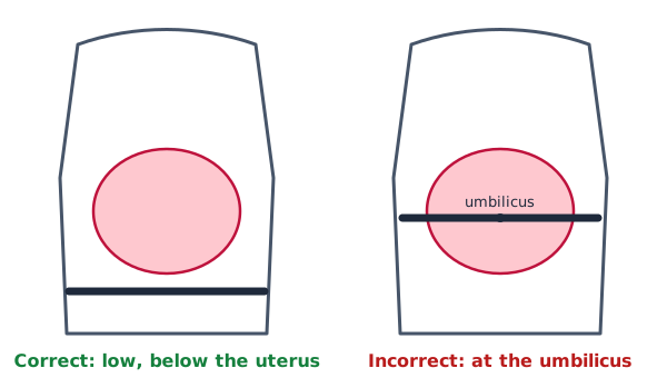
Every injured pregnant patient gets the trauma primary survey first, with the pregnancy built into it. Find the gestation in the history or a pregnancy test. If you have neither, the uterine height gives an estimate. At or above the umbilicus, the uterus means about 20 weeks. Any woman of reproductive age with a significant injury stays pregnant until a definitive pregnancy test or ultrasound says otherwise (SOGC 2015 rec 1).
Red Flags
- Maternal hypotension or signs of shock. In pregnancy, hypotension comes late with blood loss.
- Vaginal bleeding, uterine tenderness or significant abdominal pain.
- Sustained contractions (more than 1 in 10 minutes) or ruptured membranes.
- An atypical or abnormal fetal heart rate pattern in a viable pregnancy (23 weeks or more, per the SOGC 2015 guideline).
- High-risk mechanism of injury, such as ejection or a fall from height.
- Serum fibrinogen below 200 mg/dL. From 23 weeks, these adverse factors call for 24 hr admission (SOGC 2015 rec 21).
Assessment & Diagnostics
| Check | Why | Act If |
|---|---|---|
| Pregnancy status and gestation | Injury can hide a pregnancy; a uterus at or above the umbilicus is about 20 weeks | Treat as pregnant until a test or ultrasound says otherwise (see Recognition). Obstetric ultrasound if gestation is unknown and delivery may be needed |
| Type, Rh(D) and antibody screen | Sets the RhIG decision and the blood choice | Rh(D)-negative: RhIG within 72 hr of injury (SOGC 2015 rec 22); O-negative red cells until crossmatched |
| Fetal heart rate and contractions | Fetal assessment comes after the maternal primary survey | Viable fetus (23 weeks or more): electronic monitoring for at least 4 hr (SOGC 2015 rec 20); EAST 2025 conditionally recommends an observation period of at least 4 to 6 hr. Abnormal pattern or contractions: urgent obstetric consult |
| CBC and coagulation panel with fibrinogen | Hemorrhage and coagulopathy; fibrinogen is part of the SOGC trauma panel | At 23 weeks or more, fibrinogen below 200 mg/dL is one of several adverse factors that call for 24 hr admission (SOGC 2015 rec 21) |
| Kleihauer-Betke (KB) test or flow cytometry | Quantifies fetal red cells in the maternal circulation to set extra RhIG doses | Rh(D)-negative: quantify and add doses per the RhIG page. The RhIG page quantifies from 20 weeks (SOGC 2015 rec 23 sets no gestation limit). Before about 13 weeks the dose is protocol-dependent; some protocols use 50 mcg |
| FAST ultrasound | Looks for intraperitoneal bleeding in pregnancy | Positive, or the patient is unstable: surgical and obstetric teams per protocol |
| CT when indicated | Fetal radiation dose must not delay medically indicated maternal imaging | Image the mother as needed; do not delay for fetal radiation concerns, per the EAST 2025 guideline |
| Vaginal bleeding at 23 weeks or more | Placenta previa must be excluded first | Defer speculum and digital vaginal exam until ultrasound shows placental location |
Differential & Mimics
- Normal pregnancy tachycardia vs. hemorrhage: Judge the heart rate against the trend, the perfusion and the blood loss you can see or suspect. Not against a fixed number.
- Abruption vs. contractions alone: Three findings together, tenderness, bleeding and an abnormal fetal heart rate pattern, point toward abruption. Call it possible abruption until obstetrics says otherwise.
- Preeclampsia or eclampsia before the injury: A severe-range blood pressure, headache, visual change or seizure may be what caused the event. See Preeclampsia, Eclampsia and HELLP Syndrome.
- Pelvic or retroperitoneal bleeding: Pelvic fracture bleeding can be large and hidden. The source may not be the uterus at all.
BLS Management
- Scene and mechanism: note the gestational age if known, the mechanism, seat belt and airbag use, and the time of injury. That time starts the 72-hour RhIG clock. There is not enough evidence to support disabling airbags for pregnancy, per the SOGC 2015 guideline.
- Mother first: primary survey for airway, breathing and circulation. Use spinal motion restriction when indicated (Spinal Motion Restriction).
- Oxygen: keep SpO₂ above 95% (Oxygen). Low oxygen in the mother means less oxygen reaches the fetus.
- Move the uterus off the vena cava: after mid-pregnancy (about 20 weeks), use manual displacement or left lateral tilt. With possible spinal injury, manual displacement is the safer choice.
- Bleeding control: direct pressure and hemorrhage control per protocol (Hemorrhage Control).
- Do not inflate the abdominal portion of a military anti-shock trouser (MAST) or PASG garment: Inflating that section may cut placental perfusion, per the SOGC 2015 guideline.
- Early notice and priority transport: tell the receiving team the gestation and time of injury.
ALS Management
Doses vary by protocol; follow local protocols and medical direction.
- IV or IO access (Paramedic/RN/CCT): two large-bore (14 to 16 gauge) lines, placed above the diaphragm when possible. See IV Insertion and IO Access.
- Blood products (Paramedic/RN/CCT): for significant hemorrhage, give red cells, plasma and platelets in a balanced ratio per the massive transfusion protocol (Blood Transfusion). For an Rh(D)-negative mother, give O-negative red cells until crossmatched blood is available.
- Tranexamic acid (Paramedic/RN/CCT): 1 g IV over 10 min, then 1 g over 8 hr, or 2 g single dose per EMS protocol, within 3 hr of injury (off-label for trauma in the US). No pregnancy-specific trauma trial data exist, and TXA labeling warns of thromboembolic events while pregnancy is itself prothrombotic. Follow local protocol and obstetric and trauma direction. See Tranexamic Acid.
- Crystalloid: bolus per protocol. Move early to blood products when bleeding is significant, and avoid large crystalloid volumes in its place.
- Vasopressors (Paramedic/RN/CCT): reserve norepinephrine for intractable hypotension that does not respond to fluid. Vasopressors reduce uteroplacental perfusion, per the SOGC 2015 guideline.
- Airway: the pregnant patient has less oxygen reserve and desaturates faster. Plan drug-assisted airway management with that in mind (RSI), factoring in pregnancy's physiologic changes, and keep SpO₂ above 95%.
- Aspiration risk: in a semiconscious or unconscious injured pregnant patient, a nasogastric tube reduces aspiration of acidic gastric contents (SOGC 2015 rec 2), per local protocol.
- Analgesia and sedation: titrate to effect and watch for hypoventilation. Hypoxemia harms the fetus.
- RhIG: not an EMS drug. Identify Rh(D)-negative status and the time of injury, and hand them off for the RhIG decision.
ED & ICU Management
- Primary survey, then fetal assessment: fetal assessment waits until the end of the primary survey. The rapid maternal evaluation comes first, per the EAST 2025 guideline.
- Labs: type and screen with the first trauma labs, CBC, and coagulation panel with fibrinogen, per the SOGC 2015 guideline.
- Rh(D)-negative mother: give 300 mcg RhIG IM within 72 hr of injury, without waiting for quantification. From 20 weeks, KB or flow cytometry then sets any extra doses; before about 13 weeks follow local protocol (Rho(D) Immune Globulin).
- Imaging: FAST looks for intraperitoneal bleeding, per the SOGC 2015 guideline. Image the mother with CT when indicated. Fetal radiation worries should not delay that scan, per the EAST 2025 guideline. Gadolinium contrast may be considered when maternal benefit outweighs potential fetal risk (SOGC 2015 rec 16).
- Chest tube: if a thoracostomy tube is needed, place it one or two intercostal spaces higher than usual (SOGC 2015 rec 4).
- Obstetric consult: urgent for a viable fetus with contractions, suspected abruption or traumatic uterine rupture, per the SOGC 2015 guideline.
- Observation: a viable fetus gets at least 4–6 hr of monitoring after the injury, per the EAST 2025 guideline. From 23 weeks, SOGC 2015 calls for electronic fetal monitoring for at least 4 hr (rec 20). If any one of these is present, admit for 24 hr observation: uterine tenderness, significant abdominal pain, vaginal bleeding, sustained contractions (more than 1 in 10 min), ruptured membranes, atypical or abnormal fetal heart rate, high-risk mechanism, or fibrinogen below 200 mg/dL (rec 21). An obstetric ultrasound is advised before discharge after more than 4 hr of monitoring (rec 25).
- Traumatic arrest: at 20 weeks or more, during resuscitative thoracotomy for traumatic arrest, EAST 2025 conditionally recommends resuscitative hysterotomy as soon as possible. SOGC 2015 rec 31 advises cesarean no later than 4 min after maternal arrest at 23 weeks or more. The maternal arrest page below aims for delivery within 5 min. Lopez 2023 also considers hysterotomy for arrest or profound hypovolemic shock approaching 22 to 24 weeks. See Cardiac Arrest in Pregnancy and Resuscitative Hysterotomy.
- Hemorrhage: follow the Hemorrhagic Shock pathway for resuscitation, with the obstetric team involved early.
Transport Considerations
- Destination: a major injury goes to a trauma center at any gestation. A non-life-threatening injury with a viable fetus (23 weeks or more) goes to a maternity unit with obstetric triage. Under 23 weeks, or with a fetus that is not viable, the emergency department is the destination, per the SOGC 2015 guideline.
- In transit: keep the uterus off the vena cava (manual displacement, or left tilt with spinal precautions), keep SpO₂ above 95%, and reassess bleeding and vital signs at least every 15 minutes.
- Handoff: gestational age, Rh(D) status, time and mechanism of injury, bleeding, contractions, the last fetal heart rate if known, and every blood product and drug given, with times. Note the seat belt and airbag use.
- Interfacility transfer: state whether RhIG was given, the dose, the time and any KB result. If it was not given, state the 72-hour deadline.
Special Populations
- Pediatrics: pregnant adolescents follow the adult regimens above, RhIG 300 mcg IM for an Rh(D)-negative patient included. Children who are not pregnant use Weight-Based Dosing and Vital Signs. This page does not cover pediatric trauma care.
- Pregnancy: everything on this page applies to the pregnant patient. Gestation sets the approach. Uterine displacement starts from about 20 weeks. Fetal monitoring starts when the fetus is viable (23 weeks or more under the SOGC 2015 guideline). Drug choice follows the indication. Fetal effects get weighed, but they are not used to withhold necessary maternal treatment. The available RhIG evidence shows no fetal harm, so an indicated dose is not withheld (see the RhIG page).
- Intimate partner violence: ask every injured woman about domestic or intimate partner violence. In those cases, document fetal well-being carefully. The record may be used legally, per the SOGC 2015 guideline.
- Tetanus: the tetanus vaccine is safe in pregnancy. Give it when indicated, per the SOGC 2015 guideline.
Pitfalls & Safety Checks
- Error: reading normal vital signs as reassurance. Blood volume and heart rate shift in pregnancy. Judge the trend. Keep looking for hemorrhage even when the blood pressure looks normal.
- Error: waiting for ultrasound before treating abruption. Ultrasound is not sensitive enough to wait on. Treat a suspected abruption now and bring in obstetrics.
- Error: letting the 72-hour RhIG window pass. Write the injury time on the chart. Delay weakens protection, so a late dose goes in as soon as possible.
- Error: inflating the abdominal portion of a MAST or PASG garment. It may cut placental perfusion.
- Error: starting vasopressors early. Use them only for hypotension that does not respond to fluid.
- Error: discharging after minor trauma without fetal assessment. A viable fetus needs at least 4–6 hr of monitoring, per the EAST 2025 guideline. From 23 weeks, any adverse factor means 24 hr admission: uterine tenderness, significant abdominal pain, vaginal bleeding, sustained contractions (more than 1 in 10 min), ruptured membranes, atypical or abnormal fetal heart rate, high-risk mechanism, or fibrinogen below 200 mg/dL (SOGC 2015 rec 21).
- Error: performing a speculum or digital vaginal exam for bleeding before placental location is known. From 23 weeks, hold it until ultrasound rules out previa.
- Error: withholding CT out of fear of fetal radiation. EAST 2025 says radiation concern should not block medically indicated imaging.
Clinical Pearls
References
- Appelbaum RD, Yorkgitis B, Rosen J, et al. (2025). Trauma in pregnancy: A systematic review, meta-analysis, and practice management guideline from the Eastern Association for the Surgery of Trauma. J Trauma Acute Care Surg. 99(2):298–309. https://doi.org/10.1097/TA.0000000000004661
- Jain V, Chari R, Maslovitz S, Farine D; Maternal Fetal Medicine Committee, Society of Obstetricians and Gynaecologists of Canada. (2015). Guidelines for the Management of a Pregnant Trauma Patient. J Obstet Gynaecol Can. 37(6):553–574. https://doi.org/10.1016/s1701-2163(15)30232-2
- Lopez CE, Salloum J, Varon AJ, Toledo P, Dudaryk R. (2023). The Management of Pregnant Trauma Patients: A Narrative Review. Anesth Analg. 136(5):830–840. https://doi.org/10.1213/ANE.0000000000006363
- Owattanapanich N, Lewis MR, Benjamin ER, Wong MD, Demetriades D. (2021). Motor vehicle crashes in pregnancy: Maternal and fetal outcomes. J Trauma Acute Care Surg. 90(5):861–865. https://doi.org/10.1097/TA.0000000000003093
- Gutiérrez-Sánchez LA, Guzmán-López JA, Manrique-Hernández EF. (2026). Maternal-fetal outcomes following a motor-vehicle crash: systematic review and meta-analysis. Taiwan J Obstet Gynecol. 65:665–671. https://doi.org/10.1016/j.tjog.2026.01.022
- Tanaka K, Motozawa Y, Takahashi K, Maki T, Nakamura M, Hitosugi M. (2022). Severity of placental abruption in restrained pregnant vehicle drivers: correct seat belt use confirmed by finite element model analysis. Int J Environ Res Public Health. 19(21):13905. https://doi.org/10.3390/ijerph192113905
- Girard M, Marchand F, Uch R, Bretelle F. (2017). Trauma and pregnancy: Is the Kleihauer-Betke test really useful? [In French.] Gynecol Obstet Fertil Senol. 45(11):584–589. https://doi.org/10.1016/j.gofs.2017.08.009
- Kedrion Biopharma. (2025). RhoGAM Ultra-Filtered PLUS, Rho(D) immune globulin (human) [prescribing information]. DailyMed, National Library of Medicine. https://dailymed.nlm.nih.gov/dailymed/drugInfo.cfm?setid=d87e4d0b-2442-4135-b3f9-5c4f74845b87
- American College of Obstetricians and Gynecologists. (2017). Committee Opinion No. 723: Guidelines for diagnostic imaging during pregnancy and lactation. Obstet Gynecol. 130(4):e210–e216. https://doi.org/10.1097/AOG.0000000000002355
- American College of Obstetricians and Gynecologists. (2017). Practice Bulletin No. 181: Prevention of Rh D alloimmunization. Obstet Gynecol. 130(2):e57–e70. https://doi.org/10.1097/AOG.0000000000002232
Medical Disclaimer
- For Educational Purposes Only: This content is intended for educational reference and should not be used for clinical decision-making.
- Not a Substitute for Professional Judgment: Always consult your local protocols, institutional guidelines, and supervising physicians.
- Verify Before Acting: Users are responsible for verifying information through authoritative sources before any clinical application.