

At a Glance
- Represents
- The sinus node's normal P-QRS-T cycle and its variants: sinus brady, tachy, arrhythmia, pause, arrest and sick sinus syndrome.
- Normal (Adult)
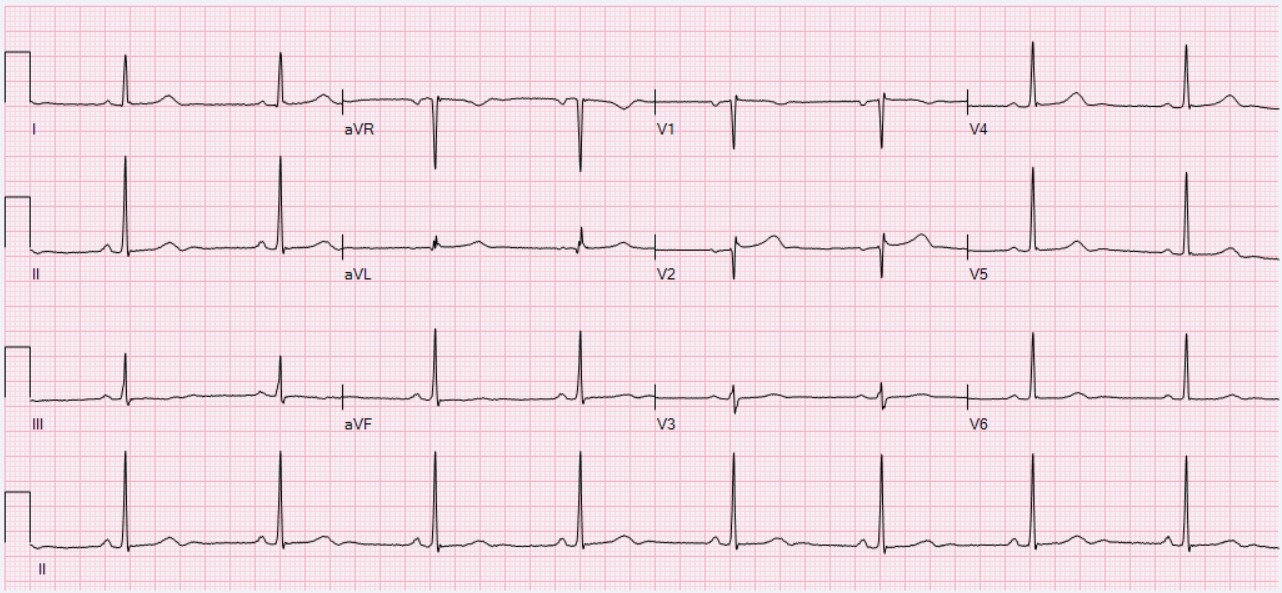
- Sinus rate 60–100 bpm; PR 120–200 ms; one P wave before every QRS.
- Measure It
- Regular: 300 ÷ large boxes between R waves. Irregular or pausing: count QRS in 6 s × 10, and look for a P wave in every gap.
- Abnormal → Think
- Under 60: sinus bradycardia. Over 100: sinus tachycardia. A gap with no P wave: sinus arrest or exit block.
- Red Flags
- Symptomatic pause or sick sinus with syncope; a slow rate with hypotension, altered mental status or ischemic chest pain.
- Bedside Action
- Unstable: atropine 1 mg IV/IO, pads on, then transcutaneous pacing or an infusion. Stable: monitor, 12-lead, treat the cause.
Physiology & What It Represents
The sinoatrial (SA) node starts every normal beat, firing on its own. The wave spreads through the atria and makes the P wave. Then it slows at the atrioventricular (AV) node, runs through the His–Purkinje system and activates the ventricles. That is the QRS. The sinus node is the fastest automatic pacemaker, so it normally sets the rhythm. When it slows or fails, a lower site takes over: about 40–60 bpm from the AV junction, or 20–40 bpm from the ventricle.
Autonomic tone sets the resting sinus rate. Vagal (parasympathetic) tone slows it. That is why the rate falls in sleep and in trained athletes. Sympathetic activity, fever, pain, hypovolemia, anemia and hypoxia push it up. Here's the key distinction: a sinus rhythm is defined by its P waves, not by its rate. Sinus P waves are upright in lead II, and one comes before every QRS.
Normal Values & How to Measure
Start with the rate and the P waves. Then decide whether the rhythm is sinus at all. Every sinus beat has a P wave before its QRS, and its PR runs 120–200 ms. Rate, regularity and any pause tell you which variant you are looking at.
| Rhythm | Rate (bpm) | Regularity | P Waves | PR | QRS | First-Line Action |
|---|---|---|---|---|---|---|
| Normal sinus rhythm | 60–100 | Regular; may vary with breathing | One before each QRS | 120–200 ms | 110 ms or less | Monitor; no treatment |
| Sinus bradycardia | Under 60 | Regular or varying with breathing | One before each QRS | 120–200 ms | 110 ms or less | Treat only if symptomatic; find the cause |
| Sinus tachycardia | Over 100 | Regular; gradual onset | One before each QRS | 120–200 ms | 110 ms or less | Treat the driver; do not slow it reflexively |
| Sinus arrhythmia | Varies with breathing | R–R shortens and lengthens with breathing | Sinus; one before each QRS | Constant | 110 ms or less | None; normal finding |
| Sinus pause or exit block | Gap, then sinus rate | Pause longer than the P-P interval | None inside the pause | Constant before and after | Escape QRS if one appears | Monitor; 12-lead; pacing evaluation if symptomatic |
| Sick sinus syndrome | Slow and fast periods | Irregular with pauses | Sinus or absent during pauses | Constant on sinus beats | Narrow | Symptomatic: pacing evaluation with cardiology |
- Rate: Use the 300 or 1500 rule for a regular rhythm, or count QRS complexes in 6 seconds for an irregular one.
- Regularity: Decide whether the R-R intervals are constant, vary with breathing, or contain a pause.
- P waves: Confirm a sinus P wave before every QRS, and look for gaps with no P wave at all.
- PR and P:QRS relationship: Measure the PR (120–200 ms) and confirm 1:1 conduction. A P wave with no QRS points to AV block, not a sinus pause.
- QRS width: A narrow QRS suggests a sinus or supraventricular origin. A wide QRS with a slow rate suggests escape from below the AV node.
- Stability: Decide stable or unstable before any drug. Unstable means hypotension, altered mental status, ischemic chest pain, shock or heart failure.
| Parameter | Normal Value | Notes |
|---|---|---|
| Sinus rate | 60–100 bpm | Resting adult rate |
| PR interval | 120–200 ms (0.12–0.20 s; 3–5 small boxes) | From P-wave onset to QRS onset |
| QRS duration | 110 ms or less | 110–119 ms is borderline; 120 ms or more is wide |
| Junctional escape | 40–60 bpm | Narrow QRS; more reliable |
| Ventricular escape | 20–40 bpm | Wide QRS; less reliable |
Sinus Bradycardia
Sinus bradycardia means a sinus rhythm slower than 60 bpm. Each QRS still has its sinus P wave in front, and the PR and QRS are normal. The node is just firing slow, so it is still sinus. Whether you treat depends on the patient in front of you, not the number on the screen.
ECG Criteria
- Rate: Under 60 bpm, regular or varying with breathing
- P waves: Upright in lead II, one before each QRS (1:1 conduction)
- PR and QRS: PR 120–200 ms; QRS 110 ms or less
Causes
- Physiologic: Sleep, endurance training and high vagal tone
- Medications: Beta-blockers, non-dihydropyridine calcium channel blockers, digoxin and clonidine
- Metabolic and endocrine: Hypothyroidism, hypothermia, hyperkalemia and hypoxia
- Neurologic: Raised intracranial pressure, which can produce a Cushing-type response
- Cardiac: Inferior MI, which can involve the AV node, and sinus node disease
Clinical Significance
- Asymptomatic: Usually, you treat nothing. Find the cause, then review the medications.
- Symptomatic: Treatment cannot wait if the patient has hypotension, altered mental status, chest pain, heart failure or syncope (see Bedside Management & Escalation).
- Inferior MI: Slow rate plus inferior ST elevation? Get a right-sided ECG. You want to know whether the right ventricle is involved.
Look-Alikes
- Sinus arrhythmia: Breathing changes the rate here, and that is normal (see Sinus Arrhythmia below).
- AV block with a slow ventricular rate: The P waves march on without touching the QRS (see Conduction Blocks).
Sinus Tachycardia
Sinus tachycardia is a regular sinus rhythm faster than 100 bpm, with a normal P wave before each QRS. Think of it as a response. Something is driving it, such as pain, fever or blood loss. It is not a primary arrhythmia.
ECG Criteria
- Rate: Over 100 bpm, typically rising and falling gradually
- P waves: Upright in lead II, one before each QRS; PR and QRS normal
- Onset: Gradual. An abrupt start or stop points to supraventricular tachycardia (SVT) instead (see Atrial Rhythms)
Causes
- Volume loss: Hypovolemia and hemorrhage
- Infection and inflammation: Sepsis and fever
- Cardiopulmonary: Heart failure, pulmonary embolism and hypoxia
- Pain, anxiety and stimulants: Including caffeine, sympathomimetic drugs and cocaine
- Endocrine and drugs: Hyperthyroidism, anemia, anticholinergic drugs and beta-agonists
Clinical Significance
- Pitfall: A regular narrow-complex rate near 150 with no visible P waves may be atrial flutter with 2:1 conduction, not sinus tachycardia. Before you call it sinus, look for flutter waves in V1 or the inferior leads.
- Outpatient follow-up: Inappropriate sinus tachycardia and postural orthostatic tachycardia syndrome (POTS) are outpatient diagnoses. Consider them when the resting rate stays high with no clear driver, or when it climbs sharply on standing.
- Treat the cause: Sinus tachycardia is a warning sign. Fix the driver, and the rate usually falls with it.
- Do not slow it reflexively: Slowing the rate can reduce cardiac output in a patient who is compensating. Don't do it on reflex.
Sinus Arrhythmia
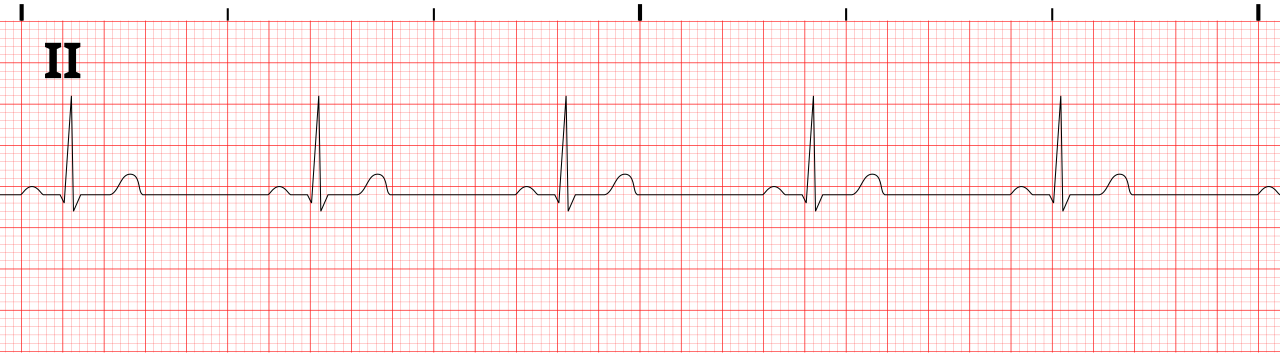
This is a normal variation. Breathing changes the sinus rate. R-R intervals get shorter on inspiration and longer on expiration. The P waves keep their sinus shape, and the PR does not move.
ECG Criteria
- R-R pattern: Intervals vary in step with breathing
- P waves: Sinus in shape, upright in I, II and aVF, with one before each QRS
- PR: Constant from beat to beat
Clinical Significance
- You will see it in children, young adults and athletes. It is normal. With sinus P waves, it needs no treatment.
- Missing it does not mean disease. Seeing it does not rule out a pause. The P waves decide that, not the variation.
Look-Alikes
- Wandering atrial pacemaker: Three or more P-wave shapes and PR intervals, usually below 100/min (see P Wave)
- Atrial fibrillation: Irregularly irregular R-R intervals with no discrete P waves
Sinus Pause, Sinus Arrest & Exit Block
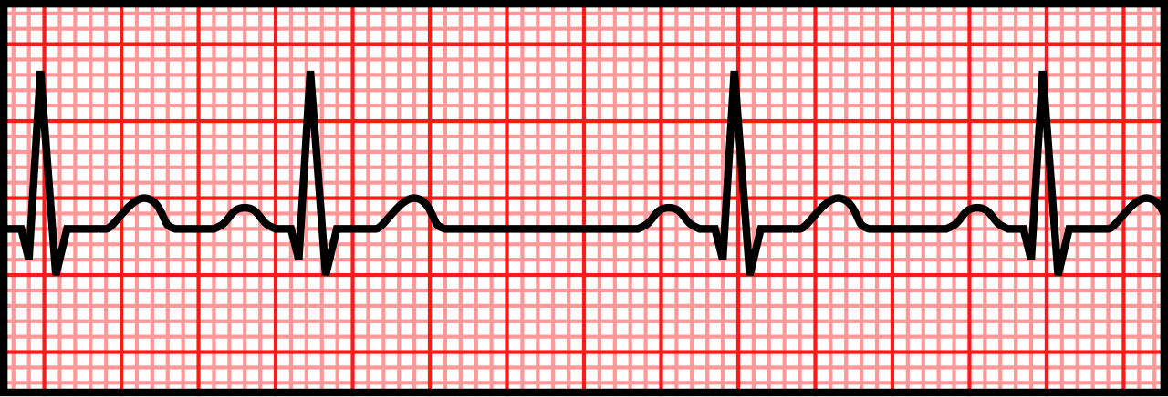
A sinus pause is a gap with no sinus P wave, and the gap runs longer than the basic P-P interval. Sinus arrest means the sinus node stops firing for a long stretch. Exit block is different: the impulse forms but never gets out to the atria. The pause then lands on a multiple of the usual P-P interval.
ECG Criteria
- Pause: A P-P gap longer than the basic P-P interval, with no P wave inside it
- Exit block: The pause is an exact multiple of the basic P-P interval
- Arrest: The pause is not a multiple of the basic P-P interval
- Escape: A narrow junctional or wide ventricular escape may end the pause, or the pause may end with no escape at all
Causes
- Sinus node disease: Fibrosis that comes with age is the most common cause
- Medications: Beta-blockers, calcium channel blockers, digoxin and antiarrhythmics
- Vagal events: Carotid sinus hypersensitivity and vasovagal reactions
- Electrolytes and ischemia: Hyperkalemia and inferior MI
Clinical Significance
- Threshold: Awake, a pause of 3 seconds or more is abnormal and needs evaluation. In sleep, especially in athletes, pauses are often benign. The number matters, but so does the setting.
- Monitor a brief pause in an asymptomatic patient, and look at drugs and electrolytes.
- Symptoms, or a pause with no escape, mean pacing and a cardiology review.
Sick Sinus Syndrome
Sick sinus syndrome is sinus node dysfunction that causes symptoms. It includes sinus bradycardia, pauses and sinus arrest. Often they alternate with a fast atrial rhythm, the tachy-brady pattern.
ECG Criteria
- Bradycardia: Sinus rate under 60 bpm that is inappropriate for the patient's activity
- Pauses or arrest: Gaps with no sinus P wave, as described above
- Chronotropic incompetence: The sinus rate does not rise appropriately with exertion
- Tachy-brady pattern: Sinus or atrial tachyarrhythmia that alternates with slow periods
Symptoms and Treatment
- Symptoms: Dizziness, presyncope, syncope, fatigue or palpitations
- Treatment: Permanent pacing is the standard treatment when symptoms line up with the bradycardia or pauses (2018 ACC/AHA/HRS bradycardia guideline).
- Drugs: Review drugs that slow the sinus node before you make the pacing decision.
Differential Diagnosis & Mimics
Separate the sinus patterns by which P waves are missing or changing. Don't stop at the rate.
| Feature | Sinus Bradycardia | Sinus Arrhythmia | Sinus Pause or Exit Block |
|---|---|---|---|
| Rate | Under 60 bpm | Varies with breathing | A gap, then the sinus rate |
| R-R pattern | Regular | Varies with breathing | Gap longer than the P-P interval |
| P waves | One before each QRS | One before each QRS | Stop during the pause |
| PR | 120–200 ms | Constant | Constant before and after the pause |
| Management | Treat only if symptomatic | None | Monitor; pacing evaluation if symptomatic |
- Sinus pause vs. AV block: In AV block the P waves keep marching, and some QRS complexes drop. In a sinus pause the P waves stop. Check the gap for P waves before you name the block (see Conduction Blocks).
- Sinus bradycardia vs. junctional rhythm: A junctional escape has no P wave, or a retrograde P wave. Its rate is typically 40–60 bpm (see Atrial Rhythms).
Artifact & Lead Misplacement
- Limb lead reversal: Reversed arm electrodes flip lead I upside down, which can mimic an abnormal axis. Check lead placement before you call a non-sinus P wave.
- Tremor and shivering: Shivering can hide P waves or make the rhythm look irregular. Check a second lead and the pulse.
- Transport vibration: Vibration in the vehicle can hide P waves. Recheck the strip once the vehicle is stopped.
Normal Variants
- Sinus arrhythmia: The rate rises with inspiration and falls with expiration. It is common and benign.
- Athletic bradycardia: Resting sinus bradycardia is a recognized adaptation in trained athletes. Symptoms and context decide whether it needs a work-up.
- Sleep: The sinus rate drops during sleep.
Bedside Management & Escalation
The algorithm behind this page is the 2025 AHA adult bradycardia with a pulse algorithm (Part 9), with the 2025 AHA pediatric version (Part 8) alongside it. The AHA adult algorithm uses a typical rate under 50/min for bradyarrhythmia. The sinus bradycardia label, though, stops at 60. Atropine is reasonable for adult bradycardia with hemodynamic compromise (COR 2a, LOE B-NR). Atropine not working? Transcutaneous pacing or an epinephrine or dopamine infusion may be effective (COR 2b, LOE C-LD).
| Situation | BLS (EMT) | ALS (Paramedic / RN; AEMT per state scope) | Critical Care / Transport |
|---|---|---|---|
| Unstable (hypotension, altered mental status, ischemic chest pain, shock, heart failure) | Oxygen if SpO₂ under 94%; monitor; 12-lead if available; call ALS early | Atropine 1 mg IV/IO; apply pacing pads now; transcutaneous pacing if atropine does not work; dopamine or epinephrine infusion as a bridge | Transcutaneous or transvenous pacing; infusion while pacing is arranged; notify cardiology |
| Stable sinus bradycardia or pause without symptoms | Monitor; 12-lead; transport | Monitor; 12-lead; check drugs and electrolytes; apply pads early if pauses are long | Monitor; cardiology review; plan for pacing if the rhythm worsens |
| Sinus tachycardia | Treat the cause (bleeding, pain, hypoxia); monitor | Treat the cause; avoid slowing the rate reflexively | Evaluate for shock and the underlying driver |
| Pulseless (pause or asystole with no pulse) | CPR and AED per the BLS sequence | Cardiac arrest algorithm; epinephrine; treat reversible causes | Arrest algorithm; pacing does not replace compressions |
| Therapy | Adult | Pediatric | Key Notes |
|---|---|---|---|
| Atropine | 1 mg IV/IO, every 3–5 min; max 3 mg total |
0.02 mg/kg IV/IO, minimum 0.1 mg, maximum 0.5 mg per dose; may repeat once (2025 AHA PALS bradycardia algorithm). The 0.1 mg minimum exceeds 0.02 mg/kg in a child under 5 kg. Part 8 states a 0.02 mg/kg dose with no minimum only for atropine premedication before intubation (COR 2b, LOE C-LD). Differs from FDA label: the label gives a usual initial range of 0.01–0.03 mg/kg with no fixed minimum. For increased vagal tone or primary AV block only (COR 1, LOE C-LD) |
First drug for symptomatic sinus bradycardia with a pulse. Often ineffective in infranodal block; do not delay pacing while waiting. Use with caution in acute coronary ischemia: atropine-induced tachycardia raises myocardial oxygen demand, and the FDA label limits total dose in coronary artery disease to 0.03–0.04 mg/kg. Not for heart transplant recipients (see the warning below) |
| Transcutaneous pacing | No pacing rate is stated in the 2025 AHA text, so none is shown (see editor flag). Start at the lowest output and increase until electrical, then mechanical, capture |
No pediatric pacing rate or output is stated in the 2025 AHA/AAP PALS text; use smaller pads. For sinus node dysfunction or complete heart block unresponsive to CPR, emergency pacing may be considered (2025 AHA PALS, COR 2b, LOE C-EO) |
Confirm mechanical capture: a pulse with each paced beat. An ECG spike without a pulse is not capture |
| Epinephrine | 2–10 mcg/min IV infusion, titrated to response |
0.01 mg/kg (0.1 mL/kg of 0.1 mg/mL) IV/IO every 3–5 min, max 1 mg per dose. No pediatric infusion dose is established in the 2025 AHA/AAP PALS text, so none is shown |
High-alert Infusion is a bridge to pacing. Check mcg/min versus mcg/kg/min on the pump. Give through a secure large vein; extravasation can cause tissue necrosis |
| Dopamine | 5–20 mcg/kg/min IV infusion, titrated to heart rate, blood pressure and perfusion; max 20 mcg/kg/min |
Not a PALS bradycardia drug; no pediatric bradycardia dose is established in the 2025 AHA/AAP PALS text, so none is shown |
High-alert Infusion only; give through a secure large vein, since extravasation can cause tissue necrosis (the FDA label names phentolamine infiltration as the antidote). Confirm mcg/kg/min and the bag concentration. Listed in the 2025 AHA adult bradycardia algorithm for use when atropine is ineffective, alongside epinephrine infusion or pacing (Part 9, COR 2b, LOE C-LD) |
AHA/AAP 2025 comes first for a pediatric dose, when it gives one. The NASEMSO National Model EMS Clinical Guidelines v3.0 (2022) come next, and then the US label. Where none gives a dose, none is shown. A pediatric maximum never exceeds the adult maximum dose (NASEMSO Universal Care Guideline, p. 13). Doses are off-label unless marked labeled.
Pediatric & Special Populations
Pediatrics
- Heart rate: Age-based norms live on the Vital Signs page. Use the age range before you call a rate abnormal.
- Usual cause: Hypoxia or increased vagal tone. Support oxygenation and ventilation first. Under the 2025 AHA PALS approach, start CPR for a heart rate under 60/min with cardiopulmonary compromise that persists despite effective ventilation with oxygen (COR 1, LOE C-EO). Give epinephrine 0.01 mg/kg IV/IO when compromise persists after you correct other factors, such as hypoxia (COR 1, LOE C-EO).
- Atropine: 0.02 mg/kg (minimum 0.1 mg, maximum 0.5 mg per dose) for increased vagal tone or primary AV block (see the table above; COR 1, LOE C-LD), not as the first drug for hypoxic bradycardia
- Tachycardia: Sinus tachycardia is common with fever, pain and dehydration. A narrow-QRS rate much faster than the age norm, or one with an abrupt onset, needs the PALS tachycardia algorithm to rule out SVT.
- Measurement: Get the strip from a calm child. A crying rate is not a baseline.
- Dosing: Weight-based doses follow Weight-Based Dosing
Other Populations
- Pregnancy: Resting sinus tachycardia is common as blood volume and output rise. Judge a new tachycardia by context. Manage unstable rhythms as you would in non-pregnant adults. If the fundal height is at or above the umbilicus, give manual left lateral uterine displacement with chest compressions (2025 AHA Part 10, COR 1, LOE C-LD). Resuscitative delivery is indicated if ROSC is not achieved with standard resuscitation (COR 1, LOE B-NR).
- Older adults: Sinus node and AV conduction disease are more common here. Pauses and bradycardia can be the first sign of ischemia or drug toxicity.
- Athletes: Resting sinus bradycardia and sinus arrhythmia are common and usually benign. Assess for symptoms, not the rate alone.
- Heart transplant: The sinus node is denervated, which means the rate is slow to respond to demand. Atropine is ineffective or harmful. Adenosine effects can be prolonged.
- Pacemaker or ICD: Know the programmed lower rate. A rate below it suggests failure to capture or failure to sense, and that needs device evaluation.
Monitoring & Transport Considerations
| Issue | What to Do |
|---|---|
| Monitoring lead | Lead II to see the P waves before each QRS |
| Pauses on the monitor | Review the strip for a P wave in every gap before treating; set the pause or asystole alarm per local protocol |
| Serial 12-leads | Repeat every 15–30 min during ongoing chest pain or after any new bradycardia or pause |
| Right-sided and posterior leads | Add V4R and V7–V9 when inferior ischemia is suspected with a slow rate |
| Artifact | Vibration, shivering and movement can hide P waves; check a second lead before calling a pause |
| Pacing pads | Apply anterior–posterior pads before moving an unstable bradycardic patient |
| Pacing capture | Check the pulse and the SpO₂ waveform with every paced beat; recheck after every move |
| Transport | Hand off the rhythm, pacing settings, drug doses and last response; recheck the pulse and capture after each transfer |
Step-by-Step Approach & Pitfalls
- Decide whether the patient is stable or unstable from perfusion before reading the strip in detail.
- Measure the rate and check whether the R-R intervals are regular, vary with breathing, or contain a pause.
- Confirm a sinus P wave before every QRS, and find any gap with no P wave.
- Measure the PR (120–200 ms) and confirm 1:1 conduction.
- Measure the QRS width in the widest lead to learn whether the escape, if any, is narrow or wide.
- Act per the table above: monitor, treat the cause, or pad and pace. Notify cardiology for symptomatic pauses or sick sinus syndrome.
Common Pitfalls
- Calling a sinus pause AV block, or the reverse, without checking the P waves in the gap
- Repeating atropine in an unstable patient instead of starting pacing or an infusion when atropine does not work
- Treating sinus arrhythmia or a rate of 45 in a well athlete as a disease
- Giving atropine to a heart transplant recipient with sinus bradycardia
- Discharging a patient with symptomatic pauses without a pacing plan
ECG Examples
Clinical Pearls
References
- Wigginton JG, Agarwal S, Bartos JA, et al. Part 9: Adult advanced life support: 2025 American Heart Association guidelines for cardiopulmonary resuscitation and emergency cardiovascular care. Circulation. 2025;152(16_suppl_2):S538–S577. Used for the adult bradycardia algorithm (Figure 8) and the atropine, pacing and vasopressor recommendations. https://doi.org/10.1161/CIR.0000000000001376
- Lasa JJ, Dhillon GS, Duff JP, et al. Part 8: Pediatric advanced life support: 2025 American Heart Association and American Academy of Pediatrics guidelines for cardiopulmonary resuscitation and emergency cardiovascular care. Circulation. 2025;152(16_suppl_2):S479–S537. Used for the pediatric bradycardia with a pulse algorithm (Figure 6). https://doi.org/10.1161/CIR.0000000000001368
- Kusumoto FM, Schoenfeld MH, Barrett C, et al. 2018 ACC/AHA/HRS guideline on the evaluation and management of patients with bradycardia and cardiac conduction delay. Circulation. 2019;140(8):e382–e482. https://doi.org/10.1161/CIR.0000000000000628
- Glikson M, Nielsen JC, Kronborg MB, et al. 2021 ESC Guidelines on cardiac pacing and cardiac resynchronization therapy. Eur Heart J. 2021;42(35):3427-3520. https://doi.org/10.1093/eurheartj/ehab364
- Drezner JA, Sharma S, Baggish A, et al. International criteria for electrocardiographic interpretation in athletes: consensus statement. Br J Sports Med. 2017;51(9):704-731. https://doi.org/10.1136/bjsports-2016-097331
- Surawicz B, Childers R, Deal BJ, et al. AHA/ACCF/HRS recommendations for the standardization and interpretation of the electrocardiogram: part III: intraventricular conduction disturbances. Circulation. 2009;119(10):e235–e240. https://doi.org/10.1161/CIRCULATIONAHA.108.191095
- Cao D, Arens AM, Chow SL, Easter SR, Hoffman RS, Lagina AT III, Lavonas EJ, Patil KD, Sutherland LD, Tijssen JA, Wang GS, Zelop CM, Rodriguez AJ, Drennan IR, McBride ME. Part 10: adult and pediatric special circumstances of resuscitation: 2025 American Heart Association Guidelines for Cardiopulmonary Resuscitation and Emergency Cardiovascular Care. Circulation. 2025;152(suppl 2):S578–S672. Used for left lateral uterine displacement and resuscitative delivery in pregnancy. doi: 10.1161/CIR.0000000000001380. https://cpr.heart.org/en/resuscitation-science/cpr-and-ecc-guidelines/adult-and-pediatric-special-circumstances-of-resuscitation
Medical Disclaimer
- For Educational Purposes Only: This content is intended for educational reference and should not be used for clinical decision-making.
- Not a Substitute for Professional Judgment: Always consult your local protocols, institutional guidelines, and supervising physicians.
- Verify Before Acting: Users are responsible for verifying information through authoritative sources before any clinical application.