

At a Glance
- Pattern
- Occlusion patterns that do not meet classic ST-elevation rules, or meet them only indirectly: posterior, de Winter, hyperacute T, Sgarbossa-positive LBBB or paced rhythm, and occlusion MI (OMI).
- Normal (Adult)
- No STE meeting the 2026 thresholds at the J point; STD under 0.5 mm; no tall symmetric T waves with upsloping depression.
- Confirm It
- Measure the J point against the local baseline in every lead. Check V1–V3, V7–V9 and V3R–V6R, and compare with any prior ECG.
- Abnormal → Think
- STD ≥1 mm V1–V3 with dominant R: posterior MI
- Upsloping STD with tall symmetric T: de Winter sign
- Concordant STE in LBBB or paced rhythm: apply Sgarbossa
- Red Flags
- De Winter sign; posterior depression with a dominant R and high suspicion; Sgarbossa-positive LBBB or paced rhythm; STE in aVR with diffuse STD (high-risk NSTE).
- Bedside Action
- Start the reperfusion pathway per protocol; emergent angiography when de Winter or posterior MI is suspected; posterior and right-sided leads; serial ECGs; withhold nitrates if RV infarction is suspected.
Physiology & What It Represents
A blocked coronary artery injures the heart muscle downstream of the blockage. That injury current shifts the ST segment and changes the T wave, but in the leads that face the injured wall. Here's the catch: the classic ST-elevation criteria were built around one pattern. They are a practical stand-in for occlusion, and they miss a share of occlusions.
Three ideas explain most STEMI equivalents.
- Mirror image: injury on the back of the heart shows up as ST depression in V1–V3. It is the reverse of posterior ST elevation.
- Proximal LAD geometry: proximal left anterior descending (LAD) occlusion can make tall, symmetric T waves with upsloping ST depression. That happens before classic ST elevation appears.
- Abnormal conduction: in LBBB or ventricular pacing, the usual thresholds don't apply. Sgarbossa criteria look for ST changes that are concordant or excessive for the QRS.
Occlusion and infarction are two different questions. Occlusion means the culprit artery is blocked. Infarction means myocardial necrosis, and that can happen with or without occlusion. The ECG can suggest occlusion. It cannot measure it.
Normal Values & How to Measure
Every threshold gets measured at the J point, where the QRS ends, against the local baseline. Amplitudes are in mm, and 1 mm is 0.1 mV.
| Parameter | Adult Threshold | Pediatric Note | Where / How to Measure |
|---|---|---|---|
| Normal ST segment | Isoelectric at the J point | Adult criteria | Every lead, against the baseline |
| STE (STEMI) | ≥1 mm in 2 contiguous leads; V2–V3: ≥2.5 mm men under 40 y, ≥2 mm men 40 y and over, ≥1.5 mm women | Adult criteria; pediatric cardiology per protocol | J point; not with LVH or bundle branch block |
| Ischemic STD | Horizontal or down-sloping ≥0.5 mm in 2 or more contiguous leads | Adult criteria | J point, every lead |
| Posterior STD | ≥1 mm in V1–V3; stronger when R is taller than S in V1 or V2 | Adult criteria | V1–V3 J point; compare R and S |
| Posterior STE | ST elevation in V7–V9 (ECDP 2022: ≥0.5 mm in at least one lead) | Adult criteria | Posterior leads placed per the Monitoring section |
| RV STE | ST elevation in V3R–V6R, especially with STE in aVR | Adult criteria | Right-sided leads |
| Sgarbossa (LBBB or paced) | Concordant STE ≥1 mm; concordant STD ≥1 mm in V1–V3; discordant STE ≥5 mm or ST/S ratio of −0.25 or lower | Adult criteria | J point against QRS onset |
- Set the baseline: use the TP or PR segment, whichever your local ECG reference uses, and keep the same reference on every tracing.
- Measure the J point in every lead, and note whether the QRS is wide or paced before applying any threshold.
- Count contiguous leads: ST elevation counts when it appears in two contiguous leads.
- Add posterior and right-sided leads when V1–V3 show depression or when RV infarction is suspected.
- Compare with the prior ECG whenever one exists; evolution matters more than one tracing.
De Winter Sign
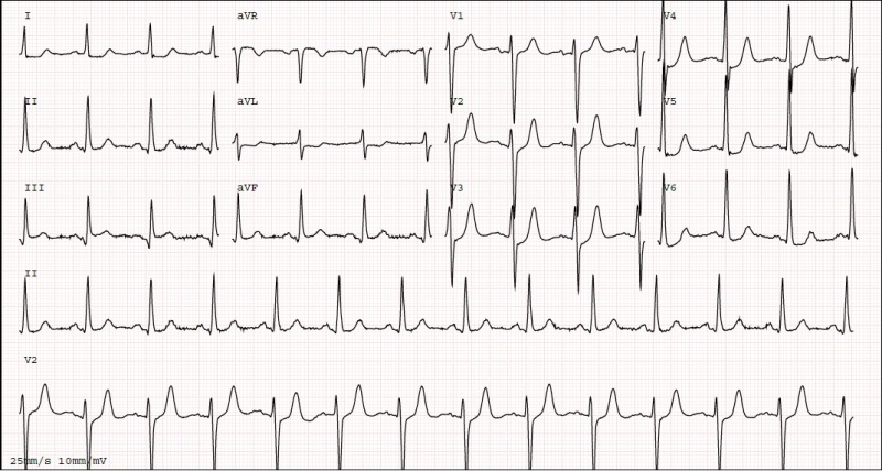
The de Winter sign is upsloping ST depression with tall, prominent, symmetric T waves in the precordial leads. It is described in proximal LAD occlusion. It is a STEMI equivalent, and it can show up with no ST elevation at all.
ECG Criteria
- ST segment: upsloping ST depression greater than 1 mm at the J point in the precordial leads (2022 ECDP)
- T waves: tall, prominent, symmetric (2022 ECDP and 2026 Universal Definition)
- Distribution: the 2026 Universal Definition places the pattern in V2–V5
- aVR: ST elevation of 0.5–1 mm may be seen in aVR (2022 ECDP)
Clinical Significance
- Action: activate the cath lab per protocol; the 2022 ECDP calls for immediate angiography
- Timing: do not wait for ST elevation to develop
Look-Alikes
- Hyperacute T waves without the upsloping depression (see below)
- Hyperkalemia with peaked T waves; see the T Wave page
Hyperacute T Waves
Early STEMI can make the T waves broad and disproportionately large for the QRS, before any ST elevation shows up. Broad T waves are a reason to repeat the ECG. They are not a diagnosis on their own.
ECG Criteria
- 2026 Universal Definition: symmetrical, broad T waves disproportionately large to the preceding QRS in two contiguous leads
- 2022 ECDP: broad, asymmetric, peaked T waves that may appear early in STEMI
- Exclusions: electrolyte imbalance and significant LVH (2022 ECDP)
The two sources disagree on symmetry. So don't judge the T-wave shape by one descriptor. Compare it with the QRS and with the next tracing.
Clinical Significance
- Serial ECGs: repeat over very short intervals when suspicion is high (2022 ECDP)
- Reciprocal change: when hyperacute T waves in V1–V2 come with reciprocal changes elsewhere, that suggests occlusion (2026 Universal Definition)
- Subtle elevation: ST elevation below 1 mm with reciprocal ST depression can represent occlusion. The 2022 ECDP says these patients should be evaluated for emergent angiography.
Look-Alikes
- Hyperkalemia: peaked T waves are the common mimic; see the T Wave page
- Left ventricular hypertrophy: the 2022 ECDP excludes significant LVH when judging hyperacute T waves
Posterior STEMI
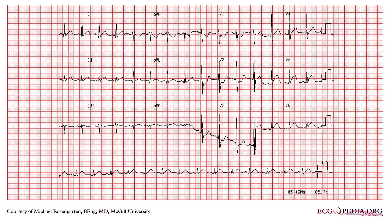
From the front, a posterior infarction looks like ST depression in V1–V3. Posterior ST elevation is what confirms it. Without posterior leads, that anterior depression is easy to misread as non-ST-elevation (NSTE) ischemia.
ECG Criteria
- Anterior clues: ST depression of 1 mm or more in V1–V3, particularly with a dominant R wave (R taller than S) in V1 or V2 (2026 Universal Definition)
- Broad R wave: a prominent, broad R wave of more than 30 ms (0.03 s), with upright anterior T waves (2022 ECDP)
- Confirmation: ST elevation in posterior leads V7–V9; the 2022 ECDP uses 0.5 mm or more in at least one lead
Lead Placement
- V7: left posterior axillary line, in the same plane as V6
- V8: tip of the left scapula
- V9: left paraspinal region, in the same plane as V6
Clinical Significance
- Action: emergent coronary angiography when posterior MI is strongly suspected; the 2022 ECDP notes that delayed reperfusion is associated with worse outcomes
- Next step: if the first ECG is non-diagnostic and suspicion is high, record a posterior ECG (2022 ECDP)
Sgarbossa and Smith-Modified Criteria
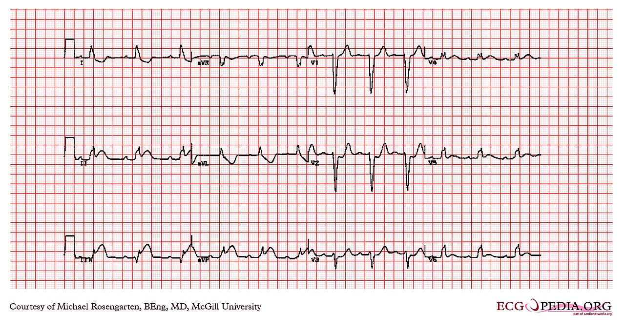
In LBBB or ventricular pacing, ST and T changes run in the expected direction for the QRS. That is why the usual thresholds don't apply here. The Sgarbossa criteria hunt for ST changes that are concordant (same direction as the QRS) or discordant and excessive.
| Criterion | Original Sgarbossa (points) | Smith-Modified (any one) |
|---|---|---|
| Concordant STE | ≥1 mm in a lead with a positive QRS (5 points) | ≥1 mm in a lead with a positive QRS |
| Concordant STD | ≥1 mm in V1–V3 (3 points) | ≥1 mm in V1–V3 |
| Discordant STE | ≥5 mm in a lead with a negative QRS (2 points) | ST/S ratio of −0.25 or lower: J-point STE ≥1 mm and at least 25% of the preceding S wave |
| Decision | Total of 3 or more points | Any one criterion positive |
- Origin: the original rule came from GUSTO-1, where 131 of 26,003 patients had acute MI with LBBB.
- Smith modification: the proportional rule was derived in 33 ECGs with coronary occlusion, with a sensitivity of 91% and specificity of 90% in that set, against sensitivities of 52% for the weighted original rule and 67% for the unweighted rule. Its specificity was lower than the weighted original rule’s, so it trades some false positives for sensitivity. It is a small derivation.
- New LBBB alone: new or presumed-new bundle branch block counts only when other signs of ischemia are present (2026 Universal Definition).
When a prior ECG exists, compare against it to pick out the new changes.
High Lateral Pattern & South African Flag
Lateral occlusion is easy to miss if you only look at the precordial leads. Under the 2026 Universal Definition, two contiguous leads outside V2–V3 with ST elevation of 1 mm or more count. That means lead I and aVL qualify when both show it.
Patterns to Recognize
- High lateral: ST elevation of 1 mm or more in I and aVL. Check V5–V6 and the inferior leads, then compare with the territory map on the STEMI Recognition page.
- South African flag: ST elevation in I, aVL and V2 with ST depression in III (2026 Universal Definition). The Universal Definition lists it among patterns that suggest acute occlusion.
Clinical Significance
- Subtle lateral elevation is often less visible than the reciprocal depression beside it (2022 ECDP). Read both.
- Subtle ST elevation under 1 mm with reciprocal ST depression can represent occlusion (see Hyperacute T Waves); compare with a prior ECG and repeat the 12-lead.
Occlusion MI vs Non-Occlusion MI
Occlusion MI (OMI) is infarction caused by an acute culprit coronary occlusion. Non-occlusion MI (NOMI) is infarction without one. A STEMI rule built on millimeters misses plenty of OMIs. And the STEMI versus NSTEMI split can leave a blocked artery without emergent reperfusion.
What the Data Show
- Missed STEMI criteria: in a 2021 retrospective review of 467 acute coronary syndrome patients, 108 had OMI and 67 of them (60%) met STEMI criteria
- Delays: median arrival-to-catheterization time was 41 minutes for STEMI-positive OMI and 437 minutes for STEMI-negative OMI
- Scale: up to 1 in 4 patients managed as NSTEMI without classic ST elevation have an acute culprit occlusion (2026 Universal Definition)
The 2021 study drew on one center. Its definition of OMI was an acute culprit with TIMI flow (thrombolysis in myocardial infarction) of 0–2, or TIMI 3 flow with a peak troponin T above 1.0 ng/mL. Read the rates as a signal, not as a population estimate.
Practical Approach
- If symptoms continue and the ECG is non-diagnostic, repeat the 12-lead, add posterior and right-sided leads, and compare with the prior ECG (2022 ECDP)
- Escalate per protocol when the clinical picture is concordant, even without STEMI criteria
- The 2026 Universal Definition also names the Aslanger pattern as suggesting occlusion. It is ST elevation isolated to III, with ST depression in V4–V6 and a positive T wave
Differential Diagnosis & Mimics
Not every ischemic ECG gets a cath lab call. The 2022 ECDP splits STEMI equivalents from ischemia patterns that are managed as high-risk NSTE acute coronary syndrome. Sort which one you have before you activate.
| Pattern | Key ECG Feature | 2022 ECDP Category |
|---|---|---|
| de Winter sign | Upsloping STD over 1 mm; tall, symmetric T waves | STEMI equivalent |
| Posterior STEMI | STD in V1–V3; R taller than S in V2; STE in V7–V9 | STEMI equivalent |
| Sgarbossa-positive LBBB or paced | Concordant STE or STD; discordant STE ≥5 mm or ST/S ≤ −0.25 | STEMI equivalent |
| Hyperacute T waves | Broad, disproportionately large T waves; may accompany STE below 1 mm | STEMI equivalent (early STEMI) |
| Wellens pattern | Biphasic or deeply inverted T waves in V2–V3; no Q waves; recent angina | Ischemia, high-risk NSTE |
| STE in aVR with diffuse STD | STE in aVR with multilead ST depression in I, II and/or V4–V6 | Ischemia, usually left main or multivessel |
Mimics
- Hyperkalemia: peaked T waves are the common mimic; see the T Wave page
- Left ventricular hypertrophy: excluded from the hyperacute T criteria by the 2022 ECDP
- Pericarditis and early repolarization: the usual STE mimics; see the STEMI Recognition page
Artifact & Lead Misplacement
- Check the posterior lead positions (V7 in the V6 plane, V8 at the scapula tip, V9 paraspinal) before calling V7–V9 normal
- Repeat the tracing when shivering, vibration or motion obscures the ST segment
- Limb-lead reversal changes the lead pattern; confirm electrode placement and repeat before dismissing the concern
Bedside Management & Escalation
Once you recognize a STEMI equivalent, it follows the reperfusion pathway. The 2022 ECDP says STEMI and STEMI equivalents identify the patients who need immediate reperfusion therapy. This page lists no doses. Use local protocols and medical direction.
| Situation | BLS (EMT) | ALS (AEMT / Paramedic / RN) | Critical Care / Transport |
|---|---|---|---|
| Equivalent pattern, stable | 12-lead acquisition and transmission where allowed; notify ALS | Transmit the 12-lead; notify the receiving PCI center per protocol; aspirin per the aspirin page after its hold check; other antithrombotic therapy per protocol | Serial 12-leads; posterior and right-sided leads when indicated; transfer per protocol |
| Equivalent pattern, unstable | Recognize shock and arrhythmia; early ALS | Follow the unstable-patient protocol; the 2022 ECDP does not apply to hemodynamic instability | Fibrinolysis decision per protocol when PCI is not available within the protocol time window, and after contraindication screening (see the Fibrinolytics page); review the RV warning before any nitrate |
Key Actions
- Activate: recognize the pattern and start the reperfusion pathway; do not wait for classic STE
- Repeat: serial 12-leads at short intervals and with any new symptom (2022 ECDP)
- Leads: posterior V7–V9 for anterior depression; V3R–V6R when RV infarction is suspected (2026 Universal Definition)
- Imaging: consider echocardiography for borderline elevation, LBBB or equivocal posterior MI (2022 ECDP)
- Aspirin: give per the aspirin page and protocol; check for aspirin or NSAID hypersensitivity, aspirin-sensitive asthma and active GI bleeding first
- Medications: follow the E3 pages for fibrinolytics, heparin and clopidogrel, and local protocol
- Oxygen: per the oxygen page and protocol
Follow local protocols and online medical direction.
Pediatric & Special Populations
Pediatrics
- Every threshold on this page is an adult criterion. Neither the 2026 Universal Definition nor the 2022 ECDP gives pediatric values.
- In children and adolescents, read ST and T changes with pediatric cardiology input and the pediatric ECG reference used locally.
Other Populations
- Women, pregnancy and postpartum: consider spontaneous coronary artery dissection (SCAD) in women under 50 and in pregnancy or the postpartum period (2026 Universal Definition)
- Older adults and people with diabetes: MI without chest pain is more common. An ECG pattern with no typical symptoms still gets the same checks (2026 Universal Definition)
- Prior bypass or complex coronary disease: an acute graft or coronary occlusion can happen without typical ST elevation (2026 Universal Definition)
- Pacemaker or LBBB: apply the Sgarbossa criteria above. The baseline ECG is already abnormal, so compare it with any stored tracing
Monitoring & Transport Considerations
| Issue | What to Do |
|---|---|
| Lead set | Standard 12-lead; add V7–V9 when V1–V3 depression is present; add V3R–V6R when RV infarction is suspected (2026 Universal Definition) |
| Serial tracings | Repeat at short intervals and whenever symptoms change (2022 ECDP); keep the prior tracing for comparison |
| Transmission | Send the 12-lead per protocol and label posterior and right-sided leads so the receiving team reads them as added leads |
| Artifact | Shivering, vibration and motion can mimic ST change; repeat with the patient still and check lead positions |
| Unstable patient | The 2022 ECDP does not apply to hemodynamic instability; use the unstable-patient protocol |
| Paced or LBBB baseline | Use the Sgarbossa criteria and compare with any stored ECG |
| Nitrates | If RV infarction is suspected, hold nitrates and follow medical direction |
Step-by-Step Approach & Pitfalls
- Confirm the QRS: note LBBB, RBBB or ventricular pacing before applying any threshold.
- Measure the J point in every lead against the local baseline.
- Read V1–V3 for ST depression of 1 mm or more and for R taller than S in V1–V2.
- Look for the de Winter pattern: upsloping depression over 1 mm with tall, symmetric T waves in the precordial leads.
- Record posterior and right-sided leads when the pattern calls for them.
- Apply the Sgarbossa or Smith-modified criteria in LBBB or paced rhythm.
- Check the T waves and subtle elevation: broad, large T waves, or elevation below 1 mm with reciprocal depression, are occlusion clues.
- Repeat the 12-lead while symptoms persist, and act on a concordant clinical picture per protocol.
Common Pitfalls
- Waiting for classic STE when a posterior or de Winter pattern is present
- Reading V1–V3 depression as NSTE ischemia without posterior leads
- Applying ST thresholds to LBBB or paced rhythm
- Treating hyperacute T waves as normal because the ST segment looks normal
- Confusing the Wellens pattern with a STEMI equivalent; it is a high-risk NSTE pattern
ECG Examples




Clinical Pearls
References
- Mills, Newby, Zaman, Almahmeed, et al. Fifth Universal Definition of Myocardial Infarction (2026). Global Heart. 2026;21:64. https://doi.org/10.5334/gh.1578
- Kontos MC, de Lemos JA, Deitelzweig SB, et al. 2022 ACC Expert Consensus Decision Pathway on the evaluation and disposition of acute chest pain in the emergency department. J Am Coll Cardiol. 2022;80(20):1925–1960. https://doi.org/10.1016/j.jacc.2022.08.750
- de Winter RJ, Verouden NJ, Wellens HJ, Wilde AA. A new ECG sign of proximal LAD occlusion. N Engl J Med. 2008;359(19):2071–2073 (landmark study). https://doi.org/10.1056/NEJMc0804737
- Sgarbossa EB, Pinski SL, Barbagelata A, et al. Electrocardiographic diagnosis of evolving acute myocardial infarction in the presence of left bundle-branch block. N Engl J Med. 1996;334(8):481–487 (landmark study). https://doi.org/10.1056/NEJM199602223340801
- Smith SW, Dodd KW, Henry TD, Dvorak DM, Pearce LA. Diagnosis of ST-elevation myocardial infarction in the presence of left bundle branch block with the ST-elevation to S-wave ratio in a modified Sgarbossa rule. Ann Emerg Med. 2012;60(6):766–776 (derivation study). https://doi.org/10.1016/j.annemergmed.2012.07.119
- de Zwaan C, Bär FW, Wellens HJ. Characteristic electrocardiographic pattern indicating a critical stenosis high in left anterior descending coronary artery in patients admitted because of impending myocardial infarction. Am Heart J. 1982;103(4):730–736 (landmark study). https://doi.org/10.1016/0002-8703(82)90480-x
- Meyers, Bracey, Lee, et al. Comparison of the ST-elevation myocardial infarction (STEMI) vs. NSTEMI and occlusion MI (OMI) vs. NOMI paradigms of acute MI. J Emerg Med. 2021;60(3):273–284. https://doi.org/10.1016/j.jemermed.2020.10.026
- Wilkinson-Stokes M, Betson J, Sawyer S. Adverse events from nitrate administration during right ventricular myocardial infarction: a systematic review and meta-analysis. Emerg Med J. 2023;40(2):108–113. https://doi.org/10.1136/emermed-2021-212294
Medical Disclaimer
- For Educational Purposes Only: This content is intended for educational reference and should not be used for clinical decision-making.
- Not a Substitute for Professional Judgment: Always consult your local protocols, institutional guidelines, and supervising physicians.
- Verify Before Acting: Users are responsible for verifying information through authoritative sources before any clinical application.