

At a Glance
- Pattern
- The STEMI pattern: new ST elevation at the J point in two or more contiguous leads, read by territory. The leads with elevation face the injured wall; the opposite leads show reciprocal depression.
- Normal (Adult)
- ST elevation <1 mm in limb leads and V4–V6; V2–V3 STE limited by sex and age (see Normal Values). No new reciprocal depression.
- Confirm It
- Read the J point against the TP (or PR) baseline in every lead. Compare contiguous leads, then check the reciprocal leads and the prior ECG.
- Abnormal → Think
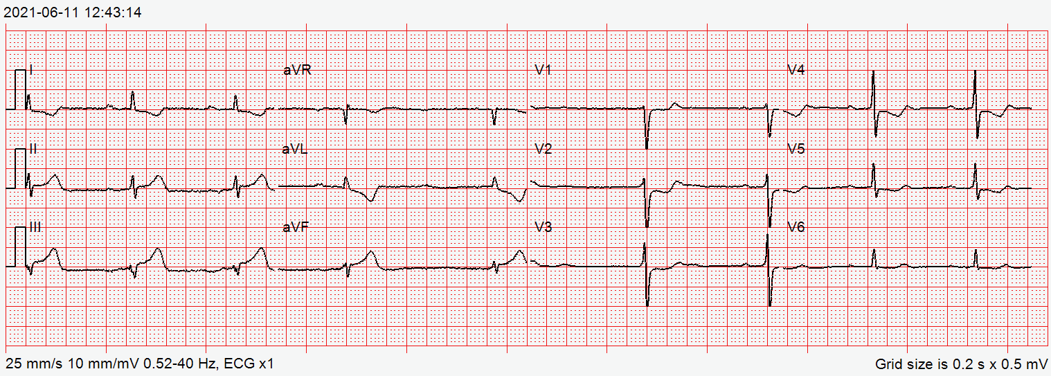
- STE II, III, aVF: inferior STEMI; add V4R and V7–V9
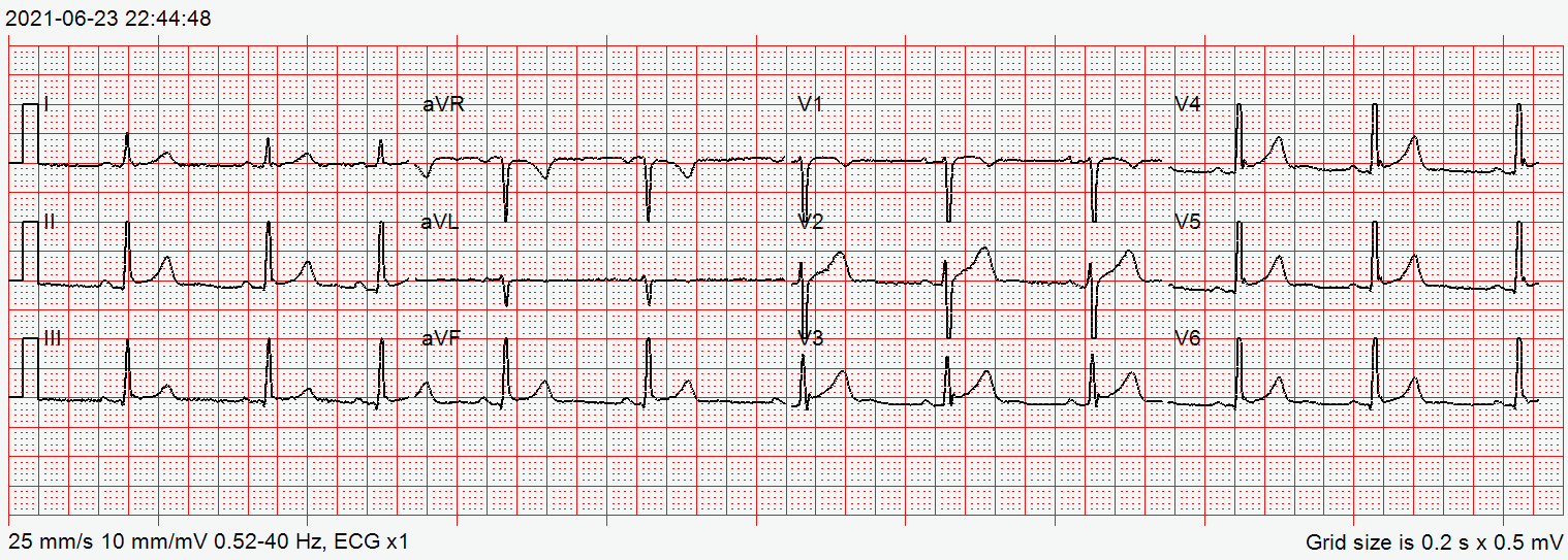
- STE V1–V4: anterior STEMI; check I and aVL
- STE I, aVL, V5–V6: lateral STEMI
- Red Flags
- Ischemic STE in contiguous leads, a STEMI equivalent (de Winter sign, posterior pattern, Sgarbossa criteria) or STE with hypotension. Do not wait for classic STE if the picture is concordant.
- Bedside Action
- If the first ECG is non-diagnostic and symptoms persist, repeat it every 15–30 min. Do not delay activation or transport when STEMI criteria are met. Notify the PCI center per protocol; hold nitrates in suspected RV infarction.
Physiology & What It Represents
In STEMI, a coronary artery occludes. The myocardium it feeds loses perfusion. Within minutes, the injured cells fail to repolarize normally. That difference between injured and healthy tissue creates a current of injury in the ST segment. Leads facing the injured wall show ST elevation. Leads facing the opposite wall show reciprocal ST depression.
The ECG shows which wall is injured, so the pattern points to the culprit vessel. A ST change is only as useful as its distribution. Look for one territory, a convex shape and reciprocal change. That is the pattern that drives reperfusion decisions.
Normal Values & How to Measure
| Parameter | Threshold | Where / How to Measure |
|---|---|---|
| Limb leads and V4–V6 | ≥1 mm at the J point in 2 contiguous leads | Read against the TP (or PR) baseline |
| V2–V3, men ≥40 years | ≥2 mm | Precordial leads run higher in normal men |
| V2–V3, men <40 years | ≥2.5 mm | Age-adjusted threshold |
| V2–V3, women | ≥1.5 mm | Sex-adjusted threshold |
| Posterior V7–V9 | ≥0.5 mm | Place leads on the back, same horizontal level as V4–V6 |
| Right-sided V4R (and V3R) | V4R ≥0.5 mm (≥1 mm in men <30 years); V3R threshold per protocol | Mirror-image placement of V4 and V3 on the right chest |
- Identify the J point in each lead as the end of the QRS, on a 12-lead at 25 mm/s with 10 mm/mV.
- Set the baseline at the TP segment, or the PR segment when the TP is not usable.
- Measure the ST deviation at the J point in each lead, applying the sex and age threshold to V2–V3.
- Map the contiguous leads that meet threshold to one territory, then check the reciprocal leads.
- Add V4R and V7–V9 when the inferior or posterior picture is present.
- Compare with the prior ECG when one exists.
STEMI Territories & Culprit Arteries
Start with the leads that meet criteria, then name the territory. The culprit vessel is typical, not certain. Coronary dominance and collateral supply change the map.
| Lead group | Wall | Reciprocal depression | Usual culprit |
|---|---|---|---|
| V1–V4 | Anterior (septal and anterior) | II, III, aVF | Proximal or mid LAD |
| V1–V2 alone | Septal | Often II, III, aVF | Proximal LAD (septal branches) |
| I, aVL, V5–V6 | Lateral | III, aVF | Circumflex or first diagonal branch |
| II, III, aVF | Inferior | I, aVL | RCA in right-dominant hearts; circumflex in left-dominant hearts |
| V7–V9 | Posterior | V1–V3 depression (mirror image) | RCA or circumflex |
| V4R | Right ventricle | Variable | Proximal RCA; often with inferior STEMI |
- Inferior STEMI: Record V4R and V7–V9 before any preload-reducing drug. You're looking for right ventricular and posterior involvement.
- Anterior STEMI: STE in I and aVL with V1–V4 points to a larger proximal LAD territory. Inferior reciprocal depression backs up the call.
- Posterior STEMI: At least 1 mm of ST depression in V1–V3, with a dominant R wave in V2, is the front view of posterior STE. Confirm with V7–V9.
Reciprocal Change
Reciprocal ST depression is the opposite wall's picture of the culprit injury. It's the main feature that separates regional occlusion from pericarditis and early repolarization. Those usually lack it.
- Inferior STE: depression in I and aVL (the classic pair)
- Anterior STE: depression in II, III and aVF
- Lateral STE: depression in III and aVF
- STE in aVR with diffuse depression: STE in aVR (often with V1) plus widespread ST depression in I, II and V4–V6 points to left main or multivessel ischemia. It doesn't meet single-territory STEMI criteria. It is high-risk NSTE-ACS. Call cardiology early and seek urgent angiography per protocol, especially with hypotension or ongoing pain.
Evolution of Acute Infarction
The ECG changes over time, and the stage changes the call. Early changes can come before classic STE. Late changes can look like old infarction.
- Hyperacute T waves: Broad, tall T waves that show up first, often before STE. The 2022 ACC pathway describes them as broad, asymmetric or peaked.
- ST elevation: Develops as the injury current forms. In the territory, it is convex (tombstone) or straight.
- Q waves: Develop in the territory during the acute phase. Their presence alone doesn't date the infarct.
- T-wave inversion: Follows the elevation. The ST segment may normalize while the T inversion stays.
- Old infarct: Persistent Q waves, resolved STE, and inverted or normal T waves. Compare with the prior ECG.
STEMI Equivalents & Occlusion Signs
Some patterns point to occlusion even though millimeter STEMI criteria are not met. The 2022 ACC Expert Consensus Decision Pathway counts posterior STEMI, LBBB or paced rhythm with Sgarbossa criteria, de Winter sign and hyperacute T waves as equivalents. For the full criteria, go to the ST Segment & T Wave page. For occlusion without classic STE, see the STEMI Equivalents & Occlusion Signs page.
- De Winter sign: Upsloping ST depression above 1 mm at the J point in precordial leads, with tall, symmetric T waves. It's described with proximal LAD occlusion.
- Posterior pattern: ST depression in V1–V3 with an upright T. Confirm with at least 0.5 mm of STE in V7–V9.
- LBBB or paced rhythm: Apply the Sgarbossa criteria. New LBBB alone is not a STEMI equivalent.
- Wellens pattern (high risk, not a STEMI): Characteristic precordial ST-T changes that point to a critical proximal LAD stenosis, without meeting STEMI criteria. The original report recommends urgent angiography and revascularization when possible.
Differential Diagnosis & Mimics
Acute pericarditis and early repolarization are the main mimics. Both can meet millimeter thresholds, so millimeters alone won't separate them. Look at distribution, morphology and reciprocal change.
| Feature | STEMI | Acute Pericarditis | Early Repolarization |
|---|---|---|---|
| ST distribution | One vascular territory | Diffuse, most leads | Lateral or inferior, often symmetric |
| ST morphology | Convex (tombstone) or straight | Concave (smiley) | Concave with J-point notch or slur |
| Reciprocal depression | Present in the opposite territory | Absent (except aVR) | Absent |
| PR segment | Normal; PR elevation in aVR may occur | PR depression in most leads | Normal |
| Evolution | Rapid; Q waves and T inversion | Days to weeks; ST normalizes | Stable on serial ECGs |
Other STEMI Mimics
- LVH: high voltage with discordant STE in V1–V3
- Brugada pattern: coved STE with a negative T wave in V1–V2
- Ventricular aneurysm: persistent STE with Q waves that does not evolve
- Takotsubo: wide-territory change after stress; echo wall motion crosses coronary territories
- Hyperkalemia: peaked T waves with QRS widening can mimic de Winter; check potassium
Artifact & Lead Misplacement
- Limb-lead reversal: changes axis and can create apparent lead I or aVR changes
- Precordial electrodes placed high: can mimic STE in V1–V2
- Tremor or motion: can mimic ST deviation or VT and VF
Bedside Management & Escalation
The ECG sets the pathway. A STEMI or a STEMI equivalent starts reperfusion. A high-risk pattern starts urgent cardiology review. Every drug and threshold below runs on local protocols and online medical direction.
| Situation | BLS (EMT) | ALS (AEMT / Paramedic / RN) | Critical Care / Transport |
|---|---|---|---|
| Unstable (hypotension, shock, pulmonary edema) | Call ALS now; 12-lead acquisition and transmission where allowed; oxygen only if SpO2 is below 90% or for respiratory distress, and not routinely at 90% or higher (DETO2X-AMI) | 12-lead; notify the PCI center; IV access; treat per protocol; hold nitrates if SBP <90 mmHg | Continuous monitoring; serial 12-leads; transport to a PCI-capable center |
| Stable with ischemic-pattern STE | 12-lead where allowed; notify ALS | 12-lead; antiplatelet and nitrate per protocol; repeat the 12-lead; notify the receiving center | Serial 12-leads every 15–30 min; cath lab team on arrival |
| Pulseless or in arrest | Start CPR and use the AED; a 12-lead does not delay CPR or shocks | Cardiac arrest protocol; treat the rhythm | Post-ROSC 12-lead; reperfusion decision per protocol |
| Therapy | Adult | Pediatric | Key Notes |
|---|---|---|---|
| Aspirin (suspected ACS) | 162–325 mg PO, chewed (non-enteric-coated; 81 mg chewable tablets, e.g. 4 × 81 mg) single loading dose per protocol | Not established for ACS in children | Give unless allergic or actively bleeding. Use non-enteric-coated tablets because they are chewed. No E3 aspirin page yet. |
| Nitroglycerin (chest pain) | 0.4 mg SL every 5 min max 3 doses in 15 min per label (nitroglycerin) | Not established in children: specialist use only | Give only if SBP ≥90 mmHg and not 30 mmHg or more below baseline. The Nitrostat label contraindicates PDE-5 inhibitors (avanafil, sildenafil, tadalafil, vardenafil) and sGC stimulators (windows on the nitroglycerin page), shock or circulatory failure, severe anemia and raised intracranial pressure; it advises caution in aortic or mitral stenosis and hypertrophic obstructive cardiomyopathy. Hold in suspected RV infarction. |
| P2Y12 inhibitor (cath-lab decision) | Ticagrelor or prasugrel preferred with primary PCI. Clopidogrel 600 mg PO load, then 75 mg daily, when those are unavailable or contraindicated (clopidogrel) | Not established in children: specialist or medical-control use only | Agent and timing set by the cath lab and protocol. EMS does not give a P2Y12 load unless protocol or medical direction says so. With fibrinolysis, clopidogrel is 300 mg load if 75 years or younger, or 75 mg with no load if older than 75 (see the clopidogrel page). |
| Fibrinolysis (when PCI is delayed) | Agent and dose per the fibrinolytics page and protocol | Systemic fibrinolysis not established in children | High-alert Considered when anticipated first-medical-contact-to-device time exceeds 120 min (non-PCI site or long transfer), if symptom onset was within 12 h and there is no contraindication; door-to-needle goal 30 min. The 90-min goal applies to direct presentation to a PCI center. Check absolute contraindications on the fibrinolytics page. |
Doses vary by protocol; follow local protocols and medical direction.
Pediatric & Special Populations
Pediatrics
- Pattern: Repolarization varies with age and heart rate. Use age-based pediatric ECG references.
- Cause: In children, ST changes are more often non-ischemic (myocarditis, pericarditis, early repolarization). Think about coronary anomalies and Kawasaki-related coronary disease when suspected.
- Action: Contact pediatric cardiology or medical direction early. Check pediatric vital signs for rate and BP norms.
- Drugs: Pediatric dosing for nitrates, antiplatelets and fibrinolytics is not established. Use weight-based dosing and specialist input.
Other Populations
- Older adults and women: Presentations are often atypical. ST depression may be the only finding.
- Pacemaker or ICD: Paced rhythms need the Sgarbossa criteria. Get a prior ECG.
- Heart transplant: Denervated hearts may have blunted symptoms and abnormal ST-T patterns. Compare with prior tracings.
- Pregnancy: ST-T changes can be normal. In pregnancy or the peripartum period, spontaneous coronary artery dissection should be considered (see reference 10). Involve cardiology and obstetrics early. Follow medical direction for reperfusion.
Monitoring & Transport Considerations
| Issue | What to Do |
|---|---|
| Monitoring lead | Use the lead that shows the abnormality; V2–V5 for anterior and lateral changes, II for inferior |
| Serial 12-leads | Repeat every 15–30 min in ongoing symptoms and transmit each tracing where allowed |
| Posterior and right-sided leads | V7–V9 for suspected posterior MI; V4R for suspected RV infarction with inferior STEMI |
| Artifact | Stop, check electrodes and limb placement, and re-record before acting on an ST change from vibration or shivering |
| Before moving an unstable patient | Place anterior-posterior pads when bradycardia or unstable tachycardia may need pacing or defibrillation |
In transport, a clean tracing is the only reliable input you have. Reduce motion before you record.
Step-by-Step Approach & Pitfalls
- Identify the J point and TP baseline in each lead before reading any shape.
- Measure the STE or STD at the J point and note its shape: convex, concave, horizontal or downsloping.
- Map the distribution to a territory using the lead-group table.
- Check the reciprocal leads and the STEMI equivalents: de Winter, posterior, Sgarbossa.
- Add V4R and V7–V9 when the inferior or posterior picture is present.
- Check the mimics: concave STE with PR depression (pericarditis), early repolarization, LVH, artifact.
- Decide the pathway and repeat the 12-lead every 15–30 min in ongoing symptoms.
Common Pitfalls
- Waiting for classic STE while a de Winter or posterior pattern is already on the tracing
- Calling isolated V1–V3 depression an NSTEMI without checking V7–V9
- Applying STEMI thresholds in LBBB or a paced rhythm instead of the Sgarbossa criteria
- Giving nitrates to a hypotensive patient with inferior changes and no V4R
- Treating one tracing as the whole answer while symptoms persist
- Missing bradyarrhythmia or heart block in inferior STEMI. Check rate and rhythm continuously, have pacing and atropine ready per protocol, and place pads early when block is likely
- Assuming a fast effect from an oral P2Y12 agent. Opioids such as morphine slow gastric emptying and can delay P2Y12 absorption. Follow protocol on timing
- Transporting to the nearest hospital. When the protocol allows prehospital activation, bypass a non-PCI hospital for a PCI-capable center
ECG Examples
Clinical Pearls
References
- Rao SV, O'Donoghue ML, Ruel M, et al. 2025 ACC/AHA/ACEP/NAEMSP/SCAI guideline for the management of patients with acute coronary syndromes. J Am Coll Cardiol. 2025;85(22):2135-2237; also published as Circulation. 2025;151(13):e771-e862. https://doi.org/10.1016/j.jacc.2024.11.009; https://doi.org/10.1161/CIR.0000000000001309
- Thygesen K, Alpert JS, Jaffe AS, et al. Fourth universal definition of myocardial infarction (2018). Circulation. 2018;138(20):e618-e651. https://doi.org/10.1161/CIR.0000000000000617
- Kontos MC, de Lemos JA, Deitelzweig SB, et al. 2022 ACC Expert Consensus Decision Pathway on the evaluation and disposition of acute chest pain in the emergency department. J Am Coll Cardiol. 2022;80(20):1925-1960. https://doi.org/10.1016/j.jacc.2022.08.750
- de Winter RJ, Verouden NJ, Wellens HJ, Wilde AA. A new ECG sign of proximal LAD occlusion. N Engl J Med. 2008;359(19):2071-2073. https://doi.org/10.1056/NEJMc0804737
- de Zwaan C, Bär FW, Wellens HJ. Characteristic electrocardiographic pattern indicating a critical stenosis high in left anterior descending coronary artery in patients admitted because of impending myocardial infarction. Am Heart J. 1982;103(4 Pt 2):730-736 (landmark study). https://doi.org/10.1016/0002-8703(82)90480-x
- Smith SW, Dodd KW, Henry TD, Dvorak DM, Pearce LA. Diagnosis of ST-elevation myocardial infarction in the presence of left bundle branch block with the ST-elevation to S-wave ratio in a modified Sgarbossa rule. Ann Emerg Med. 2012;60(6):766-776. https://doi.org/10.1016/j.annemergmed.2012.07.119
- Macfarlane PW, Antzelevitch C, Haissaguerre M, et al. The early repolarization pattern: a consensus paper. J Am Coll Cardiol. 2015;66(4):470-477. https://doi.org/10.1016/j.jacc.2015.05.033
- Adler Y, Charron P, Imazio M, et al. 2015 ESC guidelines for the diagnosis and management of pericardial diseases. Eur Heart J. 2015;36(42):2921-2964. https://doi.org/10.1093/eurheartj/ehv318
- Hofmann R, James S, Jernberg T, Lindahl B, et al. Oxygen therapy in suspected acute myocardial infarction (DETO2X-AMI). N Engl J Med. 2017;377(13):1240-1249. https://doi.org/10.1056/NEJMoa1706222
- Adlam D, Alfonso F, Maas A, Vrints C, et al. European Society of Cardiology, Acute Cardiovascular Care Association, SCAD Study Group: a position paper on spontaneous coronary artery dissection. Eur Heart J. 2018;39(36):3353-3368. https://doi.org/10.1093/eurheartj/ehy080
Medical Disclaimer
- For Educational Purposes Only: This content is intended for educational reference and should not be used for clinical decision-making.
- Not a Substitute for Professional Judgment: Always consult your local protocols, institutional guidelines, and supervising physicians.
- Verify Before Acting: Users are responsible for verifying information through authoritative sources before any clinical application.