

At a Glance
- Pattern
- Early ventricular activation over an accessory pathway: a short PR, a delta wave and a wide QRS in sinus rhythm, and pre-excited tachycardias.
- Normal (Adult)
- PR 120–200 ms, no delta wave, QRS ≤110 ms. Pre-excitation: PR <120 ms, QRS often over 110 ms.
- Confirm It
- PR in lead II; slurred QRS upstroke (delta wave) in any lead; then regular or irregular, narrow or wide, on a tachycardia strip.
- Abnormal → Think
- Short PR and delta wave: manifest pathway
- Regular narrow tachycardia: orthodromic AVRT
- Irregular, fast, wide: pre-excited AF until proven otherwise
- Red Flags
- Pre-excited AF: rapid conduction down the pathway can degenerate into VF. AV nodal blockers can precipitate it.
- Bedside Action
- Unstable: synchronized cardioversion now. Stable regular narrow: vagal, then adenosine. Stable irregular wide: IV ibutilide or procainamide. No verapamil, diltiazem, beta-blocker, digoxin or amiodarone.
Physiology & What It Represents
Normally the impulse leaves the sinus node and spreads through the atria. Then it has one gate to the ventricles: the AV node and His-Purkinje system. The AV node slows the signal. That delay gives the atria time to empty before the ventricles contract.
An accessory pathway is an extra atrioventricular connection, usually along the AV ring. It conducts fast and skips the AV node's delay. The tissue is atrial, so it can carry atrial impulses straight to the ventricle. Part of the ventricle fires early, before the AV node delay has run out.
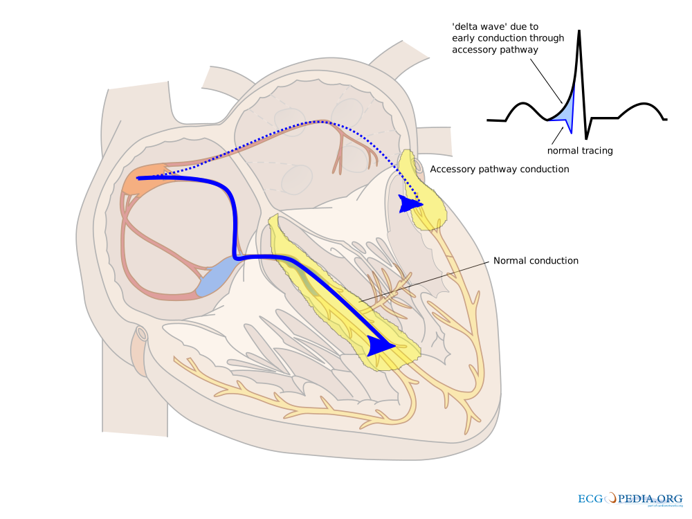
Sort pathways by what they do on the ECG. A manifest pathway conducts both ways, so pre-excitation shows in sinus rhythm. A concealed pathway conducts only backward, so pre-excitation never shows. A non-manifest pathway shows no pre-excitation, though it may still conduct both ways. WPW syndrome means a manifest pathway with recurrent tachyarrhythmias.
Normal Values & How to Measure
Measure pre-excitation on a sinus beat first. Then sort any tachycardia by regularity and QRS width. Pre-excitation changes the answer to the question "which drug is safe?"
| Parameter | Adult Normal | Where / How to Measure |
|---|---|---|
| PR interval | 120–200 ms (3–5 small boxes); pre-excited: <120 ms | From the P wave onset to the QRS onset, in lead II |
| Delta wave | Absent | Slurred first part of the QRS; polarity and best leads depend on pathway location, so look in every lead |
| QRS duration | ≤110 ms; usually over 110 ms with pre-excitation, sometimes near normal | Widest lead, from the start of the QRS to the J point |
How to Measure
- Find a sinus beat: pre-excitation shows on a sinus 12-lead or rhythm strip, so get one before judging the pattern.
- Measure the PR: lead II, from the P wave onset to the QRS onset.
- Look for the delta wave: a slurred QRS upstroke, checking several leads (the best leads vary with pathway location).
- Measure the QRS: in the widest lead.
- Classify any tachycardia: regular or irregular, narrow or wide.
- Assess stability and choose the path: stable regular narrow gets vagal then adenosine; unstable gets synchronized cardioversion; irregular wide gets no AV nodal blocker.
Tachycardia Recognition
| Rhythm | Rate and Regularity | P Waves and PR | QRS | First-Line Action |
|---|---|---|---|---|
| Orthodromic AVRT | Usually 150–250 bpm, regular | Retrograde P after the QRS | Narrow | Stable: vagal maneuver, then adenosine. Unstable: synchronized cardioversion |
| Antidromic AVRT | Usually 150–250 bpm, regular | Often hard to see | Wide, looks like VT | Treat as VT: no AV nodal blockers. Unstable: synchronized cardioversion |
| Pre-excited AF | Very fast, irregular | Fibrillatory, no clear P waves | Wide, varying width and shape | Unstable: synchronized cardioversion. Stable: IV ibutilide or procainamide. Never an AV nodal blocker |
Wolff-Parkinson-White Pattern (Pre-Excitation in Sinus Rhythm)
Pre-excitation in sinus rhythm is what a manifest accessory pathway looks like on the ECG. The sinus impulse takes the pathway and hits the ventricle early. The result: the start of the QRS slurs, and the PR interval shortens.
ECG Criteria
- Short PR: under 120 ms. The AV node delay gets bypassed.
- Delta wave: a slurred initial upstroke of the QRS.
- Wide QRS: usually over 110 ms (often 120 ms or more). Normal and pre-excited activation fuse together, so the QRS widens. With a small pathway it can be near normal.
- Where to look: delta waves change with pathway location, so check every lead. Figure 2 shows the slurring best in leads I, aVL and V2–V6. In some leads a delta wave can also be negative.
Causes
- Accessory pathway: an extra connection left behind when atrial and ventricular tissue fail to separate fully during embryologic development.
- Concealed form: a pathway that conducts only backward. It looks normal in sinus rhythm, yet it can still drive a tachycardia.
Clinical Significance
- Tachycardia risk: in WPW syndrome, AVRT is the most common arrhythmia. AF comes second.
- Sudden death risk: the one you fear is AF conducting fast over the pathway. It can degenerate into VF.
- Prophylaxis evidence: one small randomized trial (72 patients) looked at high-risk asymptomatic patients. Over five years, 2 of 37 ablation patients (5%) had an arrhythmic event. Controls fared worse: 21 of 35 (60%) did.
- Asymptomatic pre-excitation: about one in five patients will develop a pathway-related arrhythmia during follow-up (2019 ESC SVT guideline). That is why risk assessment matters.
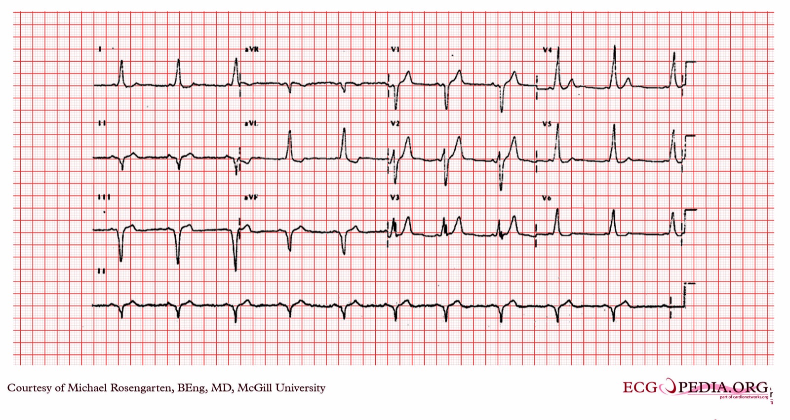
Pre-excitation becomes dangerous only once a tachyarrhythmia joins it. The sections below cover that. Pathway ablation is the definitive treatment. For the differential with a short PR and no delta wave, see PR Interval.
Orthodromic Atrioventricular Re-entrant Tachycardia (AVRT)
Orthodromic AVRT is a re-entrant circuit. It runs forward through the AV node and back up the accessory pathway. The ventricles fire through the normal conduction system, so the QRS stays narrow.
ECG Criteria
- QRS: narrow, because the ventricles are activated over the AV node and His-Purkinje system.
- Rate: usually 150–250 bpm and regular.
- P waves: retrograde, after the QRS, as the atria are activated backward over the pathway.
Causes
- Manifest pathway: it conducts both ways, so you can see pre-excitation in sinus rhythm.
- Concealed pathway: it conducts only backward. The tachycardia can still happen, with no pre-excitation on the sinus ECG.
Clinical Significance
- Frequency: in WPW syndrome, AVRT is the most common arrhythmia. In infants and young children, it is the most common SVT.
- Management: the circuit depends on the AV node. That is why stable patients can get vagal maneuvers and adenosine. For the stable-SVT approach, see Supraventricular Tachycardia.
Look-Alikes
- AV nodal re-entrant tachycardia (AVNRT): narrow and regular, and there is no pathway. See Supraventricular Tachycardia.
- Antidromic AVRT: same mechanism, run in reverse. The QRS is wide. The next section covers it.
Antidromic Atrioventricular Re-entrant Tachycardia (AVRT)
Antidromic AVRT runs the other way. The impulse goes down the accessory pathway, then back up through the AV node. Because the ventricles fire over the pathway, the QRS is wide.
ECG Criteria
- QRS: wide. Activation runs over the accessory pathway.
- Rhythm: regular, usually 150–250 bpm.
- Pattern: it often looks like ventricular tachycardia. That look is what makes it dangerous.
Causes
- Manifest pathway: it has to conduct forward. A concealed pathway cannot produce this form.
Clinical Significance
- Misdiagnosis: a regular wide-complex tachycardia that is really antidromic AVRT gets treated as VT. An unstable patient then gets synchronized cardioversion.
- Drug risk: a stable patient with a regular wide QRS should be treated as VT, per the local algorithm. Avoid AV nodal blockers until the mechanism is clear.
Pre-Excited Atrial Fibrillation
Pre-excited atrial fibrillation is AF running fast down an accessory pathway. Each beat reaches the ventricle over that pathway, which is why the QRS is wide and the rhythm is irregular. Ventricular rates can climb very high.
ECG Criteria
- Rhythm: irregularly irregular, with no organized P waves.
- QRS: wide, and varying in width and shape from beat to beat.
- Rate: expect very fast rates when the pathway conducts quickly. An RR interval of 250 ms or less, pre-excited during AF, marks high risk.
Causes
- Accessory pathway with atrial fibrillation: paroxysmal AF shows up in about half of WPW patients (2019 ESC SVT guideline). Atrial tissue is faster than the AV node, and that is what permits very rapid conduction.
- High-risk anatomy: two things push VF risk up: multiple pathways, and a short antegrade refractory period under 250 ms.
Clinical Significance
- Sudden death risk: a fast run over the pathway can degenerate into VF.
- Drugs that are contraindicated: the AV nodal blocker family, including verapamil, diltiazem, beta-blockers, digoxin, adenosine and amiodarone. Each one can drive more conduction down the pathway.
- Drugs that are recommended in stable patients: IV ibutilide or IV procainamide for pharmacologic cardioversion as an alternative to elective cardioversion.
- Definitive treatment: the definitive fix is catheter ablation of the accessory pathway. That is recommended for pre-excited AF.
Differential Diagnosis & Mimics
A wide-complex tachycardia, irregular or regular, can be one of several rhythms. Sort them with the sinus ECG, the regularity and the stability.
| Feature | Pre-Excited AF | Antidromic AVRT | Monomorphic VT |
|---|---|---|---|
| Rhythm | Irregular | Regular | Regular |
| QRS | Wide, varies beat to beat | Wide | Wide, uniform |
| Sinus ECG clue | Short PR and delta wave if the pathway is manifest | Short PR and delta wave if the pathway is manifest | Often none, unless a pathway is present |
| Drug approach | No AV nodal blockers; IV ibutilide or procainamide if stable | No AV nodal blockers until the mechanism is clear | Treat as VT per the local algorithm |
Look-Alikes
- Pseudo-infarct pattern: in some leads, delta waves can mimic old infarct Q waves, though the evidence for this is limited. Compare with a prior tracing and the sinus ECG before you call an infarct.
- Bundle branch block: a wide QRS without a short PR or delta wave. See QRS Complex.
- AF with aberrancy or bundle branch block: irregular and wide without pre-excitation. Check the sinus ECG and any prior tracing.
- Polymorphic VT and torsades: changing wide complexes, often with QT prolongation. See Ventricular Rhythms.
Artifact & Lead Misplacement
- Motion artifact: tremor, shivering and movement can fake an irregular wide-complex rhythm. Check a second lead.
- Transport vibration: the vehicle can produce a wide, irregular pattern that is not a real arrhythmia. Look again once it stops.
- Limb lead reversal: it can fake an abnormal axis or P-wave shape. Check the lead placement.
Bedside Management & Escalation
Stability comes first. Unstable? Synchronized cardioversion, not drugs. Stable with a regular narrow tachycardia gets a vagal maneuver, then adenosine. Irregular and wide gets no AV nodal blocker. Doses vary by protocol; follow local protocols and medical direction.
Stability Approach
| Situation | BLS (EMT) | ALS (AEMT / Paramedic / RN) | Critical Care / Transport |
|---|---|---|---|
| Unstable tachycardia with a pulse (any pre-excitation form) | Oxygen if SpO₂ <94%; monitor; call ALS and do not delay transport. Pads and CPR only if pulseless. | Synchronized cardioversion now, with SYNC confirmed. Sedate only if it does not delay the shock. | Synchronized cardioversion; anterior-posterior pads; continuous monitoring; notify the receiving team (Paramedic/RN/CCT). |
| Stable, regular, narrow (orthodromic AVRT) | Notify ALS; help with a vagal maneuver if trained. | Vagal maneuver, then adenosine if no pre-excitation is seen on the sinus ECG. If WPW is known or suspected, involve medical direction first and have a defibrillator ready. Verapamil is an alternative under medical direction only if adenosine fails, there is no AF or flutter, no pre-excitation on the sinus ECG, the patient is normotensive, and there is no heart failure or IV beta-blocker. | Repeat the 12-lead; watch for hypotension after verapamil. |
| Stable, irregular, wide (possible pre-excited AF) | Notify ALS. Give no drugs. | No AV nodal blocker. IV ibutilide or procainamide per protocol and online medical direction, as an alternative to elective cardioversion, after K+, Mg2+ and QTc are checked. Cardiovert if unstable. | Defibrillator at bedside; no AV nodal blockers; expert consultation. |
| Stable, regular, wide (antidromic AVRT or VT) | Notify ALS. Give no AV nodal blockers. | If WPW is known or suspected, prefer procainamide or ibutilide per protocol, or synchronized cardioversion, and avoid AV nodal blockers, adenosine and amiodarone. Otherwise, treat as VT per the ventricular rhythms algorithm. See Ventricular Rhythms. | Defibrillator at bedside; expert consultation; avoid verapamil and diltiazem. |
Therapy Table
| Therapy | Adult | Pediatric | Key Notes |
|---|---|---|---|
| Synchronized cardioversion (cardioversion) | Unstable pre-excited AF: at least 200 J biphasic. Unstable regular narrow tachycardia (orthodromic AVRT): 100 J synchronized. Escalate per the device. | 0.5–1 J/kg, then 2 J/kg if the first shock fails | Confirm a sync marker on every R wave. Sedate only if it does not delay the shock. |
| Adenosine (adenosine) | Regular narrow only: 6 mg rapid IV push, then 12 mg if no conversion (max 12 mg per dose) | 0.1 mg/kg rapid IV/IO (max 6 mg), then 0.2 mg/kg (max 12 mg) | High-alert Contraindicated in pre-excited AF (Class 3: Harm). Can provoke AF; have a defibrillator ready. Dipyridamole potentiates its effect; theophylline and caffeine blunt it. Not for wide-complex tachycardia. |
| Verapamil (verapamil) | 5–10 mg IV over at least 2 min (over at least 3 min in older patients); repeat 10 mg after 30 min if needed | 0.1–0.3 mg/kg IV over at least 2 min (max 5 mg) for ages 1–15; infants only with pediatric cardiology guidance | High-alert Label contraindications: AF or flutter with an accessory bypass tract, severe hypotension or cardiogenic shock, and IV beta-blockers. Never for pre-excited AF or wide-complex tachycardia. |
| Procainamide (procainamide) | Stable pre-excited AF: 20–50 mg/min IV (many protocols use 20 mg/min in pre-excited AF) until the arrhythmia stops, hypotension develops, QRS widens more than 50%, or a cumulative maximum of 17 mg/kg is reached (ACLS; the label loading limit is 1 g). Maintenance infusion of 1–4 mg/min only if protocol-directed. | 10–15 mg/kg IV/IO over 30–60 min, with expert consultation (see the procainamide page) | High-alert Boxed warning: positive ANA and lupus-like syndrome, blood dyscrasias, and CAST mortality. Avoid with prolonged QT, heart failure or complete heart block. |
| Ibutilide | Stable pre-excited AF: 1 mg IV over 10 min for weight 60 kg or more; a second 10-min infusion of 1 mg may follow 10 min after the first if the rhythm does not terminate | Not established in children; specialist use only | High-alert Boxed warning: polymorphic VT and torsades (1.7% in registration studies). Continuous ECG for at least 4 hours after the infusion. Correct K+ and Mg2+ first; avoid with baseline QTc over 440 ms, severe LV dysfunction or bradycardia. Under 60 kg: 0.01 mg/kg. |
| AV nodal blockers (verapamil, diltiazem, beta-blockers, digoxin, amiodarone) | Do not give to pre-excited AF | Do not give to pre-excited AF | Class 3: Harm in the 2023 AF guideline. Give no IV amiodarone in pre-excited AF. |
Follow local protocols and online medical direction.
Pediatric & Special Populations
Pediatrics
- Infants: from fetal life through the first year, accessory-pathway-mediated AVRT makes up most SVT (about 80% in one 2025 narrative review). Many of these pathways are concealed, with no delta wave. In that review the share drops to about 65% in children older than 10 years.
- Most common SVT: in infants and young children, AVRT is the SVT you will see most.
- Adenosine: first-line for stable narrow regular SVT in children when IV/IO access is available, at 0.1 mg/kg then 0.2 mg/kg. See Weight-Based Dosing.
- Verapamil: reports describe severe, sometimes fatal reactions to IV doses in infants. Use it only with pediatric cardiology guidance under age 1.
- Ablation: in asymptomatic children, consider ablation when high-risk features are present: a pre-excited RR interval during AF or atrial pacing of 250 ms or less, or multiple pathways.
- Pre-excited AF in children: consult pediatric cardiology. If unstable, synchronized cardioversion comes first.
- Cardioversion: 0.5–1 J/kg, then 2 J/kg if the first shock fails. See Synchronized Cardioversion.
Other Populations
- Pregnancy: for instability, synchronized cardioversion is the first-line treatment. For stable pre-excited AF, IV procainamide is the usual drug option, with obstetric and cardiology input. Ibutilide: oral ibutilide was teratogenic in animals and embryocidal in rat studies. The label says don't give it to a pregnant woman unless the clinical benefit outweighs the risk to the fetus. Human data are very limited. AV nodal blockers still get avoided.
- Concealed pathways: they can cause AVRT with no pre-excitation on the sinus ECG. A normal sinus ECG does not rule out the risk.
- Antiarrhythmic interactions: don't give class IA and class III drugs with IV ibutilide, or within 4 hours after it.
Monitoring & Transport Considerations
| Issue | What to Do |
|---|---|
| Monitor lead | Use a lead that shows the P waves and the QRS clearly; lead II for rhythm and V1 for wide-complex morphology. |
| Sinus ECG | Obtain a 12-lead in sinus rhythm once the patient is stable. A manifest pathway shows its pattern only in sinus rhythm. |
| Pads before moving | Place anterior-posterior pads before moving an unstable tachycardia patient. |
| Defibrillator at bedside | Keep a defibrillator ready for any irregular wide-complex transport. |
| After ibutilide | Continuous ECG monitoring for at least 4 hours after the infusion, or until QTc returns to baseline. |
| Artifact | Shivering, tremor and vehicle vibration can mimic an irregular wide-complex rhythm. Check a second lead before drugs. |
| Definitive care | Refer for electrophysiology evaluation. Catheter ablation of the accessory pathway is the definitive treatment for pre-excited AF. |
Step-by-Step Approach & Pitfalls
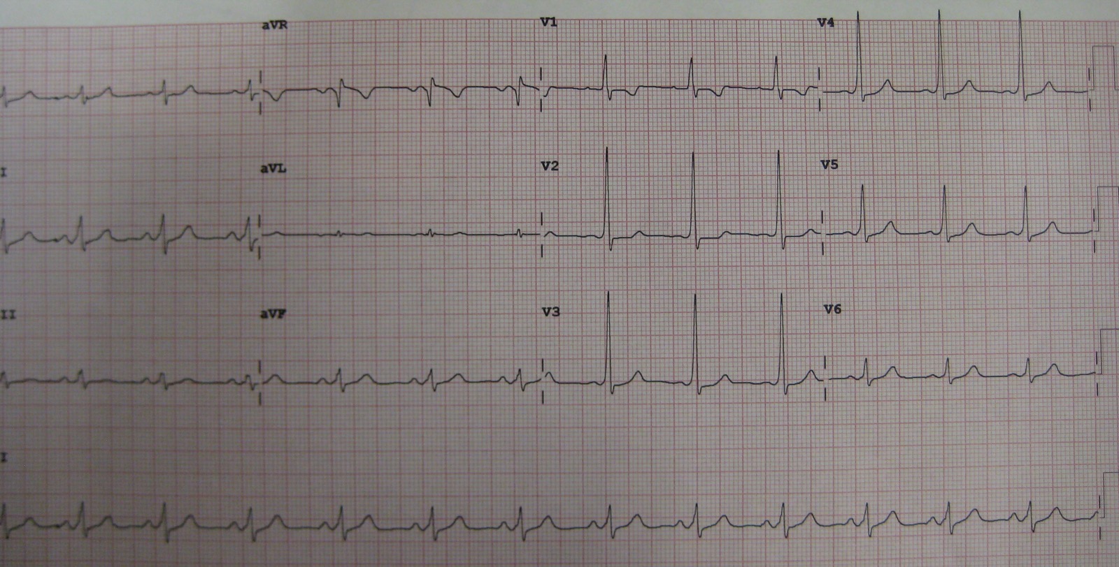
- Check the rhythm strip for pre-excitation: on sinus rhythm, look for a PR under 120 ms with a slurred QRS upstroke.
- Decide regular or irregular, then narrow or wide. Irregular and wide is pre-excited AF until proven otherwise.
- Assess stability: hypotension, altered mental status, ischemic chest pain, acute heart failure or shock.
- If unstable, shock now: confirm SYNC on every R wave, then deliver synchronized cardioversion.
- If irregular and wide and stable: give no AV nodal blocker. Check K+, Mg2+ and QTc, then use IV ibutilide or IV procainamide per protocol.
- If regular and narrow and stable: vagal maneuver, then adenosine per protocol.
- If regular and wide and stable: treat as VT and avoid AV nodal blockers until the mechanism is clear.
- Arrange definitive care: obtain a sinus 12-lead and refer for electrophysiology evaluation.
Common Pitfalls
- Treating pre-excited AF as SVT: giving adenosine, diltiazem or a beta-blocker to an irregular wide-complex rhythm.
- Adenosine for the wrong rhythm: adenosine belongs to regular narrow tachycardia. It has no place in irregular or pre-excited rhythms.
- Procainamide too fast or too much: the label caps the rate at 50 mg/min, and the loading limit is 1 g. ACLS stops at 17 mg/kg total. Slow the rate, or stop, for hypotension or QRS widening over 50%.
- Missing a subtle delta wave: compare the sinus PR and QRS upstroke across several leads before you dismiss pre-excitation.
- Stopping monitoring too early after ibutilide: keep watching for at least 4 hours.
ECG Examples
Clinical Pearls
References
- Joglar JA, Chung MK, Armbruster AL, et al. 2023 ACC/AHA/ACCP/HRS Guideline for the Diagnosis and Management of Atrial Fibrillation. Circulation. 2024;149(1):e1–e156. https://doi.org/10.1161/CIR.0000000000001193
- Wigginton JG, Agarwal S, Bartos JA, et al. Part 9: Adult Advanced Life Support: 2025 American Heart Association Guidelines for Cardiopulmonary Resuscitation and Emergency Cardiovascular Care. Circulation. 2025;152(16 suppl 2):S538–S577. https://doi.org/10.1161/CIR.0000000000001376
- Pappone C, Santinelli V, Manguso F, et al. A randomized study of prophylactic catheter ablation in asymptomatic patients with the Wolff-Parkinson-White syndrome. N Engl J Med. 2003;349(19):1803–1811 (landmark trial). https://doi.org/10.1056/NEJMoa035345
- Strangio A, Ielapi J, Sabatino J, et al. Arrhythmias in Pediatric Age: A Narrative Review. Children (Basel). 2025;12:1580. https://pmc.ncbi.nlm.nih.gov/articles/PMC12731394/
- Procainamide hydrochloride injection, USP: prescribing information. DailyMed; updated December 2, 2025. https://dailymed.nlm.nih.gov/dailymed/lookup.cfm?setid=d58751df-cd8b-45a6-8a32-af19e747e23a
- Corvert (ibutilide fumarate) injection: prescribing information. Pharmacia & Upjohn; DailyMed; published January 26, 2024. https://dailymed.nlm.nih.gov/dailymed/lookup.cfm?setid=2428969a-7431-4e94-9503-ad5c680e2ea4
- Verapamil hydrochloride injection, USP: prescribing information. Hospira, Inc. (2026). Revised July 2026; DailyMed. https://dailymed.nlm.nih.gov/dailymed/lookup.cfm?setid=1c993365-1b62-4524-e781-d307f28f63bc
- Page RL, Joglar JA, Caldwell MA, et al. 2015 ACC/AHA/HRS Guideline for the Management of Adult Patients With Supraventricular Tachycardia. Circulation. 2016;133(14):e506–e574. https://doi.org/10.1161/CIR.0000000000000311
- Brugada J, Katritsis DG, Arbelo E, et al. 2019 ESC Guidelines for the management of patients with supraventricular tachycardia. Eur Heart J. 2020;41(5):655–720. https://doi.org/10.1093/eurheartj/ehz467
Medical Disclaimer
- For Educational Purposes Only: This content is intended for educational reference and should not be used for clinical decision-making.
- Not a Substitute for Professional Judgment: Always consult your local protocols, institutional guidelines, and supervising physicians.
- Verify Before Acting: Users are responsible for verifying information through authoritative sources before any clinical application.