

Bedside Snapshot
- Normal (Adult)
- 3.5–5.0 mEq/L (3.5–5.0 mmol/L)
Some labs use an upper limit up to 5.3 - Critical Values
- <2.5 or ≥6.5 mEq/L (<2.5 or ≥6.5 mmol/L)
Typical; lab-specific - High Suggests
- Kidney failure or missed dialysis
- Crush, rhabdomyolysis, hemolysis
- ACE inhibitor, ARB, spironolactone, trimethoprim
- DKA, acidosis, adrenal insufficiency
- Low Suggests
- Loop or thiazide diuretics
- Vomiting, NG suction, diarrhea
- Insulin, albuterol, alkalosis
- Low Mg²⁺, aldosterone excess
- Act Now If
- K⁺ ≥6.5 or ECG changes: monitor, 12-lead, IV calcium for ECG changes, insulin-glucose. K⁺ <2.5, paralysis or arrhythmia: monitor, IV KCl by pump, check Mg²⁺
- Watch Out For
- A normal ECG does not exclude severe hyperkalemia. Hemolysis falsely raises K⁺: repeat the sample, but treat now if high-risk or the ECG is abnormal.
Reference & Critical Ranges
Reference ranges
| Population | Conventional | SI | Notes |
|---|---|---|---|
| Adult | 3.5–5.0 mEq/L | 3.5–5.0 mmol/L | Some labs set the upper limit as high as 5.3; serum reads slightly higher than plasma because platelets release K⁺ during clotting |
| Pregnancy | 2nd trimester about 3.3–5.0; 3rd trimester about 3.3–5.1 mEq/L | Same numbers in mmol/L | Lower limit falls slightly with hemodilution and the mild respiratory alkalosis of pregnancy |
| Older adult | Same as adult | Same as adult | Higher risk of drug-induced hyperkalemia (renin-angiotensin-aldosterone system [RAAS] blockers, trimethoprim) and of diuretic-induced hypokalemia |
| Neonate 0–28 d | 3.4–6.0 mEq/L | 3.4–6.0 mmol/L | Hyperkalemia in neonates is usually defined as >6.0; squeezed heel-stick samples hemolyze and read falsely high |
| Infant 1–12 months | 3.5–5.7 mEq/L | 3.5–5.7 mmol/L | Upper limit higher than adult |
| Child and adolescent 1–16 years | 3.5–5.0 mEq/L | 3.5–5.0 mmol/L | Same as adult range; pediatric guidelines define hyperkalemia as >5.5 |
Severity & critical values
| Level | Conventional | SI | What it means |
|---|---|---|---|
| Mild hypokalemia | 3.0–3.4 mEq/L | 3.0–3.4 mmol/L | Often no symptoms; oral replacement and find the cause |
| Moderate hypokalemia | 2.5–2.9 mEq/L | 2.5–2.9 mmol/L | Weakness, cramps, constipation; ECG changes (flat T waves, U waves) become likely |
| Severe hypokalemia | <2.5 mEq/L | <2.5 mmol/L | Paralysis, ileus, rhabdomyolysis, respiratory weakness, arrhythmia: monitored IV replacement |
| Mild hyperkalemia | 5.1–5.9 mEq/L | 5.1–5.9 mmol/L | Usually no symptoms; repeat the sample, review kidneys and drugs |
| Moderate hyperkalemia | 6.0–6.4 mEq/L | 6.0–6.4 mmol/L | 12-lead ECG; treat if unwell, rising fast or ECG changes |
| Severe hyperkalemia | ≥6.5 mEq/L | ≥6.5 mmol/L | Emergency whatever the ECG shows: continuous monitoring and K⁺-lowering treatment |
| Critical | <2.5 or ≥6.5 mEq/L | <2.5 or ≥6.5 mmol/L | Typical panic values (lab-specific): the lab calls; assess, monitor and treat now |
Thresholds: This page calls adult hyperkalemia above 5.0 mEq/L, the AHA 2025 definition. Pediatric hyperkalemia starts above 5.5 mEq/L. UK and European guidelines start the adult line at 5.5 mEq/L, which makes 5.5 to 5.9 their mild band. The moderate band (6.0–6.4) and the severe band (≥6.5) are the same in both.
Conversion: K⁺ mEq/L = mmol/L (monovalent ion; factor 1)
Causes of Abnormal Results
High (Hyperkalemia >5.0 mEq/L)
Mild 5.1–5.9, moderate 6.0–6.4, severe ≥6.5 mEq/L. Most true hyperkalemia has two parts: the kidneys are not excreting, and a drug or a sudden load pushes the number up. Rule out a spurious result. Just never let that check delay care of a high-risk patient.
| Mechanism | Common causes | Bedside clue |
|---|---|---|
| Kidney failure | Acute kidney injury (AKI), chronic kidney disease (CKD) stages 4–5, dialysis patient who missed a session | Low urine output, rising creatinine, fistula or dialysis catheter |
| Cell breakdown | Crush injury, rhabdomyolysis, tumor lysis, brisk hemolysis, large-volume or old blood transfusion | Entrapment, dark urine, high creatine kinase (CK), recent chemotherapy or transfusion |
| Shift out of cells | Metabolic acidosis, diabetic ketoacidosis (DKA) and insulin deficiency, digoxin toxicity, non-selective β-blockers | Kussmaul breathing, high glucose, digoxin use |
| Succinylcholine in at-risk patients | Given after the acute phase of major burns, multiple trauma, denervation or upper motor neuron injury, or with myopathy | Arrest minutes after rapid sequence intubation (RSI) |
| Drugs that block K⁺ excretion | Angiotensin-converting enzyme (ACE) inhibitors, angiotensin receptor blockers (ARBs), mineralocorticoid receptor antagonists (MRAs) such as spironolactone, K⁺-sparing diuretics, trimethoprim, nonsteroidal anti-inflammatory drugs (NSAIDs), heparin, tacrolimus | Recent start or dose change; risk stacks when combined, in CKD, diabetes and old age |
| Low aldosterone | Adrenal insufficiency (Addison disease), type 4 renal tubular acidosis (common in diabetes) | Hypotension and low Na⁺; diabetic with CKD |
| Excess intake | K⁺ supplements, salt substitutes, IV potassium chloride (KCl) error | Rarely causes hyperkalemia unless excretion is impaired |
| Spurious (pseudohyperkalemia) | Hemolysis, difficult draw or fist clenching, EDTA contamination, delayed or cold samples, very high platelets, white cells or red cells | Well patient, normal ECG and kidneys; repeat the sample |
Low (Hypokalemia <3.5 mEq/L)
Mild 3.0–3.4, moderate 2.5–2.9, severe <2.5 mEq/L. Symptoms usually appear below 3.0 mEq/L, or sooner if the level falls fast. Most cases are losses, through the kidneys or the gut. A shift into cells can drop K⁺ just as fast, and nothing has left the body.
| Mechanism | Common causes | Bedside clue |
|---|---|---|
| Shift into cells | Insulin (including DKA treatment), β₂-agonists (albuterol), alkalosis, refeeding, thyrotoxic or familial periodic paralysis | Recent insulin or nebulizers; sudden weakness or paralysis with a normal body K⁺ store |
| Renal loss: diuretics and magnesium | Loop and thiazide diuretics, osmotic diuresis, low Mg²⁺, amphotericin B | Diuretic use; low Mg²⁺ makes K⁺ hard to correct |
| Upper GI loss | Vomiting, nasogastric (NG) suction | Metabolic alkalosis; most K⁺ is lost in the urine (bicarbonaturia and aldosterone), not in gastric fluid; urine Cl⁻ low |
| Lower GI loss | Diarrhea, laxative abuse, villous adenoma | Low urine K⁺, often metabolic acidosis |
| Mineralocorticoid excess | Primary aldosteronism, Cushing syndrome, licorice, Liddle syndrome | Hypertension with high urine K⁺ |
| Renal tubular disorders | Renal tubular acidosis types 1 and 2, Bartter and Gitelman syndromes | Lifelong or recurrent low K⁺; acidosis (RTA) or alkalosis (Bartter, Gitelman) |
| Low intake | Malnutrition, anorexia, alcohol use disorder | Rarely the only cause; usually adds to losses |
Interpretation & Bedside Approach
Step-by-step approach
- Is it real? Hemolysis, a difficult draw, a delayed or cold sample, very high platelets or white cells, or a draw above a K⁺-containing infusion can all fake it. Repeat a surprising result on a free-flowing sample. A blood-gas analyzer gives a whole-blood K⁺ in minutes. If the patient is high-risk or the ECG is abnormal, don't wait for the repeat.
- How sick is the patient? Get a 12-lead ECG at K⁺ ≥6.0 mEq/L. Monitor continuously at ≥6.5 mEq/L, or with any ECG change, weakness, bradycardia or arrhythmia. Both extremes can cause weakness that climbs upward, then respiratory failure.
- How fast, and which way? Pull up the earlier results. A fast rise (AKI, rhabdomyolysis, crush, missed dialysis) is more dangerous than the same number sitting stable in CKD. The trend tells you more than any single value.
- Check the partners. Creatinine and urine output, glucose, bicarbonate and pH, Mg²⁺ and Ca²⁺, CK after crush or prolonged down time, and a digoxin level if the patient takes it.
- Classify. High: kidneys, drugs, low aldosterone, shift, load or spurious. Low: shift, renal loss or GI loss, using urine K⁺ and the acid-base status (table below).
- Escalate or dispose. K⁺ ≥6.5 mEq/L or ECG changes: monitored bed, renal or critical care input; dialysis patients need urgent dialysis. K⁺ <2.5 mEq/L or symptoms: monitored IV replacement.
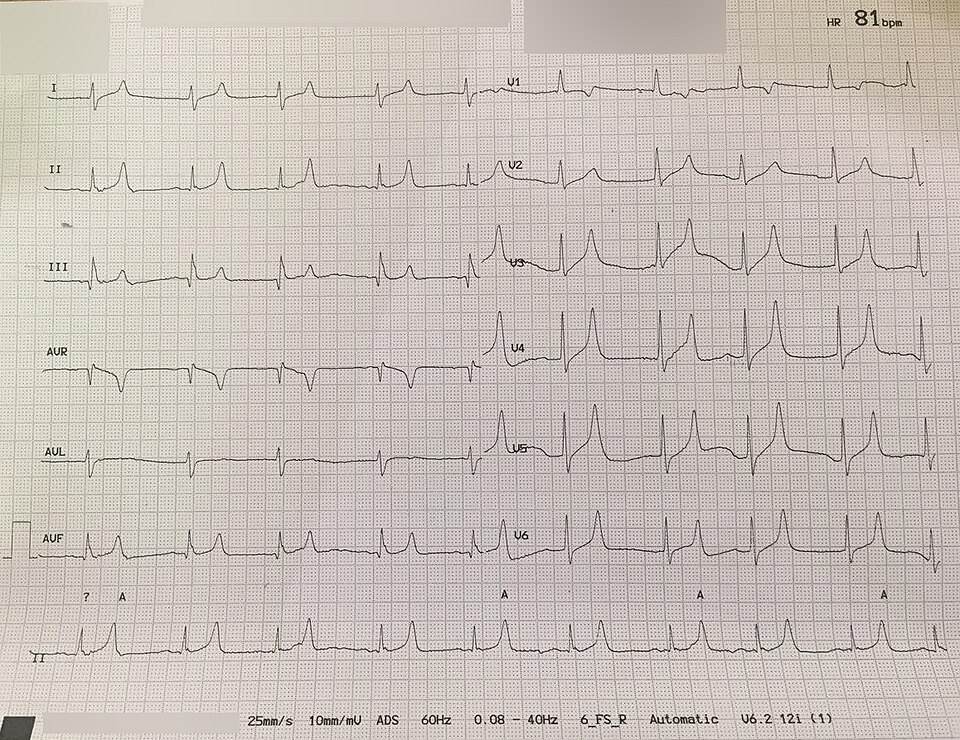
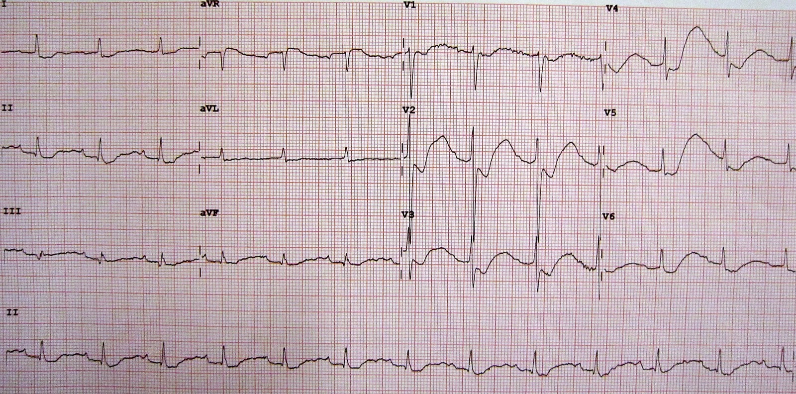
ECG changes
| Disorder | Typical findings | Caveat |
|---|---|---|
| Hyperkalemia, early | Tall, narrow, symmetric (peaked) T waves; small or flat P waves; PR prolongation | Peaked T waves occur in only about 1 in 3 patients with moderate to severe hyperkalemia; hyperacute T waves of early ST-elevation myocardial infarction (STEMI) look similar |
| Hyperkalemia, advanced | QRS widening, bradycardia, junctional rhythm, AV block, sine wave, VT, VF, asystole | Stages are often skipped. QRS widening, bradycardia (HR <50) and junctional rhythm best predict adverse events. May mimic STEMI or a Brugada pattern; pacing thresholds rise |
| Hypokalemia | T-wave flattening or inversion, ST depression, prominent U waves (V2–V3), apparent QT prolongation, ectopy | Ventricular arrhythmias, worse with digoxin, low Mg²⁺ or QT-prolonging drugs |
Hypokalemia: urine and acid-base clues
| Pattern | Suggests |
|---|---|
| Low urine K⁺ (<15 mEq/day, or spot K⁺/creatinine <13 mEq/g) with metabolic acidosis | Lower GI loss: diarrhea, laxatives, villous adenoma |
| Low urine K⁺ with metabolic alkalosis | Past vomiting or diuretic use (losses have stopped) |
| High urine K⁺ with metabolic acidosis | DKA, renal tubular acidosis types 1 and 2 |
| High urine K⁺, alkalosis, normal BP, low urine Cl⁻ | Ongoing vomiting or NG suction |
| High urine K⁺, alkalosis, normal BP, normal urine Cl⁻ | Current diuretic use, Bartter or Gitelman syndrome |
| High urine K⁺, alkalosis, hypertension | Primary aldosteronism, Cushing syndrome, licorice, Liddle syndrome |
Management Priorities
Emergency: Severe hyperkalemia (K⁺ ≥6.5 mEq/L or ECG changes)
- Monitor: continuous ECG, 12-lead, IV access, pacing pads ready; stop K⁺ sources and K⁺-raising drugs.
- Protect the heart (any ECG change): calcium gluconate 3 g (30 mL of 10%) IV over 10 min, or calcium chloride 1 g (10 mL of 10%) over 5 min in arrest or peri-arrest; repeat if ECG changes persist.
- Shift: regular insulin 10 units IV with dextrose 25 g (plus D10 50 mL/hr for 5 hr if glucose is under 126 mg/dL), plus nebulized albuterol 10–20 mg as an add-on.
- Remove: dialysis (urgent for dialysis patients at K⁺ ≥6.5 mEq/L; any life-threatening or treatment-resistant case); sodium zirconium cyclosilicate as an add-on.
- Monitor & repeat: K⁺ at 1, 2, 4, 6 and 24 hr; glucose before insulin, every 30 min for 2 hr, then hourly to at least 6 hr.
Emergency: Severe hypokalemia (K⁺ <2.5 mEq/L, or paralysis or arrhythmia)
- Monitor: continuous ECG; watch breathing for respiratory muscle weakness.
- Replace IV: KCl by pump (rates in the table); never IV push.
- Replace magnesium: check Mg²⁺ and correct it, or K⁺ will not stay up.
- Stop the cause: treat vomiting or diarrhea, review diuretics; switch to oral K⁺ when safe.
- Recheck: K⁺ during and after each replacement; smaller doses in kidney impairment.
Medications
| Drug | Adult dose | Pediatric dose (max) | Onset / key notes |
|---|---|---|---|
| Calcium gluconate 10% (ECG changes) | 3 g (30 mL) IV over 10 min; repeat if ECG changes persist | Not established in children; specialist use only | Onset 1–3 min, lasts 30–60 min; does not lower K⁺. Peripheral IV acceptable. With digoxin toxicity: use only with medical or toxicology direction (RCH advises against calcium) Differs from FDA label: the US labels list only hypocalcemia and cap the rate (calcium gluconate 200 mg/min for adults, so 3 g takes at least 15 min; calcium chloride 1 mL/min, central or deep vein, no bolus). The hyperkalemia rate here (gluconate 30 mL over 10 min, chloride 10 mL over 5 min) is from UKKA 2026 and is off-label. |
| Calcium chloride 10% (arrest or peri-arrest) | 1 g (10 mL) IV over 5 min (peri-arrest or central/deep access); IV/IO push in arrest; repeat if changes persist | 20 mg/kg (0.2 mL/kg) IV/IO, max 1 g | About 3 times the calcium of gluconate per mL; extravasation causes necrosis. Differs from FDA label: the label caps the rate at 1 mL/min (central or deep vein). |
| Regular insulin with dextrose | 10 units IV (give over 5–15 min, mixed in the dextrose syringe) with dextrose 25 g (50 mL D50); pre-dose glucose <126 mg/dL: add D10 50 mL/hr for 5 hr | 0.1 units/kg IV (max 10 units) with dextrose 0.5 g/kg (D10 5 mL/kg) | High-alert K⁺ falls within 15 min, peaks at 30–60 min, lasts about 2 hr. Hypoglycemia peaks 1–3 hr after the dose. Insulin dose: some US protocols use 5 units or 0.1 units/kg. A pooled analysis of 10 retrospective studies found less hypoglycemia with lower doses and no difference in K⁺ fall; a later study found 10 units lowered K⁺ more (0.9 vs 0.63 mmol/L); UKKA 2026 states more study is needed before 5 units can be recommended. This site uses 10 units with the glucose rules above. |
| Albuterol (nebulized) | 10–20 mg nebulized (four to eight 2.5 mg vials); 10 mg with ischemic heart disease; single dose | Not established in children; studies in ages 5–18 used 2.5–5 mg nebulized (no labeled or guideline pediatric dose) | Onset within 30 min, lasts at least 2 hr. Adjunct, not monotherapy; not used in cardiac arrest (AHA 2025: 3, No Benefit). Differs from FDA label: the label nebulized dose is 2.5 mg for asthma; the 10–20 mg dose is from UKKA 2026 and is off-label. |
| Sodium bicarbonate 8.4% | Not routine. Metabolic acidosis: per protocol. Hyperkalemic arrest: 50 mEq IV/IO, repeat by blood gas | 1 mEq/kg IV slowly (max 50 mEq), only with metabolic acidosis or arrest | Does not reliably lower K⁺ alone; do not give through the same line at the same time as calcium (RCH); use separate lines or flush thoroughly between them |
| Sodium zirconium cyclosilicate (Lokelma) | 10 g PO three times daily for up to 48 hr, then 10 g once daily; dialysis patients 5 g on non-dialysis days | Not established in children | Onset about 1 hr; not emergency monotherapy. About 400 mg sodium per 5 g (edema); other oral drugs 2 hr apart. Differs from FDA label: the label says SZC should not be used as emergency treatment for life-threatening hyperkalemia because of its delayed onset. UKKA 2026 recommends it as an add-on to calcium and insulin-glucose in severe hyperkalemia and allows up to 72 hr. |
| Patiromer (Veltassa) | 8.4 g PO once daily; increase by 8.4 g at weekly intervals (max 25.2 g/day) | Age 12–17 yr: 4 g PO once daily, titrate by 4 g weekly (max 25.2 g/day); under 12 yr: not established | Onset about 7 hr; not for emergencies. Other oral drugs 3 hr apart; lowers Mg²⁺ |
| Sodium polystyrene sulfonate (Kayexalate, Kionex) | 15 g PO 1–4 times daily, or 30–50 g PR every 6 hr | Not established in children; specialist use only; contraindicated in neonates | Not part of the acute regimen: onset hours to days, unpredictable; intestinal necrosis reported, highest with sorbitol, ileus or after surgery. Label says it alone may be insufficient in severe hyperkalemia. |
| Potassium chloride IV (hypokalemia) | K⁺ >2.5 mEq/L: up to 10 mEq/hr (max 200 mEq/24 hr). K⁺ 2.0–2.5 mEq/L: rate per protocol (the label gives none). K⁺ <2 mEq/L with ECG changes or paralysis: up to 40 mEq/hr (max 400 mEq/24 hr); rates above 10 mEq/hr need continuous cardiac monitoring. Peripheral: up to 40 mEq/L; 40–60 mEq/L needs senior review; above 60 mEq/L central line only | Not established in children; specialist use only | High-alert Infusion pump only; never IV push. Central line for concentrations above 60 mEq/L (RCH); continuous monitoring above 10 mEq/hr (label). Use saline, not dextrose, as the carrier |
| Potassium chloride PO (hypokalemia) | Mild to moderate: 40–100 mEq/day or more PO in divided doses; no single dose above 20 mEq (labeling); adjust to the individual and recheck K⁺ | Not established in children; specialist use only | Preferred when K⁺ ≥2.5 mEq/L and the gut works; give with food and plenty of water |
| Magnesium sulfate (low Mg²⁺) | IV replacement per institutional protocol; use caution in renal impairment, where magnesium excretion is reduced | Not established in children; specialist use only | High-alert Replace with K⁺; hypokalemia is often refractory until Mg²⁺ is corrected |
Hyperkalemic cardiac arrest
- Suspect it: dialysis, AKI, crush, rhabdomyolysis, DKA, or a pre-arrest wide QRS or bradycardia; no lab result is needed to treat.
- Resuscitate: high-quality CPR and ACLS or PALS. AHA 2025 Part 10 (hyperkalemia recommendations): IV calcium, COR 2b (effectiveness not well established; C-LD); sodium bicarbonate, COR 2b (C-EO); insulin and glucose, COR 2b (C-EO); inhaled β₂-agonists, 3: No Benefit (C-EO). Calcium chloride 1 g IV/IO is the calcium dose used here, per the UKKA 2026 guideline.
- Doses differ: AHA 2025 Table 3 gives insulin 10 units IV over 15–30 min with dextrose 50 g (child 0.1 units/kg, max 10 units, with D10 5 mL/kg). Differs from UKKA 2026 and ERC: those give 25 g (plus D10 50 mL/hr if glucose is under 126 mg/dL).
- Dialysis patients: disconnect the dialysis machine before defibrillation unless it is defibrillator-proof. Consider dialysis during CPR if hyperkalemia resists medical therapy and staff and equipment are available, per the UKKA 2026 guideline.
Definitive care & disposition
- Hyperkalemia: monitored bed for K⁺ ≥6.5 mEq/L or ECG changes; nephrology for dialysis; critical care for airway, breathing, circulation or consciousness problems. Hold RAAS blockers during acute illness; review K⁺-raising drugs before restarting.
- Hypokalemia: monitored care for IV replacement, K⁺ <2.5 mEq/L, ECG changes or weakness; investigate recurrent or unexplained cases.
- Digoxin toxicity with hyperkalemia: digoxin immune Fab (DigiFab) is labeled for life-threatening digoxin toxicity; give per poison center or toxicology advice. Potassium can fall rapidly after DigiFab, so recheck K⁺.
Doses are typical references. Follow local protocols, medical direction and institutional policy.
Field & Transport Considerations
Point-of-care testing
Cartridge blood-gas and chemistry analyzers give a whole-blood K⁺ in minutes. They cannot see hemolysis, so a high value in a well patient still needs a lab repeat. Agreement with the lab is best above 3 mEq/L. No K⁺ result? The 12-lead ECG is your field screen. It can confirm hyperkalemia. It cannot rule it out.
Before and during interfacility transport
| Setting | Consideration |
|---|---|
| Dialysis patient | Weakness, bradycardia or a wide QRS after a missed session: hyperkalemia until proven otherwise; ask about the last dialysis |
| Crush or prolonged entrapment | Expect K⁺ to rise as tissue is released; monitor ECG continuously |
| Treated hyperkalemia | Record K⁺ values and times, and when calcium, insulin and albuterol were given; calcium fades in 30–60 min and the insulin shift in about 2 hr, so recheck K⁺ and glucose en route or on arrival |
| Refractory hyperkalemia | Needs a dialysis-capable destination; stabilize (calcium, shift) before departure |
| KCl infusion running | Pump only; confirm bag concentration and rate; continuous ECG; never bolus |
| RSI planned | Avoid succinylcholine with known or suspected hyperkalemia or after the acute phase of burns, crush or denervation; use protocol alternatives |
Scope notes
Paramedics, RNs and critical care transport (CCT) teams give IV calcium, insulin-dextrose, nebulized albuterol and KCl infusions. Each one runs per local protocol and medical direction.
Special Populations
| Population | What changes | Clinical implication |
|---|---|---|
| Neonates & infants | Higher normal range; heel-stick samples often hemolyze | Confirm a high K⁺ on a free-flowing sample; never give sodium polystyrene sulfonate to neonates |
| Children & adolescents | Same range as adults from age 1; hyperkalemia defined as >5.5 mEq/L (neonates >6.0) | Weight-based calcium, insulin and dextrose; succinylcholine can cause hyperkalemic arrest in undiagnosed myopathy |
| Pregnancy | Lower limit falls slightly (about 3.3 mEq/L); hyperemesis causes hypokalemia | Use pregnancy-specific ranges; check Mg²⁺ with prolonged vomiting |
| Older adults | Same range; more RAAS blockers, diuretics, NSAIDs and trimethoprim; lower kidney reserve | Highest risk for drug-induced hyperkalemia and for hypoglycemia after insulin-glucose |
| CKD / dialysis | Little capacity to excrete a K⁺ load; constipation and missed sessions raise K⁺ | Dialysis is definitive; up to 40% do not respond to albuterol; insulin lasts longer, so hypoglycemia comes late |
| Liver disease | Spironolactone use and hepatorenal syndrome raise K⁺; diuretics and alkalosis lower it | Check K⁺ often when diuretics change |
| Other: periodic paralysis | Sudden K⁺ shift into cells (thyrotoxic or familial) | Replace slowly and recheck often: K⁺ returns from cells and rebound hyperkalemia occurs |
Interfering Factors & Pitfalls
Collection & handling
| Factor | Effect | Why | Avoid / fix |
|---|---|---|---|
| Hemolysis (difficult draw, small needle, pneumatic tube) | ↑ | K⁺ leaks from damaged red cells | Free-flowing draw; send a larger sample; repeat |
| Fist clenching, long tourniquet time | ↑ | Muscle releases K⁺ locally | Short tourniquet, no pumping |
| EDTA (lavender-top) contamination | ↑ K⁺ with ↓ Ca²⁺ | Potassium EDTA carried over | Follow the order of draw; redraw |
| Drawn above a K⁺-containing infusion | ↑ | Sample mixes with the infusion | Draw from the other arm |
| Delayed processing or cold storage | ↑ | Cells leak K⁺ when not spun or kept cold | Prompt delivery; label the draw time |
| Warm storage | ↓ | Cells take K⁺ back up | Prompt delivery |
Physiologic / biologic
| Factor | Effect | Why | Avoid / fix |
|---|---|---|---|
| Very high platelets, white cells or red cells | ↑ serum | Cells release K⁺ during clotting | Paired lithium-heparin plasma and serum: a gap >0.4 mmol/L confirms it |
| Insulin, albuterol or alkalosis before the draw | ↓ (true shift) | K⁺ moves into cells | Interpret with the treatment timeline; expect rebound |
Assay & device-specific
| Factor | Effect | Why | Avoid / fix |
|---|---|---|---|
| Blood-gas (whole blood) vs lab plasma | Small differences; hemolysis hidden | Whole blood cannot be checked for hemolysis | Use for speed; confirm in the lab |
Common misinterpretations
- Don't wait for a normal-looking ECG before treating K⁺ ≥6.5 mEq/L.
- Writing off a hemolyzed high K⁺ in a dialysis, AKI or crush patient.
- A normal K⁺ in DKA does not mean a normal body K⁺ store.
- Calcium or albuterol alone will not fix hyperkalemia.
Physiology & Test Basics
What it measures
This is the serum or plasma K⁺ concentration: the 2% of body potassium that sits outside cells. About 98% is inside cells. So the blood level reflects the balance between total body stores and the shift across cell membranes. It does not show the store itself.
Physiology & regulation
- The Na⁺/K⁺-ATPase pump keeps K⁺ high inside cells. That sets the resting membrane potential of nerve, muscle and heart.
- Insulin and β₂-adrenergic stimulation pull K⁺ into cells within minutes.
- Acidosis tends to push K⁺ out of cells. Alkalosis pulls it in.
- The kidneys excrete about 90% of daily intake, mostly in the collecting duct under aldosterone. The colon handles most of the rest.
- The kidneys are better at excreting a K⁺ load than at saving K⁺. So poor intake plus any loss leads to depletion.
- Each 1 mEq/L drop in serum K⁺ stands for a deficit of roughly 200–400 mEq.
How it is measured
- Specimen: Serum (gold or red top) on the basic or comprehensive metabolic panel, or lithium-heparin plasma (green top). The lab can run the plasma faster, because it needs no clotting time.
- Point of care: arterial or venous whole blood in a blood-gas syringe on a blood-gas or cartridge analyzer.
- Method: ion-selective electrode; result in minutes on a blood-gas analyzer.
- Related tests: creatinine, glucose, bicarbonate and pH, Mg²⁺, Ca²⁺, CK, urine K⁺ and Cl⁻.
Why it matters
High or low, potassium can stop a heart. Either way the muscles go weak and the rhythm can turn fatal. Hyperkalemia is on the Hs list: a reversible cause of cardiac arrest. The K⁺ result drives time-critical decisions in kidney failure, DKA, crush injury, digoxin toxicity and RSI.
Clinical Pearls
References
- Alfonzo, A., Harrison, A., Baines, R., Chu, A., Mann, S., & MacRury, M. (2026). Clinical practice guidelines: Treatment of acute hyperkalaemia in adults (updated July 2026). UK Kidney Association. https://www.ukkidney.org/sites/default/files/documents/FINAL%20VERSION%20-%20UKKA%20CLINICAL%20PRACTICE%20GUIDELINE%20-%20MANAGEMENT%20OF%20HYPERKALAEMIA%20IN%20ADULTS%20-%20UPDATED%20JULY%202026_0.pdf
- Cao, D., Arens, A. M., Chow, S. L., Easter, S. R., Hoffman, R. S., Lagina, A. T., Lavonas, E. J., et al. (2025). Part 10: Adult and pediatric special circumstances of resuscitation: 2025 American Heart Association Guidelines for Cardiopulmonary Resuscitation and Emergency Cardiovascular Care. Circulation, 152(16 Suppl 2), S578–S672 (hyperkalemia section, Table 3). https://doi.org/10.1161/CIR.0000000000001380
- Sun Pharmaceutical Industries, Inc. (2024). Potassium chloride extended-release tablets: prescribing information (updated January 2024). DailyMed, U.S. National Library of Medicine. https://dailymed.nlm.nih.gov/dailymed/drugInfo.cfm?setid=8ed8ea55-8b15-f718-e053-2a95a90a587a
- Hospira, Inc. (2026). Potassium Chloride for Injection Concentrate, USP: prescribing information (revised January 2026). DailyMed, U.S. National Library of Medicine. https://dailymed.nlm.nih.gov/dailymed/drugInfo.cfm?setid=559a0a8c-a8fe-40a5-b196-21f9308780ab
- AstraZeneca Pharmaceuticals LP. (2025). LOKELMA (sodium zirconium cyclosilicate) for oral suspension: prescribing information. DailyMed, U.S. National Library of Medicine. https://dailymed.nlm.nih.gov/dailymed/drugInfo.cfm?setid=90bf8e28-748d-4e4b-a19f-9cf483370eff
- Vifor Pharma, Inc. (2025). VELTASSA (patiromer) for oral suspension: prescribing information. DailyMed, U.S. National Library of Medicine. https://dailymed.nlm.nih.gov/dailymed/drugInfo.cfm?setid=bf002984-d6c9-46df-aecb-a07733f763c1
- ANI Pharmaceuticals, Inc. (2025). Kionex (sodium polystyrene sulfonate suspension, USP): prescribing information. DailyMed, U.S. National Library of Medicine. https://dailymed.nlm.nih.gov/dailymed/drugInfo.cfm?setid=676c3d81-d575-43f6-8748-e04b7c0cafeb
- The Royal Children's Hospital Melbourne. (2024). Clinical practice guidelines: Hyperkalaemia (updated August 2024) and Hypokalaemia (updated October 2024). https://www.rch.org.au/clinicalguide/guideline_index/Hyperkalaemia/; https://www.rch.org.au/clinicalguide/guideline_index/Hypokalaemia/
- Moussavi, K., et al. (2021). Reduced alternative insulin dosing in hyperkalemia: a meta-analysis of effects on hypoglycemia and potassium reduction. Pharmacotherapy. https://pubmed.ncbi.nlm.nih.gov/33993515/
- Finder, et al. (2022). 5 versus 10 units of intravenous insulin for hyperkalemia in patients with moderate renal dysfunction. Journal of Emergency Medicine. https://pubmed.ncbi.nlm.nih.gov/35058093/
- Fresenius Kabi USA. (2026). Calcium gluconate injection: prescribing information. DailyMed, U.S. National Library of Medicine. https://dailymed.nlm.nih.gov/dailymed/drugInfo.cfm?setid=8b77c3d2-992d-4261-8421-9cfd07328fbf
- Hospira, Inc. (2026). Calcium chloride injection 10%: prescribing information. DailyMed, U.S. National Library of Medicine. https://dailymed.nlm.nih.gov/dailymed/drugInfo.cfm?setid=c4c65e48-85f8-4dcf-6281-06e40959cc79
Medical Disclaimer
- For Educational Purposes Only: This content is intended for educational reference and should not be used for clinical decision-making.
- Not a Substitute for Professional Judgment: Always consult your local protocols, institutional guidelines, and supervising physicians.
- Verify Before Acting: Users are responsible for verifying information through authoritative sources before any clinical application.