

Bedside Snapshot
- Indications
- Refractory K⁺ >6.5 mEq/L or hyperkalemic ECG change
- Severe acidosis, pH <7.1, not responding to medical therapy
- Dialyzable toxin (e.g., lithium above 4 mEq/L with impaired kidney function, or with decreased consciousness)
- Pulmonary edema refractory to diuretics
- Contraindications / Abort
- Systolic BP <90 mmHg or symptomatic hypotension: reduce UF, call nephrology
- Air in the circuit: clamp lines and stop the pump
- Active bleeding or HIT: heparin-free plan only, per nephrology
- Critical Steps
- Confirm UF goal, Qb, dialysate K⁺ and heparin plan
- Check access; no IV or BP on the fistula or graft arm
- Prime and de-air before connecting
- Start Qb low and ramp; UF never above prescribed rate
- Confirm & Monitor
- Post-treatment weight near dry weight
- Thrill and bruit before and after the run
- Vitals and symptoms at the interval set by unit protocol
- Potassium and ECG after emergency treatment
Key Numbers
Limits and emergency doses for hemodialysis (HD). The nephrologist's prescription governs every session.
| Parameter | Value |
|---|---|
| Urgent HD: potassium | K⁺ >6.5 mEq/L or any hyperkalemic ECG change, not controlled by medical therapy |
| Urgent HD: acidosis | pH <7.1 not responding to medical therapy |
| UF rate ceiling | ≤13 mL/kg/h. Cohort data show rising risk above 10 mL/kg/h |
| Blood flow (Qb) | 300–500 mL/min in chronic HD, per prescription |
| Calcium gluconate 10% | 1 g (10 mL) IV; repeat if no ECG response in 5–10 min |
| Regular insulin + dextrose | Adult: 5–10 units IV with 25 g dextrose Peds: per pediatric protocol |
| Albuterol, nebulized | Adult: 10–20 mg in 4 mL; onset about 30 min Peds: per pediatric protocol |
| Protamine (heparin reversal) | 1 mg per 100 units of heparin still active in the circulation; max 50 mg; give by slow IV injection over 10 min |
| Fistula or graft arm | No IV, BP cuff or blood draw on this arm |
Overview & Principles
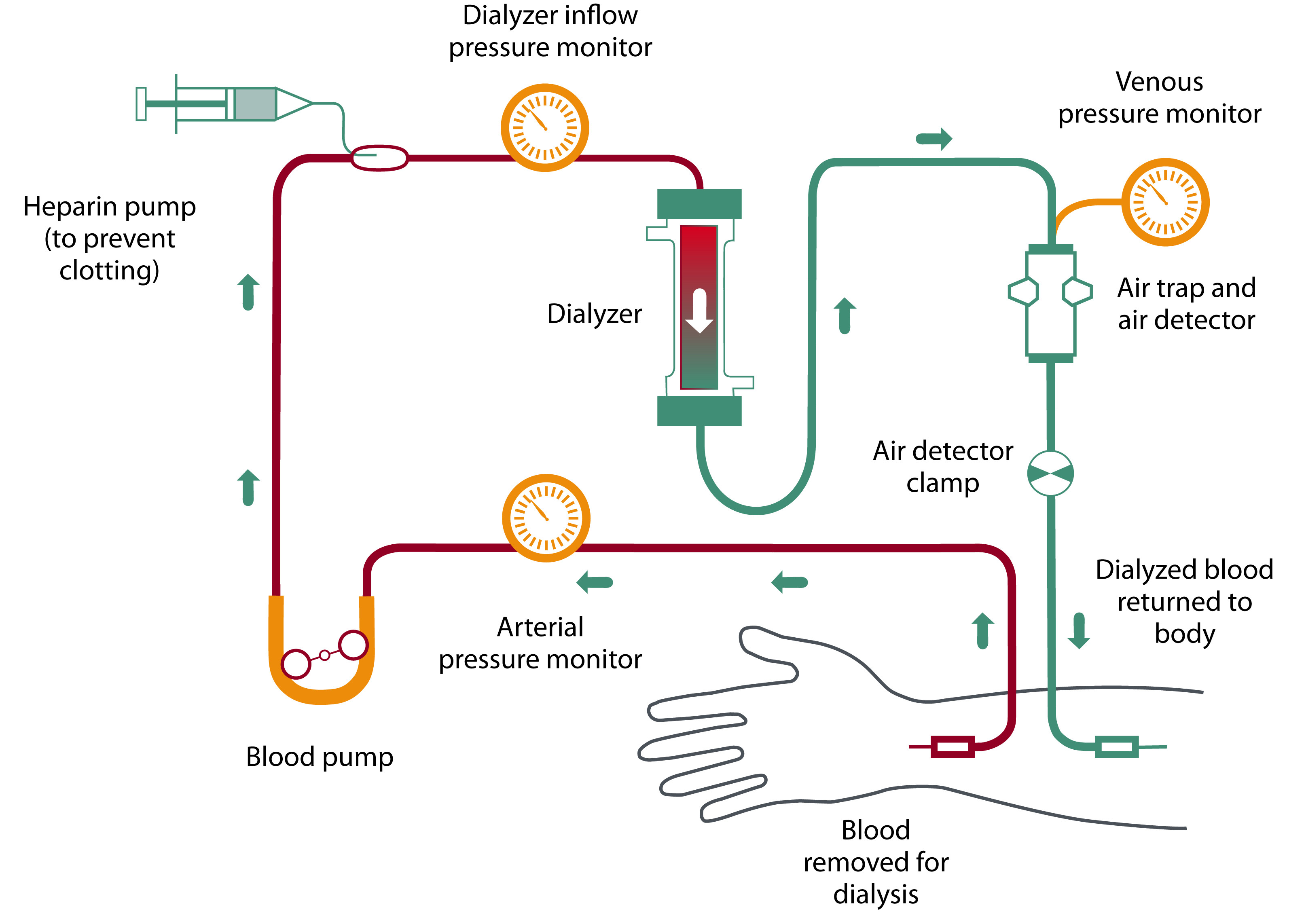
Hemodialysis (HD) replaces kidney function outside the body. Blood runs through an extracorporeal circuit and a semipermeable dialyzer. The circuit removes solutes, corrects potassium and acid–base disturbances, and takes off excess fluid.
How It Works
- Diffusion: Small solutes (urea, potassium, creatinine) drift down a concentration gradient into the dialysate.
- Convection: Pressure pulls water across the membrane. The water drags solutes along with it.
- Ultrafiltration (UF): the planned fluid removal. It's set as a total volume and a rate in mL/kg/h.
Where It Fits
Chronic HD is outpatient care for end-stage kidney disease (ESKD). In the ICU, intermittent HD treats acute kidney injury (AKI) or ESKD in a critically ill patient. It's the choice when continuous renal replacement therapy (CRRT) is unavailable or not required.
| Modality | Setting | Hemodynamic Tolerance |
|---|---|---|
| Chronic HD | Outpatient unit, usually 3 times per week | Best in stable patients |
| Intermittent HD (ICU) | Hospital, session by session | Variable; needs longer, gentler runs when unstable |
| CRRT | ICU, continuous | Usually better tolerated when hemodynamically unstable |
Indications & Contraindications
Indications & Patient Selection
Remember the urgent indications with the mnemonic AEIOU. Each one generally means medical therapy has failed or a life threat is present. Some toxins, lithium among them, also use level-based criteria.
- A: Acidosis. Severe metabolic acidosis (pH <7.1) not responding to medical therapy.
- E: Electrolytes. Hyperkalemia with K⁺ >6.5 mEq/L or an ECG change, despite medical therapy.
- I: Intoxication. Dialyzable toxins. For lithium, EXTRIP recommends extracorporeal treatment if kidney function is impaired and the level is above 4 mEq/L, or with decreased consciousness, seizures or life-threatening dysrhythmias at any level. It suggests treatment for a level above 5 mEq/L, significant confusion, or an expected time above 36 h to reach 1 mEq/L. Toxic alcohols and salicylate are a call for toxicology or the poison center (US Poison Help: 1-800-222-1222).
- O: Overload. Pulmonary edema or hypertension refractory to diuretics and medical therapy.
- U: Uremia. Pericarditis, encephalopathy, bleeding from uremic platelet dysfunction, or refractory vomiting.
Missed or shortened sessions are the most common emergency presentation. Read Special Populations.
Absolute Contraindications
There are none when HD is clearly indicated for a life threat.
Relative Contraindications & Cautions
- Hemodynamic instability: Systolic BP <90 mmHg or a vasopressor need. HD is poorly tolerated. Discuss CRRT.
- Active bleeding, recent surgery, trauma, GI or intracranial bleeding, coagulopathy: heparin-free or minimal anticoagulation, per nephrology.
- Heparin-induced thrombocytopenia or heparin allergy: heparin-free circuit, per nephrology.
- Protamine: Watch for a fish allergy, prior protamine, or protamine-zinc insulin. Each one raises the reaction risk.
- No usable access or an infected catheter site: Nephrology decides access placement and any catheter use.
Consider an Alternative
- CRRT for unstable patients: Continuous renal replacement therapy.
- Peritoneal dialysis for stable patients on a planned home program: Peritoneal dialysis.
Abort / Stop Criteria
- Systolic BP <90 mmHg or symptomatic hypotension (typical threshold; follow unit protocol): reduce or stop UF and notify nephrology.
- Air detected in the circuit: clamp and stop the pump at once.
- Chest pain, new dysrhythmia or severe dyspnea: stop, reassess and follow ACLS or the ED chest-pain pathway.
- Bleeding from access or a circuit disconnection: clamp, stop and apply pressure per protocol.
Scope & Team Roles
| Provider Level | Typical Role | Notes |
|---|---|---|
| EMT | Not in scope | Not in national model: state/agency credentialing. Recognize life threats and transport per protocol. |
| AEMT | Not in scope | Not in national model: state/agency credentialing. Monitor and treat per protocol. |
| Paramedic | Not in scope | Not in national model: state/agency credentialing. Treat hyperkalemia or pulmonary edema per protocol; never use the access arm. |
| RN (ED/ICU) | Performs (protocol/credentialing required) | Credentialed ICU RNs run intermittent HD; outpatient dialysis RNs run routine sessions. |
| Critical Care Transport (CFRN / CTRN / FP-C / CCP-C) | Monitors & maintains | Monitor the patient, clamps and access during transfer. Do not start or stop the circuit in transport. |
| Physician / APP | Performs | Nephrology orders the prescription, access and anticoagulation; ED or ICU physicians manage emergencies. |
Team Roles During the Procedure
- Operator: the credentialed RN or nephrology clinician running the circuit.
- Access and safety: watches the access, lines, clamps and air detector.
- Medication clinician: heparin, protamine and emergency potassium therapy, per order.
- Monitor and recorder: vitals, pressures, UF volume, times and symptoms.
Pre-Procedure Assessment
Pre-Dialysis Assessment
- Vitals: BP, heart rate, respiratory rate, SpO₂ and temperature, compared with the last session.
- Weight: Compare it with dry weight (the target post-dialysis weight). That gap estimates the fluid to remove.
- Volume status: work of breathing, lung sounds, JVD, edema and orthopnea.
- Access: inspect for redness, swelling, bleeding or drainage. Feel the thrill and listen for the bruit.
- Symptoms: dyspnea, chest pain, confusion, nausea, cramps and pruritus.
- Labs: potassium, bicarbonate, BUN/creatinine, calcium and glucose. Get a 12-lead ECG when K⁺ is high or unknown.
- Medications: antihypertensives taken before the run, and any anticoagulant or antiplatelet drug.
Physiologic Risk
Any one of these means call nephrology before you start. Systolic BP is <90 mmHg. SpO₂ is below the unit's protocol threshold. K⁺ is >6.5 mEq/L, or there is any hyperkalemic ECG change. Don't start an elective run in an unstable patient.
Site / Device Check
Before the first needle or connection, confirm the catheter's caps, clamps and dressing. For a fistula or graft, confirm the thrill and bruit.
Anatomy & Landmarks
| Site | Best For | Landmark | Angle / Depth | Avoid If |
|---|---|---|---|---|
| AV fistula | Long-term maintenance HD | Forearm, artery joined to a vein | Per dialysis team | Clotted, infected or absent thrill |
| AV graft | HD when native veins are unsuitable | Synthetic conduit, arm | Per dialysis team | Infected graft or thrill loss |
| Tunneled catheter (IJ) | Urgent or bridging HD | Internal jugular vein | Ultrasound-guided, per operator | Central vein stenosis or catheter infection |
| Non-tunneled catheter (IJ or femoral) | Short-term urgent HD | Internal jugular or femoral vein | Ultrasound-guided, per operator | Infected skin at site or severe coagulopathy |
AV Fistula
- Feel and listen: confirm thrill and bruit before every run.
- Follow the cannulation map: rotate needle sites as the unit prescribes.
- Secure and check: tape or secure the needles and recheck thrill after connection.
Pediatric
Same protection rules. Pediatric nephrology or surgery handles placement and sizing. See pediatric vascular access.
AV Graft
- Feel and listen: check thrill and bruit over the graft before every run.
- Use the graft sites only: cannulate only the prescribed zones, away from the graft's anastomoses, per the unit's map.
- Watch for hematoma: check the graft site and needle sites after the run.
Pediatric
Protection is the same as for adults. Pediatric surgery and nephrology decide graft placement.
Tunneled Catheter (Internal Jugular)
- Check the exit site: dressing, sutures and signs of infection before use.
- Use only the dialysis lumens: no IV medication or lab draws unless ordered.
- Keep caps and clamps on: secure the catheter so it cannot pull out.
Pediatric
Pediatric nephrology sets catheter size and site by weight. See pediatric vascular access.
Non-Tunneled Catheter (IJ or Femoral)
- Confirm the placement note: site, lumens and tip position as documented.
- Protect the site: sterile dressing; hub caps on.
- Plan removal: short-term catheters are removed as soon as the team no longer needs them.
Pediatric
Same principles. The pediatric team sets size and site.
Equipment & Preparation
Equipment
| Item | Spec / Notes |
|---|---|
| HD machine | Blood pump, dialysate delivery, UF control, pressure and air monitors |
| Dialyzer | Semipermeable membrane; blood and dialysate flow on opposite sides |
| Blood lines | Arterial and venous lines with air trap and air detector on the venous side |
| Dialysate | Electrolyte and bicarbonate solution, mixed to the prescribed K⁺ and bicarbonate |
| Access needles or catheter | Matched to the access; catheter lumens for dialysis only |
| Saline | Priming and return volumes per unit protocol |
| Heparin and reversal | Heparin per prescription; protamine on the unit for reversal |
| Emergency supplies | Oxygen, suction, crash cart, calcium gluconate, insulin, dextrose and nebulized albuterol per protocol |
| Scale | Pre- and post-treatment weights |
Sizing
Nephrology matches needle gauge, catheter lumens and blood flow to the access. There is no universal size.
Patient Positioning & Preparation
The patient sits semi-reclined in the dialysis chair, with the access arm supported and exposed. Keep the catheter field clean and protected.
Pre-Procedure Checklist
- Nephrology has signed off on the order: UF goal, Qb, dialysate K⁺, heparin plan
- Check the patient's identity with two identifiers
- Review the last session, the dry weight and today's potassium result
- Check the access for thrill and bruit, caps, clamps and dressing
- Run the machine self-test and set up the dialysate per protocol
- Prime the circuit and check for air before you connect
- Know where the heparin and protamine are
- Emergency drugs, oxygen and suction are at the bedside
- Wash your hands, gear up, and use aseptic technique for catheter access
- Have the scale and charting ready
Medications & Settings
Treat these as typical adult and pediatric reference points. Cross-check each one against local protocol and current drug labeling. Doses vary by protocol; follow local protocols and medical direction.
Prescription (Set by Nephrology)
| Parameter | Typical Setting |
|---|---|
| Blood flow (Qb) | 300–500 mL/min in chronic HD; lower when access or hemodynamics limit it |
| Dialysate flow (Qd) | About 500–800 mL/min |
| Dialysate potassium | Lower bath for severe hyperkalemia; higher bath to avoid hypokalemia in chronic treatment |
| Dialysate bicarbonate | 32–38 mEq/L |
| UF rate limit | ≤13 mL/kg/h. Higher rates were linked to higher mortality in a cohort analysis |
| Session length | Chronic HD usually 3–5 h |
| Anticoagulation | Heparin bolus then infusion per prescription; heparin-free circuit when bleeding risk is high, per nephrology |
Emergency and Reversal Drugs
| Drug | Adult Dose | Pediatric Dose | Onset / Duration | Notes |
|---|---|---|---|---|
| Calcium gluconate 10% | 1 g (10 mL) IV; repeat if no ECG response in 5–10 min. Titrate to ECG: no fixed max |
Per pediatric protocol; see weight-based dosing |
IV 1–2 min / 10–30 min |
Preferred for peripheral IV; calcium chloride carries about 3 times the elemental calcium and can cause tissue necrosis if it extravasates. In the hemodynamically unstable patient, calcium chloride may be preferred through a secure line. Do not mix with bicarbonate; flush between them. See calcium chloride. |
| Regular insulin with dextrose | 5–10 units IV with 25 g dextrose (50 mL D50). Check glucose before dosing; dextrose and dose choice per protocol. Titrate to glucose: no fixed max |
Per pediatric protocol |
IV, effect within about 15 min / several hours |
|
| Albuterol, nebulized | 10–20 mg in 4 mL, nebulized; single dose |
Per pediatric protocol |
Onset about 30 min / about 2 h |
Adjunct only; reported potassium drop varies (0.3–1 mEq/L). Watch heart rate. See albuterol. |
| Sodium bicarbonate | 50–100 mEq IV per dose; no fixed cumulative max |
Per pediatric protocol |
IV, onset about 30 min / 2–6 h |
Use for acidosis; weak potassium-lowering effect on its own. See sodium bicarbonate. |
| Furosemide | 40–80 mg IV, only if the patient makes urine |
Per pediatric protocol |
IV, onset within about 5 min / about 2 h (peak within 30 min) |
Not useful in anuric patients. See furosemide. |
| Heparin | Per HD prescription (bolus, then infusion); no fixed max on this page |
Per pediatric protocol |
IV, immediate / variable |
High-alert Avoid in HIT and active bleeding; use a heparin-free circuit per nephrology. See heparin. |
| Protamine | 1 mg per 100 units of heparin still active (dose to residual heparin, not the total given, because heparin disappears rapidly; adjust for time since the last dose and infusion); doses not to exceed 50 mg. Dose per nephrology or pharmacy order |
Per pediatric protocol |
Slow IV over 10 min |
High-alert Can cause severe hypotension and anaphylactoid reactions. Fish allergy raises risk. See protamine. |
Step-by-Step Procedure
Phase 1: Prepare
- Confirm the prescription: UF goal, blood flow, dialysate potassium, heparin plan and dry weight with nephrology.
- Check the access: thrill and bruit for a fistula or graft; caps, clamps and dressing for a catheter.
- Weigh and assess: compare with dry weight and the last session; check vitals, symptoms and recent potassium.
- Prime and de-air the circuit: fill and rinse lines per protocol and confirm no air before connection.
Phase 2: Perform
- Connect with aseptic technique: hand hygiene and PPE; connect the arterial and venous lines to the access. For a catheter, remove caps and scrub hubs per protocol.
- Start the blood pump slowly: begin at low flow and increase to the prescribed Qb while watching arterial and venous pressures.
- Start heparin: bolus then infusion, exactly as prescribed.
- Start ultrafiltration: set the UF rate at or below the prescribed rate. Never exceed the prescribed rate.
- Monitor throughout: vitals, symptoms and access at the interval set by unit protocol, and document each check.
Phase 3: Confirm & Secure
- Confirm effective flow: pressures within the usual range for this access, and thrill and bruit still present.
- Return the blood and end the run: return blood with saline per protocol, then stop UF and the pump.
- Remove the access and secure hemostasis: apply pressure per protocol for a fistula or graft; for a catheter, cap, dress and lock per protocol.
- Record and hand off: post-treatment weight, UF volume, vitals, complications and the access state.
Confirmation & Monitoring
Primary Confirmation
The post-treatment weight tells you. It should sit near dry weight, with the planned UF volume removed. This is the only objective success check for each session.
Secondary Confirmation
- Thrill and bruit present at the access after the run.
- Arterial and venous pressures in the usual range for the access. No air detected.
- Post-dialysis potassium and glucose when ordered after emergency treatment.
| Finding | Success | Failure / Malposition |
|---|---|---|
| Weight | Near dry weight; UF volume achieved | Still above dry weight, or UF stopped early |
| Thrill and bruit | Present before and after the run | Absent: possible clotting or stenosis; call nephrology |
| Arterial pressure | Within the usual range for the access | Increasingly negative: needle position or access problem |
| Venous pressure | Stable | Rising: kink, clot or clamp problem |
| Air detector | No air | Air detected: clamp and stop the pump |
Ongoing Monitoring
- Vitals and symptoms at the interval set by unit protocol, and with any change.
- Use continuous ECG for patients with hyperkalemia, arrhythmia risk or unstable hemodynamics. The hyperkalemic ECG changes are peaked T waves, PR and QRS prolongation, a sine-wave pattern and ST depression. Any new one is an urgent HD trigger.
- Check blood glucose after insulin with dextrose, at the interval set by protocol. Hypoglycemia can recur for hours.
- Thrill and bruit at each check of a fistula or graft.
Troubleshooting
| Problem | Likely Cause | Fix |
|---|---|---|
| Intradialytic hypotension | Fast UF, blunted vascular response, limited cardiac output | Reduce or pause UF; Trendelenburg; saline bolus per orders; cool dialysate or a dialysate sodium change only per nephrology order |
| Muscle cramps | Rapid volume removal and electrolyte shifts | Reduce UF; stretch the muscle; saline bolus per orders; review electrolytes with nephrology |
| Arterial pressure very negative | Needle position, kink or access flow problem | Check needle position and tubing; reduce Qb per nephrology |
| Venous pressure rising | Kink, clot in the venous line or needle migration | Check for kinks and clots; notify nephrology; never force flush |
| Air detected | Leak, disconnection or air in the line | Clamp lines, stop pump, follow the air embolism plan |
| No thrill or bruit | Clotted or stenotic access | Stop; notify nephrology for urgent declotting or new access |
| Bleeding at the access | Heparin, needle site or loose catheter connection | Apply pressure per protocol; notify team; heparin-free plan per nephrology |
| Dyspnea or hypoxemia during HD | Fluid overload or pulmonary edema | Stop UF per team; oxygen; escalate per ACLS or ED protocol |
| Hyperkalemic ECG change | Potassium not cleared fast enough | Calcium, insulin with dextrose, nebulized albuterol per protocol; notify nephrology for urgent HD |
| Hypoglycemia after insulin | Insulin effect outlasts dextrose | Check glucose; treat per protocol; keep checking for hours |
If It Still Fails
- Stop: stop UF and the blood pump; clamp the lines.
- Call: nephrology and the dialysis unit for urgent direction.
- Treat: life threats per ACLS or ED protocol; arrange CRRT or transfer if unstable.
Complications
| Complication | Recognition | Prevention | Management |
|---|---|---|---|
| Immediate Intradialytic hypotension | Dizziness, nausea, cramps, low BP, syncope | Limit UF rate to ≤13 mL/kg/h; hold BP medications per orders | Reduce UF; Trendelenburg; saline per orders; notify nephrology |
| Immediate Air embolism | Sudden dyspnea, chest pain, hypotension, arrhythmia | Air trap and detector working; clamp before any disconnection | Clamp, stop pump, follow the emergency box below |
| Immediate Arrhythmia | Ectopy, bradycardia, VT or VF, ECG change | Continuous ECG in high-risk patients; avoid rapid electrolyte shifts | ACLS per protocol; treat hyperkalemia; stop UF if hypotensive |
| Immediate Anaphylactoid reaction | Itching, flushing, wheeze, hypotension | Know dialyzer, heparin and drug allergies; ask about ACE inhibitor use | Stop dialysis at once and clamp both lines; do not return the blood through the circuit. Give epinephrine IM, airway support and fluids per anaphylaxis protocol; notify nephrology, preserve the dialyzer and lot number, and report the reaction. |
| Delayed Dialysis disequilibrium syndrome | Headache, nausea, confusion, seizures, coma | Slower, shorter early runs, especially with very high pre-HD BUN | Supportive care; rule out stroke, bleeding and infection |
| Delayed Access infection or bacteremia | Fever, redness, drainage, catheter-site pain | Aseptic technique; caps on; minimize catheter use | Blood cultures and antibiotics per protocol; catheter removal per team |
| Delayed Access thrombosis or stenosis | No thrill or bruit; high venous pressure; poor flow | Regular access checks; fistula preferred over catheter | Urgent nephrology or interventional referral for declotting or revision |
| Rare Bleeding on heparin | Bleeding at needle sites, GI bleed, hematuria | Heparin-free or minimal heparin in high-risk patients | Pressure; hold heparin; protamine only per team order and label |
Air Embolism: Act Now
- Clamp and stop: clamp the lines and stop the blood pump.
- Position: left lateral decubitus; head-down (Durant maneuver) only if your protocol directs it and it can be done safely.
- Oxygen and help: 100% O₂, call for expert help and a hyperbaric consult where available.
Special Populations
Pediatrics
- Prescription: Pediatric nephrology sets blood flow, UF and anticoagulation, and scales them to weight.
- Circuit volume: The extracorporeal prime is a large share of a small child's blood volume. Plan it with the team before you connect.
- Emergency drugs: dose by weight and stay within the maximum in the pediatric protocol. See Broselow for emergency sizing.
- Access: Pediatric nephrology sizes it by weight. See pediatric vascular access.
- Doses: see weight-based dosing and equipment sizing.
Pregnancy
- Nephrology and obstetrics set the dialysis schedule and prescription together. Avoid intradialytic hypotension.
Older Adults
- Older adults face a higher risk of intradialytic hypotension and falls. Reduce the UF rate before symptoms appear.
Critical Care and Transport
- Emergency catheter use: If no other access is possible, a dialysis catheter may be used only on medical direction. Document the decision.
- CRRT versus intermittent HD: Confirm the modality at handoff. Don't start or stop either one in transport.
Dialysis Patients: Missed or Incomplete Sessions
- Common presentation: dyspnea, hyperkalemia, edema, hypertension, confusion and uremic symptoms.
- Ask first: "When was your last dialysis session, and what is your usual schedule?"
- Life threats first: ABCs, a 12-lead ECG and potassium. Treat hyperkalemia per the Medications table and pulmonary edema per local protocol.
- Call nephrology early if HD is urgent. An unstable patient may need CRRT instead.
- Arrest: Don't delay lifesaving access. Use the other arm or intraosseous access. Keep the fistula or graft arm free of IV and BP cuffs unless no other route exists. Hyperkalemia is a reversible cause of arrest, and one of the special circumstances in the 2025 AHA guidelines (Part 10). In an unstable patient, calcium chloride may be preferred. It carries about three times the elemental calcium of gluconate, per the expert-panel consensus in References. Give sodium bicarbonate for acidosis. That consensus does not support it as a potassium-lowering agent without acidosis. While potassium results are pending, follow current AHA guidance and local protocol for agent, dose and sequence.
Post-Procedure Care & Transport
Immediate Aftercare
- Hemostasis at the fistula or graft needle sites, or cap and dressing check for a catheter.
- Recheck vitals, weight and symptoms. Stay at the bedside if BP is low.
- Potassium after emergency treatment, when ordered.
Ongoing Maintenance
- Catheter: caps and clamps on. Lock and dressing changes per unit protocol.
- Fistula or graft: thrill and bruit checks each day. No BP cuffs, IVs or blood draws on the arm.
Transport Considerations
- Clamp and cap catheter lumens before moving the patient. Secure the catheter to prevent dislodgement.
- Keep the access arm free of cuffs, IVs and tight restraints.
- Reassess vitals at every move. Post-dialysis hypotension can show up during transport.
- Confirm the next dialysis session before discharge from the ED.
Handoff & Documentation
Document & Hand Off
- Log the time, access type, site and lumens
- Pre- and post-treatment weight, and the UF volume achieved
- Heparin given, plus any protamine with its dose and time
- The potassium result, and each emergency treatment with its time
- Any complications, and how the patient responded
- The next dialysis session and the nephrology contact
Clinical Pearls & Pitfalls
Pearls
- Protect the access: That fistula or graft is the patient's lifeline. Keep IVs, BP cuffs and blood draws off that arm.
- Missed dialysis is a potassium emergency: Hyperkalemia, pulmonary edema and uremia cluster in patients who skipped a run.
- Dry weight is the target: Above it, there's fluid to remove. Below it, you've over-removed.
- Slow UF is safer: Cohort data tie high UF rates to more cardiovascular risk and death.
- Heparin is the bleeding risk: Recent surgery, trauma, GI or intracranial bleeding each call for a heparin-free plan.
Common Pitfalls
- BP cuff or IV on the access arm: mark the arm and tell every team member.
- Insulin alone, then discharge: recheck potassium and ECG, and arrange urgent HD.
- Calcium chloride through a peripheral IV: Use calcium gluconate unless the patient is unstable. Calcium chloride can cause tissue necrosis if it extravasates. It may be preferred for hemodynamic instability through a secure line.
- Stopping glucose checks too early: After insulin, hypoglycemia can return for hours in kidney failure.
- Starting with air in the lines: prime and de-air before connecting.
- Fixed UF volume without a weight: set UF from weight against dry weight.
Education & Skill Maintenance
Skill Maintenance
- Rehearse circuit priming, de-airing and clamp drills in simulation at intervals set by your unit.
- Debrief air-embolism and access-disconnection scenarios.
- Use a written checklist for every connection and disconnection.
Patient & Family Teaching
- Attend every session. Missed runs let fluid and potassium build up to dangerous levels.
- Protect the access: no BP cuffs, blood draws or tight clothing on that arm.
- Report access problems: loss of thrill or bruit, redness, drainage or fever.
- Seek emergency care for shortness of breath, chest pain, confusion, severe weakness or a racing or irregular heartbeat.
- Follow the potassium, phosphorus and fluid limits set by the dialysis team.
- Keep a list of medications, dialysis schedule and nephrology contact information.
References
- American Heart Association. Part 10: Adult and Pediatric Special Circumstances of Resuscitation: 2025 American Heart Association Guidelines for Cardiopulmonary Resuscitation and Emergency Cardiovascular Care. Circulation. 2025. https://doi.org/10.1161/cir.0000000000001380
- Lok CE, Huber TS, Lee T, et al. KDOQI Clinical Practice Guideline for Vascular Access: 2019 Update. Am J Kidney Dis. 2020;75(4 suppl 2):S1-S164. https://doi.org/10.1053/j.ajkd.2019.12.001
- Flythe JE, Kimmel SE, Brunelli SM. Rapid fluid removal during dialysis is associated with cardiovascular morbidity and mortality. Kidney Int. 2011;79:250-257. Secondary analysis of the HEMO randomized trial. https://doi.org/10.1038/ki.2010.383
- Rafique Z, Peacock F, Armstead T, et al. Hyperkalemia management in the emergency department: an expert panel consensus. J Am Coll Emerg Physicians Open. 2021;2:e12572. https://doi.org/10.1002/emp2.12572
- Kreitzer N, Albert NM, Amin AN, et al. EMCREG-International Multidisciplinary Consensus Panel on Management of Hyperkalemia in Chronic Kidney Disease and Heart Failure. Cardiorenal Med. 2025;15:133-152. https://doi.org/10.1159/000543385
- Kelley D, Piccuirro S, Gandhi P, Kim H. Intravenous regular insulin for treatment of hyperkalemia in overweight patients. Pharmacotherapy. 2025;45:794-800. https://doi.org/10.1002/phar.70080
- Decker BS, Goldfarb DS, Dargan PI, et al. Extracorporeal treatment for lithium poisoning: systematic review and recommendations from the EXTRIP Workgroup. Clin J Am Soc Nephrol. 2015;10:875-887. https://doi.org/10.2215/cjn.10021014
- Protamine sulfate injection, USP [prescribing information]. Fresenius Kabi USA, LLC. DailyMed; 2025. Set ID c76876da-b9a8-45d0-9278-7df3288d3a06. https://dailymed.nlm.nih.gov/dailymed/drugInfo.cfm?setid=c76876da-b9a8-45d0-9278-7df3288d3a06
Medical Disclaimer
- For Educational Purposes Only: This content is intended for educational reference and should not be used for clinical decision-making.
- Not a Substitute for Professional Judgment: Always consult your local protocols, institutional guidelines, and supervising physicians.
- Verify Before Acting: Users are responsible for verifying information through authoritative sources before any clinical application.