

Bedside Snapshot
- Normal (Adult)
- 65–75% SvO₂ on a pulmonary artery sample. ScvO₂ from a central line is a surrogate: strongly correlated, not interchangeable.
- Critical Values
- No universal critical value. Typical teaching flags: <65% SvO₂ or <70% ScvO₂; interpret by trend and clinical context.
- High Suggests
- Oxygen not extracted: sepsis maldistribution or tissue injury
- Less oxygen used: sedation, paralysis, hypothermia
- High cardiac output (hyperdynamic sepsis)
- Low Suggests
- Low cardiac output: cardiogenic, hypovolemic or obstructive shock
- Anemia or hypoxemia (low Hb or SaO₂)
- More oxygen used: fever, shivering, agitation, seizure
- Act Now If
- ScvO₂ <70% or SvO₂ <65% with hypotension or lactate >2 mmol/L: check lactate, Hb and SaO₂; restore perfusion per protocol; look for bleeding, low output and fever; repeat with lactate after each change; notify the team.
- Watch Out For
- A normal or high value does not exclude hypoperfusion: sepsis maldistribution can hold it up. Pair it with lactate, capillary refill and mentation.
Reference & Critical Ranges
Reference ranges
| Population | Conventional | SI | Notes |
|---|---|---|---|
| Adult, mixed venous (SvO₂) | 65–75% | 65–75% | Blood from the pulmonary artery. Range as stated in van Beest 2011; ranges differ between references and analyzers. |
| Adult, central venous (ScvO₂) | Use the SvO₂ range as a guide; ScvO₂ usually reads a few points higher in shock. No separate normal range is set here. | Same | Blood from a central catheter in the superior vena cava. Strongly correlated with SvO₂ but not interchangeable. |
| Children | No pediatric range in the sources used | — | Use the monitor or laboratory pediatric reference. The pediatric resuscitation target (2026 SSC, conditional) is under Special Populations. |
| Pregnancy | No pregnancy-specific range in the sources used | — | Use the obstetric protocol; do not assume adult thresholds apply unchanged. |
Severity & critical values
| Level | SvO₂ / ScvO₂ | What it means |
|---|---|---|
| Within reference | 65–75% | Global oxygen delivery is probably meeting global demand; regional hypoperfusion is not excluded. |
| Below 2012 SSC goal | <65% SvO₂ or <70% ScvO₂ | Delivery is falling behind demand. The 2012 guideline used 65% and 70% as resuscitation goals; the 2026 adult summary sets no venous saturation target. |
| Critical | No validated number | Read the trend. A falling value with rising lactate, hypotension or altered mentation means delivery is failing; act now. |
| High | >75% SvO₂ | Either oxygen use has fallen or tissues are not extracting oxygen (maldistribution, cell injury). Not reassuring on its own. |
Units: saturation is a percentage. You don't need a conversion factor.
Take SaO₂ 98% and SvO₂ 65–75%. The extraction ratio lands at about 24–34%. A rising ratio means tissues are pulling more oxygen out of each unit of blood.
Units first. VO₂ is in mL/min, cardiac output (CO) in L/min and hemoglobin (Hb) in g/dL. The 100 turns the oxygen fraction into a percentage. The 1.34 is the mL of oxygen one gram of fully saturated hemoglobin binds. The dissolved-oxygen term is left out. Lower CO, lower Hb, lower SaO₂ or higher VO₂ each pull SvO₂ down, with the other inputs held constant.
Worked example: Plug in VO₂ 250 mL/min, CO 5 L/min, Hb 15 g/dL and SaO₂ 98%: 100 × 250 ÷ (5 × 15 × 1.34 × 10) = 24.9, so SvO₂ ≈ 98 − 25 = 73%. Run it to see which variable moved. Don't use it to replace a measured value.
Causes of Abnormal Results
Low SvO₂ means demand is outrunning delivery. Tissues pull more oxygen out, so the blood coming back carries less. A low value is a signal, not a diagnosis. A normal or high value can still sit alongside failing tissue.
Low (below 65% SvO₂ or 70% ScvO₂)
| Mechanism | Common causes | Bedside clue |
|---|---|---|
| Low cardiac output | Cardiogenic shock, hypovolemia, obstructive shock | Cool or mottled skin, slow capillary refill, low urine output. Low SvO₂ is typical in cardiogenic shock but also occurs in hypovolemic and obstructive shock. |
| Low oxygen content | Anemia, including acute hemorrhage; hypoxemia | Pallor, a bleeding source, low SpO₂ or PaO₂, a falling Hb on serial labs. Trauma studies link low ScvO₂ to blood loss and transfusion need. |
| Higher oxygen use | Fever, shivering, agitation or seizure; work of breathing; sepsis with a raised metabolic rate | Temperature rise, rigors, restlessness, labored breathing, or a fall in SvO₂ with a stable blood pressure. |
High (above 75% SvO₂)
| Mechanism | Common causes | Bedside clue |
|---|---|---|
| Lower oxygen use | Sedation, neuromuscular blockade, hypothermia | A calm, paralysed or cooled patient whose SvO₂ rises without clinical improvement. |
| Tissues not extracting | Cyanide and other mitochondrial poisons (hydroxocobalamin page); cell injury; maldistribution of blood flow in sepsis | High venous saturation alongside high lactate. Falsely reassuring. Consider cyanide after smoke inhalation or a known exposure. |
| Left-to-right shunt | Ventricular septal defect or other shunt that returns oxygen-rich blood to the right heart | A pulmonary artery sample reads high. Known VSD, or a step-up in saturation between vena cava and pulmonary artery samples. |
| High cardiac output | Hyperdynamic sepsis and other high-output states | Warm, flushed, bounding pulses. A high cardiac output in sepsis is associated with worse outcomes. |
Interpretation & Bedside Approach
Step-by-step approach
- Is it real? Confirm the sampling site (pulmonary artery or central line), the catheter position and that a co-oximeter measured the sample. A pulse oximeter does not measure venous saturation.
- How sick is the patient? Check blood pressure, mentation, capillary refill, skin and urine output.
- Trend, not one value. Compare with the last reading from the same site. A fall matters more than a single number.
- Calculate. Work out the extraction ratio and check Hb and SaO₂. With normal Hb and SaO₂, a low SvO₂ points to cardiac output or oxygen use.
- Pair with lactate and the pCO₂ gap. A low ScvO₂ with rising lactate means oxygen transport is inadequate. A normal or high ScvO₂ with high lactate can hide local hypoperfusion; a widened venoarterial pCO₂ gap suggests under-resuscitation.
- Escalate. Treat the cause, involve the team, and reassess after each change.
The 2014 ESICM consensus suggests measuring ScvO₂ and the venoarterial pCO₂ difference in patients with a central venous catheter. The point is to read the pattern of hypoperfusion and whether cardiac output is adequate (Level 2, moderate quality). ScvO₂ is a guide to the cause here. It is not a target on its own.
Read the pair, not either value alone. Venous saturation asks whether oxygen supply meets consumption across the body. Lactate signals whether cells are short of oxygen or clearing lactate less well. When the two disagree, the disagreement is the finding. Recheck the sample and the site first. Then look for a local or microcirculatory cause.
Pattern and what it suggests
| Pattern | Suggests |
|---|---|
| Low SvO₂ or ScvO₂ with high lactate | Inadequate oxygen transport: low output, anemia or hypoxemia. Act now. |
| Low SvO₂ or ScvO₂, normal lactate, normal Hb and SaO₂ | Rising oxygen use or falling output. Check temperature, agitation and cardiac output. |
| Normal or high SvO₂ or ScvO₂ with high lactate | Local or microcirculatory hypoperfusion, or maldistribution. Check the pCO₂ gap and capillary refill. |
| High SvO₂ in a sedated, paralysed or cooled patient | Lower oxygen use. Not a sign of recovery by itself. |
| Falling SvO₂ after a therapy | The therapy did not match the cause. Reassess the cause before repeating it. |
Management Priorities
A venous saturation number does not pick the drug or the fluid. Treatment follows the cause the numbers point to. Protocol-driven resuscitation to a venous saturation goal is not supported as a standard. Three randomized trials of early goal-directed therapy did not reduce mortality (table below).
Restore delivery, then match demand
- Volume and blood: give fluid when the patient is underfilled, and transfuse for anemia per protocol.
- Pressure: keep MAP at the goal in your protocol. The 2026 adult guideline recommends norepinephrine as the first-line vasopressor in septic shock.
- Output: add an inotrope only when output stays low despite volume and pressure. Mechanical circulatory support is considered when shock persists.
- Demand: treat fever, shivering, pain and agitation. Sedation and temperature decisions belong to the treating team.
Drugs named by this pattern
| Drug | When low SvO₂ points to it | Dosing and detail |
|---|---|---|
| Norepinephrine | Hypotension with a low SvO₂; pressure first | |
| Dobutamine | Low output with cardiac dysfunction, added to a vasopressor | |
| Milrinone | Low output; a PDE-3 inhibitor that can lower blood pressure |
Doses are on the linked medication pages. Follow local protocols and medical direction.
Evidence for venous-saturation-guided resuscitation
Five randomized trials, including ARISE, ProCESS and ProMISe, were pooled together. Early goal-directed therapy made no difference in mortality. The pooled OR was 1.01 in the 2015 Angus meta-analysis.
| Trial | Patients | Result |
|---|---|---|
| Rivers 2001 (single-center emergency department) | 263 | Goal ScvO₂ 70%; baseline ScvO₂ about 49%. In-hospital mortality 30.5% with goal-directed therapy vs 46.5% standard care. |
| ProCESS 2014 (31 US emergency departments) | 1,341 | 60-day mortality 21.0% with protocol-based EGDT vs 18.9% usual care. No difference. |
| ARISE 2014 (51 centers, mostly Australia and New Zealand) | 1,600 | 90-day mortality 18.6% vs 18.8%. No difference. |
| ProMISe 2015 (56 English hospitals) | 1,260 | 90-day mortality 29.5% vs 29.2%. No difference; more fluids, vasoactive drugs and transfusions in the intervention arm. |
Field & Transport Considerations
Point-of-care and monitoring devices
Venous saturation needs a sample run on a co-oximeter (a lab or ICU blood-gas analyzer) or a catheter-based oximetry system. Pulse oximetry reads arterial saturation at the finger or ear. It cannot stand in for venous saturation. In the field you judge perfusion from blood pressure, mentation, skin, capillary refill and, where you have it, lactate.
Before and during interfacility transport
- Obtain before moving: the most recent SvO₂ or ScvO₂ with its time and sampling site, plus Hb, SaO₂ and lactate.
- Protect the sampling route: document the catheter position and the port used. Sample only from the port the protocol names, and not through an infusion lumen unless the protocol allows it.
- Recheck: repeat with lactate after any change in vasoactive drugs, fluid, transfusion or sedation, and after a move that changes the patient's position.
- Escalate: falling value with hypotension? Escalate under your protocol before transport continues.
Mechanical circulatory support
On VA-ECMO, drainage (pre-oxygenator) saturation is measured continuously. It shows how delivery balances demand. ELSO's 2021 interim VA guideline advises adjusting flow and hemoglobin to keep it above 66%. That keeps DO₂:VO₂ above 3. The value depends on the cannulation site and circuit flow, so read it with the ECMO team. A falling value means delivery is slipping behind demand. Adjust flow and hemoglobin per the ECMO protocol, maintain MAP and oxygenation, and reduce metabolic demand where the team indicates. No pre-oxygenator target for VV support was verified for this page. Follow the ECMO center.
Special Populations
| Population | What changes | Clinical implication |
|---|---|---|
| Children & adolescents | The 2026 pediatric SSC suggests targeting ScvO₂ of 70% or higher when central venous access is available (conditional recommendation, very low certainty). ScvO₂ is ideally measured from a central catheter with its tip near the right atrium. The 2012 cardiac index target (3.3–6.0 L/min/m²) is historical and is not restated in the 2026 summary. | Use the PICU protocol and the monitor's pediatric range. Treat the ScvO₂ goal as one protocol option with very low certainty; check the full 2026 pediatric guideline for detail. |
| Pregnancy | No pregnancy-specific threshold was located in the sources used for this page. | Use the obstetric resuscitation protocol and involve obstetric teams. Do not apply an adult threshold without local guidance. |
| Older adults | No age-specific venous saturation threshold was found. Current adult perfusion advice names lactate and capillary refill. | Use the trend and the same perfusion markers as for other adults. |
| Cardiogenic shock | Low SvO₂ is typical but not specific; it also occurs in hypovolemic and obstructive shock. | Read it with lactate, blood pressure, urine output and cardiac output when measured. |
| Mechanical circulatory support | ELSO 2021 VA guideline: keep drainage saturation above 66% by adjusting flow and hemoglobin. No VV pre-oxygenator target was verified here. | Follow the trend; a falling value warrants escalation to the ECMO or shock team. |
Interfering Factors & Pitfalls
Collection and sampling
| Factor | Effect | Why | Avoid / fix |
|---|---|---|---|
| Sampling site | ScvO₂ usually reads higher than SvO₂ in ICU patients, by about 1 to 8 points across studies; the gap varies | Site, shunts and regional extraction (renal, splanchnic) change the difference | Trend one site; document which site each value came from; do not read a 70% ScvO₂ as a 70% SvO₂ |
| Catheter tip position | Misleading value if the tip is off target | A tip near or within the right atrium reduces measurement error | Confirm tip position per protocol |
| Infusion lumen | Drug contamination can distort the value | Infusate mixes with the sample | Sample from the protocol port (practice rule) |
Physiologic and biologic
| Factor | Effect | Why | Avoid / fix |
|---|---|---|---|
| Anemia | ↓ | Less hemoglobin means less oxygen per unit of blood (Fick relationship) | Check Hb before reading the value as failing perfusion |
| Hypoxemia | ↓ | Less arterial oxygen reaches the tissues | Check SaO₂ and PaO₂ |
| Fever, shivering, agitation | ↓ | Raise oxygen use | Treat the cause; trend after treatment |
| Sedation, paralysis, hypothermia | ↑ | Lower oxygen use | Do not read a sedated value as recovery |
| Sepsis maldistribution | Looks normal while tissues fail | Blood flow redistributes; extraction changes | Pair with lactate and capillary refill |
Assay and device
| Factor | Effect | Why | Avoid / fix |
|---|---|---|---|
| Pulse oximetry used in place of co-oximetry | Wrong measure; cannot give venous saturation | Pulse oximetry reads arterial saturation at the periphery | Use a co-oximeter on a venous sample |
Common misinterpretations
- A normal ScvO₂ feels like good news. It is not proof. Tissues can fail with a normal number, so check lactate and capillary refill.
- ScvO₂ and SvO₂ are cousins, not twins. They track each other closely, but they come from different places and are different measurements.
- Is a 70% ScvO₂ goal still current adult practice? No. Current adult guidance names lactate and capillary refill instead.
- Do protocol goals alone improve survival? No. Three randomized trials of early goal-directed therapy showed no mortality benefit.
Physiology & Test Basics
What it measures
SvO₂ is the hemoglobin saturation of blood coming back to the heart, sampled from the pulmonary artery. ScvO₂ is the same measure taken from the superior vena cava, usually through a central venous catheter with its tip near the right atrium. Both reflect the balance between oxygen delivery and oxygen consumption. That balance is why they give you a window on tissue oxygenation.
Physiology & regulation
- Oxygen delivery (DO₂) is cardiac output times arterial oxygen content: DO₂ = CO × CaO₂ × 10, where CaO₂ = 1.34 × Hb × SaO₂ + 0.003 × PaO₂ (mL/dL). Shock is global tissue hypoxia. It is an imbalance between DO₂ and oxygen consumption (VO₂).
- Tissues take oxygen from arterial blood. The venous blood leaving them carries whatever they didn't use.
- When delivery falls behind demand, extraction climbs and SvO₂ drops.
- Hemoglobin carries most of the oxygen. Lower Hb means lower SvO₂ at the same delivery.
- Sedation, cooling and paralysis lower demand. SvO₂ rises with them.
- A normal SvO₂ is no guarantee that every tissue is oxygenated.
How it is measured
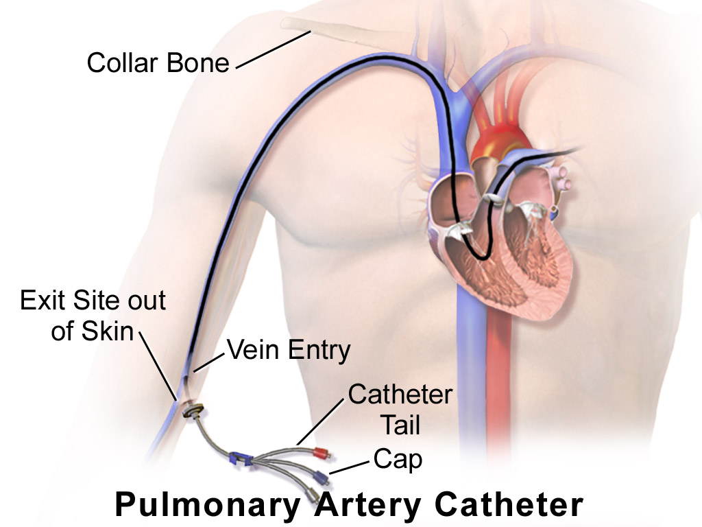
- Specimen: whole blood from the pulmonary artery catheter (SvO₂) or a central venous catheter (ScvO₂), collected in a heparinized blood-gas syringe or tube per local protocol.
- Analysis: co-oximeter, which measures saturation directly; analyze promptly.
- Continuous or intermittent: fiber-optic catheters give a running value and flag changes fast. Intermittent co-oximetry gives one sampled value. Continuous SvO₂ is described as particularly useful and nurse-driven. Its therapeutic value and cost-effectiveness are still debated, so check trends against a sample.

Why it matters
The circulation either keeps up with the body's oxygen demand or it doesn't. SvO₂ and ScvO₂ show which. That is the core question in shock. Trends help in septic shock, cardiogenic shock and on mechanical circulatory support. As a resuscitation target, though, they did not improve survival across three large trials.
Clinical Pearls
References
- Prescott HC, Antonelli M, Alhazzani W, Møller MH, Alshamsi F, Azevedo LC, et al. Surviving Sepsis Campaign: international guidelines for management of sepsis and septic shock 2026. Crit Care Med. 2026;54:725–812. https://doi.org/10.1097/CCM.0000000000007075
- Surviving Sepsis Campaign international guidelines for the management of sepsis and septic shock in children, 2026. Pediatr Crit Care Med. 2026. https://doi.org/10.1097/PCC.0000000000003927 (also Intensive Care Med, https://doi.org/10.1007/s00134-026-08360-2).
- Dellinger RP, Levy MM, Rhodes A, Annane D, Gerlach H, Opal SM, et al. Surviving Sepsis Campaign: international guidelines for management of severe sepsis and septic shock, 2012. Intensive Care Med. 2013;39(2):165–228. https://doi.org/10.1007/s00134-012-2769-8
- Cecconi M, De Backer D, Antonelli M, et al. Consensus on circulatory shock and hemodynamic monitoring. Task force of the European Society of Intensive Care Medicine. Intensive Care Med. 2014;40(12):1795–1815. https://doi.org/10.1007/s00134-014-3525-z
- van Beest P, Wietasch G, Scheeren T, Spronk P, Kuiper M. Clinical review: use of venous oxygen saturations as a goal: a yet unfinished puzzle. Crit Care. 2011;15(5):232. https://doi.org/10.1186/cc10351
- Lorusso R, Shekar K, MacLaren G, Schmidt M, Pellegrino V, Meyns B, et al. ELSO Interim Guidelines for Venoarterial Extracorporeal Membrane Oxygenation in Adult Cardiac Patients. ASAIO J. 2021;67(8):827–844. https://doi.org/10.1097/MAT.0000000000001510
- Rivers E, Nguyen B, Havstad S, Ressler J, Muzzin A, Knoblich B, et al. Early goal-directed therapy in the treatment of severe sepsis and septic shock. N Engl J Med. 2001;345(19):1368–1377. https://doi.org/10.1056/NEJMoa010307
- Angus DC, Barnato AE, Bell D, Bellomo R, Chong CR, Coats TJ, et al. A systematic review and meta-analysis of early goal-directed therapy for septic shock: the ARISE, ProCESS and ProMISe Investigators. Intensive Care Med. 2015;41:1549–1560. https://doi.org/10.1007/s00134-015-3822-1
Medical Disclaimer
- For Educational Purposes Only: This content is intended for educational reference and should not be used for clinical decision-making.
- Not a Substitute for Professional Judgment: Always consult your local protocols, institutional guidelines, and supervising physicians.
- Verify Before Acting: Users are responsible for verifying information through authoritative sources before any clinical application.