pH
Normal: 7.35–7.45 (arterial)
Hydrogen ion concentration and overall acid-base status. Acidemia below 7.35, alkalemia above 7.45. Direction only: the cause is found in PaCO₂ and HCO₃⁻.
Learn More

One arterial sample for acid-base, oxygenation and ventilation in critically ill, ventilated and transported patients.
The arterial blood gas (ABG) panel measures pH, PaCO₂, PaO₂ and SaO₂ from one arterial sample. HCO₃⁻ and base excess are calculated, and most analyzers also report lactate. Co-oximetry models add carboxyhemoglobin (COHb) and methemoglobin (MetHb). Six core values (pH, PaCO₂, PaO₂, SaO₂, HCO₃⁻ and base excess) are reported from the gas. Lactate is a seventh value on most analyzers.
The panel is drawn in emergency departments, ICUs, operating rooms and during transfers. Venous blood gas, ionized calcium and anion gap are common add-ons: they do not replace the arterial sample when oxygenation or ventilation is in question.
Hydrogen ion concentration and overall acid-base status. Acidemia below 7.35, alkalemia above 7.45. Direction only: the cause is found in PaCO₂ and HCO₃⁻.
Learn MoreRespiratory component of acid-base balance and a direct measure of ventilation. Raised in hypoventilation, low in hyperventilation.
Learn MoreMetabolic component of acid-base balance. Low in metabolic acidosis; high in metabolic alkalosis or chronic respiratory acidosis.
Learn MoreCalculated metabolic component independent of PaCO₂. Negative values indicate a metabolic acidosis or deficit; positive values an alkalosis or excess.
Learn MoreOxygen dissolved in arterial blood; reflects gas exchange. Interpret with the FiO₂ used. Below 60 mmHg is respiratory failure in most adults.
Learn MorePercentage of hemoglobin saturated with oxygen, measured by co-oximetry. It is not the same as pulse-oximetry SpO₂, and it misses CO and methemoglobin effects.
Learn MorePaO₂ divided by fractional inspired oxygen. Lower values mean worse oxygenation. The 2024 global ARDS definition uses 300 or less, with its support criteria.
Learn MoreMarker of tissue hypoperfusion. Raised above 2 mmol/L (1.5–2.0 sits above the textbook 0.5–1.5 range but below the sepsis flag; many lab ranges run to 2.2); 4 mmol/L or more means shock. Trend it during resuscitation.
Learn MoreHemoglobin bound to carbon monoxide. Pulse oximetry cannot separate it from oxyhemoglobin. Draw co-oximetry early; severity is judged clinically as well as by value.
Learn MoreHemoglobin with oxidized iron that cannot carry oxygen. Causes a low SaO₂ with a normal PaO₂. Treat if symptomatic or above 30% (common threshold; protocol-dependent). Methylene blue can make SpO₂ read falsely low.
Learn MoreNa⁺ minus (Cl⁻ plus HCO₃⁻), from a chemistry panel. Raised in high-anion-gap acidosis: lactic acidosis, ketoacidosis, toxic alcohols, salicylate, uremia.
Learn MoreVenous pH and HCO₃⁻ track arterial values closely; venous pCO₂ runs about 6 mmHg higher and agrees poorly with PaCO₂ in individuals. Not a substitute for PaO₂.
Learn MorePhysiologically active calcium. Low values cause tetany, seizures and arrhythmia; check it with K⁺ in acidemia, alkalemia and transfusion.
Learn MoreVenous saturation from a central line (ScvO₂) or pulmonary artery catheter (SvO₂). A low value warns that delivery is not meeting demand, and a normal one does not prove perfusion.
Learn More| Test | Arterial Normal | Venous Normal | SI Units | Critical | Notes |
|---|---|---|---|---|---|
| pH | 7.35–7.45 | 7.31–7.41 | [H⁺] 35–45 nmol/L (arterial) | <7.20 or >7.60 | Venous about 0.04 lower |
| PaCO₂ / PvCO₂ | 35–45 mmHg | 41–51 mmHg | 4.7–6.0 kPa (arterial) | <20 or >70 mmHg | Venous poorly predicts arterial |
| HCO₃⁻ | 22–26 mmol/L | About the same as arterial | 22–26 mmol/L | <10 or >40 mmol/L | Calculated on arterial samples |
| Base excess | −2 to +2 mmol/L | Similar to arterial | −2 to +2 mmol/L | — | Lab-dependent for venous |
| PaO₂ | 80–100 mmHg (room air) | Not used | 10.7–13.3 kPa | <40 mmHg | Falls with age; record FiO₂ |
| SaO₂ | 95–100% | Not used | 0.95–1.00 | — | Co-oximetry measured value |
| Lactate | 0.5–1.5 mmol/L | 0.5–1.5 mmol/L (venous plasma) | 0.5–1.5 mmol/L | ≥4 mmol/L | Raised above 2 mmol/L |
| COHb | <3% (smoker up to 10%) | Not used | <0.03 fraction | >25% (typical; severe) | Pregnancy: lower threshold |
| MetHb | About 1% | Not used | About 0.01 fraction | Treat if symptomatic or >30% (protocol) | Co-oximetry; SpO₂ reads low after dose |
| Ionized Ca²⁺ | 1.12–1.32 mmol/L | Lab-dependent | 1.12–1.32 mmol/L | <0.8 or >1.6 mmol/L (typical) | Calibrate to pH |
| Anion gap | 8–12 mEq/L | Not used | 8–12 mmol/L | — | Analyzer-dependent; no K⁺ |
The normal compensation band runs from 1.5 × HCO₃⁻ + 6 to + 10 mmHg. Above that band, think concurrent respiratory acidosis. Below it, think concurrent respiratory alkalosis.
At sea level on room air, the expected A–a gradient is about age ÷ 4 + 4 mmHg. Wider than that? Think shunt, ventilation–perfusion mismatch or diffusion limitation.
The Berlin definition grades ARDS by P/F ratio, with PEEP of at least 5 cmH₂O. Mild: above 200 to 300. Moderate: above 100 to 200. Severe: 100 or less mmHg. The 2024 global definition is different. It uses a P/F of 300 or less, or a SpO₂/FiO₂ of 315 or less when SpO₂ is 97% or less. Support counts too: HFNO at 30 L/min or more, CPAP or NIV at 5 cmH₂O or more, or PEEP of at least 5 cmH₂O when intubated.
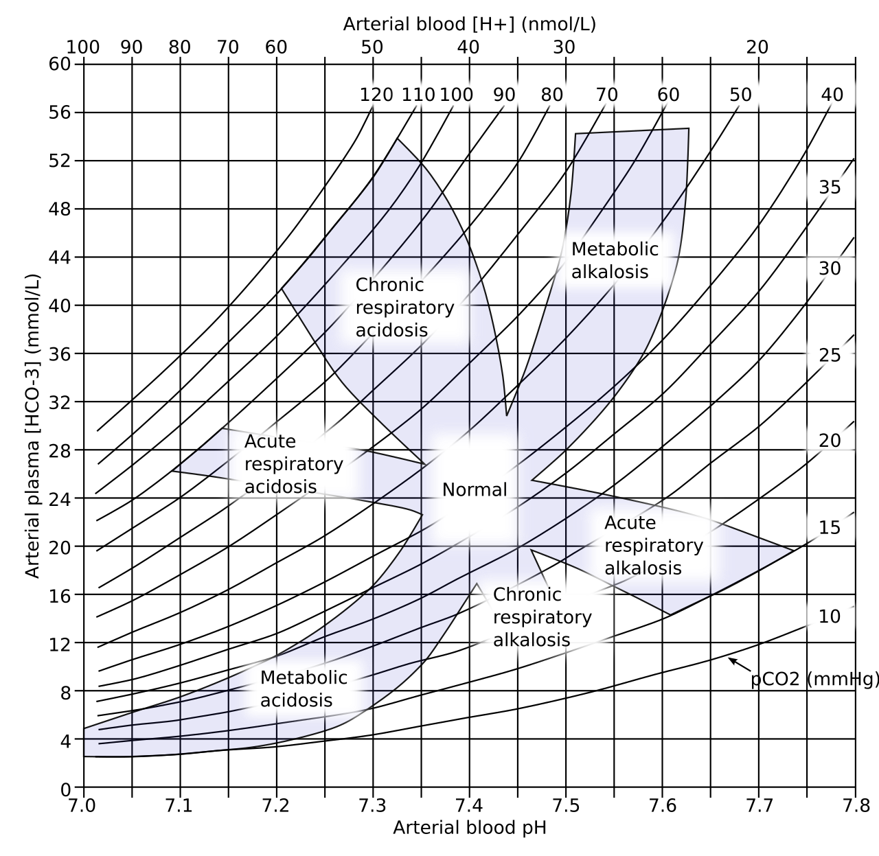
Check each pattern against pH, PaCO₂, HCO₃⁻, the anion gap and PaO₂. Mixed disorders are common in critical illness. Start with the simple patterns in the table, then look for the second one.
| Pattern | Key Findings | Common Causes | Next Step |
|---|---|---|---|
| Acute respiratory acidosis | pH low, PaCO₂ high, HCO₃⁻ near normal | Opioid or sedative overdose, COPD or asthma exacerbation, neuromuscular weakness, airway obstruction | Support ventilation; find and treat the cause; naloxone if opioid toxicity |
| Chronic respiratory acidosis | PaCO₂ high, HCO₃⁻ high, pH near normal | COPD, obesity hypoventilation, chronic neuromuscular disease | Correct ventilation slowly; a rapid PaCO₂ fall causes alkalemia |
| High-anion-gap metabolic acidosis | pH low, HCO₃⁻ low, gap raised | Lactic acidosis (sepsis, shock), DKA, toxic alcohols, salicylate, uremia | Lactate, ketones, glucose, toxicology, creatinine |
| Normal-gap metabolic acidosis | pH low, HCO₃⁻ low, gap normal, chloride high | Diarrhea, renal tubular acidosis, large-volume chloride-rich fluid | Urine electrolytes, stool losses, fluid review |
| Metabolic alkalosis | pH high, HCO₃⁻ high, PaCO₂ compensating upward | Vomiting or NG suction, diuretics, low K⁺ | Volume status, K⁺, chloride, medication review |
| Respiratory alkalosis | pH high, PaCO₂ low | Hyperventilation from hypoxemia, PE, sepsis, pain, anxiety; early salicylate toxicity | Treat the trigger; check PaO₂ and lactate |
| Hypoxemia with normal PaCO₂ | PaO₂ low, PaCO₂ normal or low | Ventilation–perfusion mismatch, shunt, diffusion limitation | Check P/F ratio, chest imaging, PEEP and FiO₂ |
| Low SaO₂ with normal PaO₂ | SaO₂ low, PaO₂ normal | Carboxyhemoglobin (reads as oxyhemoglobin on pulse oximetry), methemoglobin | Co-oximetry for COHb and MetHb; oxygen for CO; methylene blue for MetHb per protocol |
| Analyte | Rises or Changes | Peaks | Repeat Interval | Monitoring Target |
|---|---|---|---|---|
| PaO₂ / SaO₂ | Changes within minutes of an FiO₂, PEEP or ventilator change | — | After each change, per unit protocol | Target set by the ventilator or ARDS protocol |
| PaCO₂ and pH | Move with minute ventilation; a rate or tidal volume change shifts both | — | After each ventilator change, per unit protocol | Stable or improving pH while PaCO₂ falls gradually |
| Lactate | Rises with hypoperfusion | — | Repeat when the initial value is above 2 mmol/L, per sepsis protocol | Falling lactate guides resuscitation |
| MetHb | Falls after methylene blue | — | One hour after the first dose if MetHb stays above 30% or symptoms persist (label repeat dose 1 mg/kg) | Below 30% and symptoms resolved |
| COHb | Falls with oxygen; the rate depends on FiO₂ | — | Repeat only if the result will change management | Clinical recovery; severity is judged clinically |
Recognize: pH below 7.20 with falling blood pressure, dysrhythmia, or altered mental status.
Recognize: PaCO₂ rising while pH falls, somnolence, COPD exacerbation, or sedative or opioid overdose.
Recognize: PaO₂ falling despite higher FiO₂, low P/F ratio, or SpO₂ that does not respond to oxygen.
Recognize: lactate of 4 mmol/L or more, or hypotension with a rising lactate.
Recognize: headache, dizziness, nausea or confusion in several people, or an exposure source. SpO₂ can read normal.
Recognize: SaO₂ or SpO₂ low with a normal PaO₂ that does not rise with oxygen; chocolate-brown blood; dyspnea or altered mental status.
| Device or Method | Analytes | Specimen | Limitations |
|---|---|---|---|
| Handheld blood gas analyzer (cartridge) | pH, PCO₂, PO₂ and calculated HCO₃⁻, base excess, SO₂; lactate or iCa on some cartridges | Arterial or venous whole blood | Confirm which analytes the cartridge reports; check expiry and storage temperature; follow medical direction for use |
| Handheld lactate meter | Lactate | Capillary or venous whole blood | Results differ from laboratory plasma values; use the trend and the protocol cut-off |
| Pulse CO-oximetry (SpCO, SpMetHb) | COHb and MetHb estimates | Noninvasive, fingertip | Screening only; cannot confirm CO poisoning; motion and poor perfusion reduce accuracy; confirm with blood co-oximetry |
Past the newborn period, the pH, PaCO₂ and HCO₃⁻ ranges sit close to adult values. Neonates and infants use the lab's age-specific range. A capillary sample can screen pH and PCO₂, but it does not give a reliable PaO₂. When an oxygenation decision depends on that number, draw an arterial sample.
Pediatric dosing: methylene blue 1 mg/kg IV over 5–30 minutes. The label gives no separate pediatric dose. It cites two pediatric case series with the same safety profile. Repeat only as in the label. See weight-based dosing and pediatric vital signs.
Progesterone drives hyperventilation, and PaCO₂ falls to about 27–32 mmHg. The compensating HCO₃⁻ sits around 18–22 mmol/L. Near 40 mmHg in pregnancy? That suggests CO₂ retention. Maternal COHb underestimates fetal exposure to carbon monoxide. Keep a low threshold for hyperbaric consultation.
Think about COPD or obesity hypoventilation. These patients may carry a high HCO₃⁻ and a breathing drive that leans on hypoxemia. Keep the 88–92% oxygen target until a blood gas is available (BTS oxygen guideline). Re-check the gas after every oxygen change.
| Error or Interferent | Effect on Results | Prevention |
|---|---|---|
| Air bubbles | PO₂ falsely high, PCO₂ falsely low | Expel bubbles immediately and cap |
| Venous contamination | PO₂ falsely low, PCO₂ falsely high | Confirm arterial sampling; repeat if the result does not fit the patient |
| Delayed analysis | PO₂ falls; PCO₂, lactate and potassium rise; pH falls | Analyze promptly; follow laboratory stability time |
| Excess heparin | Dilution lowers HCO₃⁻ and PCO₂; ionized calcium falsely low | Use lyophilized heparin; fill to the required volume |
| Very high WBC count | In-vitro oxygen consumption lowers PO₂ | Analyze promptly; chill a delayed sample only as laboratory policy directs |
| Carbon monoxide or methemoglobin | Calculated SO₂ and SpO₂ overstate oxygen content | Use co-oximetry when exposure is possible |