

Bedside Snapshot
- Recognize
- Cardiac pump failure with hypoperfusion: SBP below 90 mmHg for 30 min or more, or a pressor, inotrope or MCS needed to hold SBP at 90, plus lactate above 2 mmol/L, altered mentation, cool skin or low urine output. Grade SCAI A to E.
- Red Flags
- SBP below 90 mmHg despite a fluid bolus
- Crackles or falling SpO₂ during fluids
- Lactate 4 mmol/L or higher
- ST elevation, new arrhythmia or heart block with low output
- First Actions
- 12-lead ECG, lactate and two IV or IO lines; norepinephrine to hold MAP; add an inotrope for low output with adequate pressure; emergency PCI for STEMI-related shock.
- Key Doses
- Norepinephrine 8–12 mcg/min IV initial, adjusted to effect (label)
- Dobutamine 0.5–1 mcg/kg/min IV, usual 2–20 mcg/kg/min
- Milrinone 50 mcg/kg IV over 10 min (omit if BP low), then 0.375–0.75 mcg/kg/min
- Don't Miss
- Tension pneumothorax, tamponade, massive PE and new VSD or papillary muscle rupture that look like cardiogenic shock; nitrates given with SBP below 90 mmHg.
- Escalate & Transport
- Escalate early for SBP below 90 mmHg despite norepinephrine, rising lactate or a rising pressor need: call the PCI and cardiac-ICU team and plan MCS per SCAI stage. STEMI-related shock goes to a PCI center with cardiac surgery and MCS.
Definition & Pathophysiology
Cardiogenic shock is end-organ hypoperfusion from primary pump failure. The organs pay for it. Registry and trial work now uses the Shock Academic Research Consortium (SHARC) definition. It names a cardiac disorder that drops systolic pressure below 90 mmHg for 30 minutes or more, or one that needs vasopressors, inotropes or mechanical circulatory support (MCS) to hold systolic pressure at 90 mmHg or higher. You also need evidence of hypoperfusion.
Output falls and filling pressures climb. Catecholamines then clamp the vessels down. The body is trying to hold pressure, but that squeeze pulls flow away from the kidneys, gut and skin. Ischemia weakens the pump further, and the cycle goes round again (Figure 1). Blood pressure can look nearly normal from compensation alone. The patient can still be hypoperfused, so perfusion markers matter as much as the pressure.
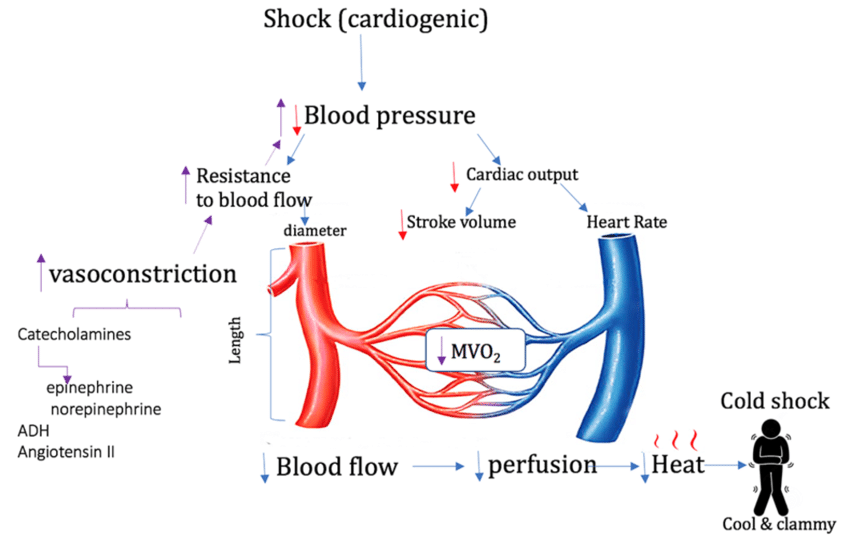
In the SHARC cohort of cardiac intensive care unit patients (2017 to 2023), 59% of cardiogenic shock was heart-failure related. 27% was acute MI-related, and 65% of those were STEMI. The mix shifts by setting. Read these figures as context, not as bedside probabilities.
- Acute MI: most often a large anterior STEMI takes out contracting muscle. Mechanical complications do it too: ventricular septal rupture, papillary muscle rupture and free-wall rupture.
- Acute decompensated heart failure: the most common cause in the SHARC cohort, usually acute-on-chronic.
- Myocarditis and cardiomyopathy: including peripartum and stress-related states.
- Arrhythmia: sustained tachycardia or bradycardia that cuts output.
- Acute valve failure: acute severe mitral regurgitation or aortic stenosis.
- Drug toxicity: beta-blocker or calcium channel blocker overdose (see Toxidromes).
Recognition & Red Flags
SCAI Stages A to E
The SCAI stages grade severity and tell you when to escalate. They name the stage. They are a clinical framework, not a validated prognostic tool.
| Stage | Meaning | Escalate When |
|---|---|---|
| A: At risk | Risk factors present, no shock yet | Monitor closely; plan early if MI or decompensation |
| B: Beginning | Relative hypotension or tachycardia without hypoperfusion | Start support; watch for hypoperfusion |
| C: Classic | Hypoperfusion; needs one intervention (pressor, inotrope or device) beyond volume | Pressor and inotrope; call cardiology and the PCI team |
| D: Deteriorating | Failure of initial support to restore perfusion: rising lactate, worsening hemodynamics, or more than one vasoactive agent or device | Escalate MCS and drug therapy; expert team |
| E: Extremis | Highly unstable, often with cardiovascular collapse | Immediate MCS or ECMO decision and resuscitation |
Record the worst stage on admission, then reassess at every change in support. Shock moves. Don't mix up the arrest modifier (A) with stage A (At risk). The modifier tags a patient who has had cardiac arrest. Its exact criteria and time window are set out in Naidu et al. They were not checked against the full text for this page, so read that source before you apply it. Escalate by stage. Stage B: start support and watch perfusion. Stage C: one intervention beyond volume, so call the PCI and cardiac-ICU team. Stage D: more than one drug or device, or a rising need, so escalate MCS and bring in the expert team. Stage E: an immediate MCS or ECMO decision, with resuscitation. Stage definitions follow the 2022 SCAI SHOCK update (Naidu et al., JACC 2022).
Red Flags
- SBP below 90 mmHg for 30 minutes or more, or a pressor or inotrope needed to hold SBP at 90 mmHg or higher
- Lactate above 2 mmol/L; 4 mmol/L or higher is critical (see Lactate)
- Altered mentation, cool or mottled skin, or low urine output despite treatment
- Pulmonary edema: new crackles or falling SpO₂ during resuscitation
- ST elevation or a new arrhythmia or heart block with low output
Assessment & Diagnostics
Treat the perfusion problem while the work-up runs. Each row gives the threshold for action.
| Check | Why | Act If |
|---|---|---|
| SBP and MAP, trend | Defines the shock and the response to treatment | SBP below 90 mmHg for 30 min or more: shock treatment. Re-check every 2 min during titration |
| 12-lead ECG (see STEMI Recognition) | Ischemia, arrhythmia, heart block, right heart strain | STEMI or STEMI equivalent: emergency reperfusion pathway. Arrhythmia: treat per ACLS |
| Lactate (see Lactate) | Hypoperfusion, not pressure | Above 2 mmol/L: hypoperfusion. 4 mmol/L or higher: escalate. Repeat to confirm clearance |
| Point-of-care ultrasound (see Hemodynamic Monitoring) | LV and RV function, pericardial effusion, IVC | Effusion with chamber collapse: obstructive shock, urgent drainage. Dilated RV: RV infarction or PE |
| BNP and troponin (see BNP, Troponin) | Heart failure load and myocardial injury | Use with the ECG and echo; neither value sets the type alone |
| Potassium, glucose, creatinine (see Potassium, Glucose, Creatinine) | Arrhythmia risk, low glucose mimic, renal dosing | Correct potassium before arrhythmia. Creatinine rising: reduce milrinone per label |
| Hemoglobin (see Hemoglobin) | Excludes bleeding as the cause or a contributor | Falling value or ongoing loss: hemorrhage pathway |
Differential & Mimics
- Obstructive shock: tension pneumothorax (one silent side, shifted trachea; see Pneumothorax and Hemothorax), tamponade (muffled heart sounds, pulsus paradoxus) and massive PE (see Pulmonary Embolism). Fluids and pressors won't fix any of these.
- Hypovolemic shock: bleeding shows up as a falling hemoglobin and flat neck veins. Cold, wet and crackling points to a cardiac cause. See Hemorrhagic Shock.
- Septic shock with myocardial depression: sepsis can depress the heart. A fever doesn't rule out a cardiogenic component (see Sepsis and Septic Shock).
- Rhythm-caused low output: a slow or fast rhythm can drop output all by itself. See Adult Bradycardia and Adult Tachycardia.
- Anaphylaxis: hives, swelling or wheeze, with hypotension, after an exposure. Give IM epinephrine first (see Anaphylaxis).
BLS Management
- Scene safety and primary assessment: airway, breathing, circulation and mental status. Look for chest pain, dyspnea and sudden weakness. Treat as high risk.
- Position of comfort: sit the patient up if breathing is hard. Do not force a flat position when breathing is labored.
- Oxygen only when SpO₂ is below the protocol target. Watch for worsening crackles.
- Aspirin: give chewable aspirin for suspected ACS if your protocol allows (see Aspirin).
- Call ALS and notify the receiving PCI center early, reporting the suspected cause, the time of symptom onset and the SBP trend.
- Cardiac arrest: start CPR and use the AED per the current AHA guideline (see Adult Cardiac Arrest).
ALS Management
Doses vary by protocol; follow local protocols and medical direction. Begin with the cause and perfusion. Drugs come in the order below, and after every step, reassess MAP, perfusion and lung water.
- Access and monitoring (Paramedic/RN/CCT): two large-bore IV lines, or IO when IV fails (see IO Access). Run continuous ECG, SpO₂ and blood pressure. Get a 12-lead ECG at once. The arterial line goes in once the patient reaches the ED or ICU.
- Fluids (Paramedic/RN/CCT): give fluid only when the patient is not congested. Use small boluses per protocol. Stop at the first sign of pulmonary edema: new crackles, falling SpO₂ or rising work of breathing.
- Norepinephrine (Paramedic/RN/CCT): first-line vasopressor to hold MAP. The label starts at 8–12 mcg/min IV (a fixed rate; weight-based protocols start differently, so confirm the unit and dosing weight on the pump), adjusted to effect, with maintenance usually 2–4 mcg/min. Titrate to the MAP target set by protocol, not to the label’s generic 80–100 mmHg systolic range, which is not a shock target. Correct hypovolemia first: the label warns that pressor use in hypovolemia can cause severe vasoconstriction and reduced perfusion. Infuse into a large, well-functioning vein or a central line, and check the site often for free flow. For extravasation, stop the infusion and follow local protocol; the label regimen is infiltration with phentolamine 5–10 mg in 10–15 mL of normal saline, ideally within 12 hours. Taper gradually: abrupt cessation can cause marked hypotension. Monitor blood pressure every 2 min until the effect is reached, then every 5 min. See Norepinephrine.
- Dobutamine (RN/CCT, per local protocol): add for low output with adequate perfusion pressure. The label starts at 0.5–1 mcg/kg/min IV, titrated every few minutes; usual range 2–20 mcg/kg/min, rarely up to 40. Watch for hypotension, ventricular ectopy and tachycardia. Dobutamine can speed AV conduction: in atrial fibrillation with rapid response, the label calls for a digitalis preparation first. The label contraindicates it in idiopathic hypertrophic subaortic stenosis, and it says experience after MI is insufficient, with concern that it may intensify ischemia or enlarge infarct size. Run it with norepinephrine when pressure is low. See Dobutamine.
- Milrinone (RN/CCT, per local protocol): an alternative or add-on inotrope with vasodilating effect. The label loading dose is 50 mcg/kg IV over 10 min, then 0.375–0.75 mcg/kg/min (standard 0.5); omit the load when pressure is low or borderline (per the Milrinone page), and reduce the rate in renal impairment using the label table: 0.20 mcg/kg/min at creatinine clearance 5 mL/min/1.73 m², 0.23 at 10, 0.28 at 20, 0.33 at 30, 0.38 at 40 and 0.43 at 50. Label warnings include hypotension and ventricular arrhythmias, including nonsustained VT. Correct hypokalemia (the label notes potassium loss with diuresis can predispose to arrhythmia), and watch the rhythm and blood pressure throughout. See Milrinone.
- Epinephrine (RN/CCT, per local protocol): add it when MAP or output stays inadequate on norepinephrine and an inotrope. The infusion range is on the Epinephrine IV page. In a small randomized trial in post-MI cardiogenic shock that stopped early (OptimaCC; Levy et al., JACC 2018), refractory shock hit 10 of 27 patients (37%) on epinephrine. It hit 2 of 30 (7%) on norepinephrine. Epinephrine also brought more tachycardia and lactic acidosis. Use it with caution. Vasopressin is a second-line pressor per local protocol (see Vasopressin).
- Dopamine (second line): the alternative vasopressor per protocol, for when norepinephrine is unavailable (see Dopamine). It caused more arrhythmic events than norepinephrine (24.1% vs 12.4%). Overall 28-day mortality did not differ significantly. In the SOAP II cardiogenic-shock subgroup (280 patients), though, dopamine went with higher mortality (De Backer et al., NEJM 2010). Reach for it only when norepinephrine is unavailable.
- Nitrates (RN/CCT, only with adequate pressure): IV nitroglycerin for pulmonary edema or ongoing ischemia, but only when SBP is at least 90 mmHg. Do not give nitrates with SBP below 90 mmHg, when RV infarction is suspected, or after recent or current PDE-5 inhibitor use. The label contraindicates combining nitrates with sildenafil, tadalafil or vardenafil. Nitrate-induced hypotension can cause paradoxical bradycardia, so watch the heart rate (see Nitroglycerin).
- Diuresis: IV furosemide for congestion, once perfusion is restored (see the Furosemide page). Too much diuresis drops preload and output.
- Rhythm (Paramedic/RN/CCT): treat VT, VF and symptomatic bradycardia per ACLS. Use transcutaneous pacing (see Transcutaneous Pacing) and cardioversion (see Cardioversion) per protocol.
- Antithrombotics for STEMI: aspirin, heparin and a P2Y12 inhibitor per the ACS protocol and cath lab (see Heparin, Clopidogrel).
- Airway (Paramedic/RN/CCT): peri-intubation hypotension and arrest are common in shock. Have a vasopressor running before induction, and plan drug choice with the team (see RSI). Positive-pressure ventilation lowers preload. Noninvasive ventilation for pulmonary edema is per protocol (see Noninvasive Ventilation).
ED & ICU Management
- Confirm the cause and stage: bedside echocardiography for LV and RV function, pericardial effusion and IVC; serial lactate; SCAI stage at each reassessment.
- Reperfusion for STEMI-related shock: emergency PCI. In the SHOCK trial, early revascularization did not significantly lower 30-day mortality (46.7% vs 56.0%, P=0.11). 6-month mortality was lower, though (50.3% vs 63.1%, P=0.027). In CULPRIT-SHOCK (706 patients with multivessel disease and cardiogenic shock), culprit-lesion-only PCI, with the non-culprit lesions treated later in stages, gave a lower 30-day rate of death or renal replacement therapy than immediate multivessel PCI (45.9% vs 55.4%, RR 0.83; Thiele et al., NEJM 2017). If PCI is out of reach within the system time target, the regional STEMI pathway decides on fibrinolysis (see Tenecteplase).
- Mechanical circulatory support: the choice depends on the team, the SCAI stage and local availability. In DanGer Shock (Impella CP plus standard care vs standard care alone), death from any cause at 180 days was 45.8% vs 58.5% (HR 0.74). Every patient had STEMI-related shock. The substudy describes the criteria this way: reduced LVEF (below 45%), SBP below 100 mmHg or on vasopressors, and lactate above 2.5 mmol/L. Patients in coma after cardiac arrest, and those with mechanical complications or right-ventricular failure, were excluded. The result does not show benefit for non-MI shock or for patients outside these criteria (Møller et al., NEJM 2024; substudy, JAMA Cardiology 2025). Severe bleeding, limb ischemia, hemolysis, device failure or worsening aortic regurgitation occurred in 24.0% vs 6.2%, and renal replacement in 41.9% vs 26.7%. See Impella.
- IABP and VA-ECMO evidence: in IABP-SHOCK II, 30-day mortality was 39.7% with IABP vs 41.3% without (RR 0.96, P=0.69). ECLS-SHOCK put it at 47.8% with VA-ECMO vs 49.0% without. Neither trial showed a significant 30-day gap. See Intra-Aortic Balloon Pump and ECMO.
- Pressor and inotrope titration: Titrate to perfusion: lactate trend, mentation, skin and urine output. Titrate to the MAP target set by protocol too. Wean gradually once perfusion recovers.
- Renal and volume care: Follow creatinine and urine output. Diurese once perfusion is restored. Start renal replacement per indications and local protocol.
- Consults: cardiology, interventional cardiology, cardiac surgery for mechanical complications, and advanced heart failure.
Right-ventricular shock
- Confirm it: right-sided leads (V4R) for inferior STEMI, and an echo showing a dilated, poorly contracting RV.
- Preload dependence: RV infarction is the exception to the usual no-fluid rule. Give a cautious fluid trial and reassess lung congestion after each bolus.
- Avoid preload reduction: nitrates, diuretics and opioids can drop output in RV infarction.
- Rhythm: treat bradycardia or AV block. Both are common with RV infarction.
- Reperfusion and support: early reperfusion, and RV support options chosen by the team and local availability.
Transport Considerations
- Destination: STEMI-related shock to a PCI-capable center with cardiac surgery and MCS. A patient already on a device goes to the center that placed it, per protocol.
- Monitoring in transit: Run continuous ECG, SpO₂, EtCO₂ and arterial or non-invasive BP. Reassess pressure and perfusion every 5 min or per protocol. Recheck the pump library before you leave.
- Infusion safety: every time the rate changes, confirm the drug, concentration, dosing weight and units (mcg/min vs mcg/kg/min). Keep a second pump or syringe for pressors, plus an independent double-check. Do not bolus a vasoactive infusion. Check the IV site for free flow and extravasation at each check. Never stop a pressor abruptly.
- Mechanical support in transit: move a patient on IABP, Impella or ECMO only with a trained team and device-specific checks, per protocol and the device pages.
- Equipment: defibrillator with pacing, suction, airway kit, arrest drugs, spare vasoactive drugs and a pressure cuff or arterial line kit.
- Handoff: symptom onset, STEMI time, SCAI stage, lactate trend, every drug with dose and start time, fluids given, device settings and alarms, and the reason for each escalation.
Special Populations
- Pediatrics: in children, myocarditis, cardiomyopathy or congenital heart disease is more often the cause. Dose by weight (see Weight-Based Dosing) and use age-specific vital signs (see Vital Signs). The adult labels above don't set pediatric doses. Follow the Pediatric Shock page and pediatric cardiology or PICU advice.
- Pregnancy: Late in pregnancy or after delivery, peripartum cardiomyopathy and MI can cause cardiogenic shock. Aortocaval compression lowers output in late pregnancy. Left lateral tilt is used to relieve it, and fluids and drugs need reassessment. The AHA 2015 scientific statement on cardiac arrest in pregnancy (Jeejeebhoy et al., Circulation 2015) describes left uterine displacement for arrest. Drug, fluid, reperfusion and MCS decisions are made with the obstetric team, per local pathway.
- Older Adults: in DanGer Shock the median age was 67 years. When you weigh MCS, use the SCAI stage and goals of care. Expect a longer recovery.
- Renal Impairment: milrinone builds up in renal impairment, so reduce the rate per label. With Impella in DanGer Shock, renal replacement was more frequent (41.9% vs 26.7%).
- Hepatic Impairment: No specific adjustment is established for norepinephrine, dobutamine or milrinone in this summary. Follow lactate and mentation closely.
- Obesity: Name the dosing weight (actual, ideal or adjusted) on every mcg/kg/min infusion. Pick one convention per patient and stick to it.
- Patients on Anticoagulants: Heparin, PCI access and MCS all raise bleeding risk. Severe bleeding was a safety event in DanGer Shock.
- Patients on Beta-Blockers: They may not mount a tachycardia, and their skin may not look cold early. Judge by lactate, mentation and urine output.
Pitfalls & Safety Checks
- Error: fluid boluses given into cardiogenic shock with no reassessment. Stop at crackles, falling SpO₂ or rising filling pressure.
- Error: nitrates given with SBP below 90 mmHg, in suspected RV infarction, or after recent PDE-5 inhibitor use. Hold them and support pressure first.
- Error: a full milrinone load given to a hypotensive patient. Omit or reduce the load per the Milrinone page.
- Error: mcg/kg/min entered as mcg/min. At 0.1 mcg/kg/min, an 80 kg adult gets 8 mcg/min. Confirm units and dosing weight at every change.
- Error: dobutamine and dopamine get mixed up. Both run in mcg/kg/min. Dobutamine is an inotrope, though, and dopamine is a pressor. Read the bag and the pump library.
- Error: missing an obstruction or a mechanical complication. Tension pneumothorax, tamponade, massive PE, and new VSD or papillary muscle rupture all look like cardiogenic shock. They need different treatment.
- Error: assuming MCS fixes everything. DanGer Shock showed more safety events with Impella (bleeding, limb ischemia and hemolysis among them). Monitor the device, the limb and the access site.
- Error: norepinephrine given through a site with no free-flow check, or extravasation missed. Check the site often. For extravasation, stop the infusion and treat per local protocol (phentolamine per label).
Clinical Pearls
References
- Naidu SS, et al. SCAI SHOCK stage classification expert consensus update: a review and incorporation of validation studies. Journal of the American College of Cardiology. 2022;79(9):933–946. https://doi.org/10.1016/j.jacc.2022.01.018
- Berg DD, Bohula EA, Patel SM, et al. Epidemiology of cardiogenic shock using the Shock Academic Research Consortium (SHARC) consensus definitions. European Heart Journal: Acute Cardiovascular Care. 2024;13(10):709–714. https://doi.org/10.1093/ehjacc/zuae098
- Rao SV, O'Donoghue ML, Ruel M, et al. 2025 ACC/AHA/ACEP/NAEMSP/SCAI Guideline for the Management of Patients With Acute Coronary Syndromes. Circulation. 2025;151(13):e771–e862. https://doi.org/10.1161/CIR.0000000000001309. Its shock-specific recommendations were not verified against the full text, so this page does not restate them.
- Hochman JS, Sleeper LA, Webb JG, et al. Early revascularization in acute myocardial infarction complicated by cardiogenic shock (SHOCK trial; landmark trial). New England Journal of Medicine. 1999;341(9):625–634. https://doi.org/10.1056/NEJM199908263410901
- Thiele H, Zeymer U, Neumann FJ, et al. Intraaortic balloon support for myocardial infarction with cardiogenic shock (IABP-SHOCK II; landmark trial). New England Journal of Medicine. 2012;367(14):1287–1296. https://doi.org/10.1056/NEJMoa1208410
- Møller JE, Engstrøm T, Jensen LO, et al. Microaxial flow pump or standard care in infarct-related cardiogenic shock (DanGer Shock). New England Journal of Medicine. 2024;390(15):1382–1393. https://doi.org/10.1056/NEJMoa2312572. Substudy with inclusion criteria and hemodynamic data: JAMA Cardiology. 2025. https://doi.org/10.1001/jamacardio.2024.4197
- Thiele H, Zeymer U, Akin I, et al. Extracorporeal life support in infarct-related cardiogenic shock (ECLS-SHOCK). New England Journal of Medicine. 2023;389(14):1286–1297. https://doi.org/10.1056/NEJMoa2307227
- Thiele H, et al. PCI strategies in patients with acute myocardial infarction and cardiogenic shock (CULPRIT-SHOCK). New England Journal of Medicine. 2017;377(25):2419–2432. https://doi.org/10.1056/NEJMoa1710261
- De Backer D, et al. Comparison of dopamine and norepinephrine in the treatment of shock (SOAP II). New England Journal of Medicine. 2010;362(9):779–789. https://doi.org/10.1056/NEJMoa0907118
- Prescribing information (DailyMed), each accessed 10/2026: norepinephrine bitartrate injection, Baxter Healthcare, version 6 (setid e375a0b9); dobutamine in dextrose injection, Hospira, version 28 (setid 9943a961); milrinone lactate injection, Hikma, version 8 (setid 4c2f052b).
Medical Disclaimer
- For Educational Purposes Only: This content is intended for educational reference and should not be used for clinical decision-making.
- Not a Substitute for Professional Judgment: Always consult your local protocols, institutional guidelines, and supervising physicians.
- Verify Before Acting: Users are responsible for verifying information through authoritative sources before any clinical application.