

Bedside Snapshot
- Recognize
- Abdominal pain that does not match the exam (mesenteric ischemia), peritonitis (guarding, rebound, rigidity), or obstruction (distension, vomiting, no flatus). Fever with jaundice suggests cholangitis.
- Red Flags
- Pain out of proportion to exam; lactate >2 mmol/L with abdominal pain; shock; peritonitis; free air or portal venous gas on imaging; Charcot triad with hypotension or confusion
- First Actions
- IV access; crystalloid for hypoperfusion; nothing by mouth; NG decompression for obstruction or ischemia per protocol; analgesia; antibiotics for peritonitis or septic shock; early surgical consult; call ALS and transport
- Key Doses
- Crystalloid at least 30 mL/kg within 3 hr for hypoperfusion per SSC 2026; ceftriaxone 2 g IV every 24 hr plus metronidazole for mild community-acquired infection (severe: per protocol); fentanyl 1 mcg/kg IV (usual 25–100 mcg), titrated
- Don't Miss
- Mesenteric ischemia with a benign exam and a normal plain film. Metoclopramide is contraindicated when obstruction or perforation is possible; ondansetron may mask progressive ileus or gastric distension.
- Escalate & Transport
- Early surgical consult. Transfer to a center with surgery, CT angiography, interventional radiology and ICU. Altitude expands bowel gas; keep the NG tube working and reassess in flight.
Definition & Pathophysiology
An acute abdomen is sudden and severe, and it needs a decision fast: surgery, drainage, medical treatment or a timed reassessment. Five pathways look alike at the bedside, and the provider has to pull them apart. Each one has a different first action, so a working diagnosis early beats a complete one late.
- Mechanical bowel obstruction: something blocks the bowel and it stops emptying. Adhesions are the most common cause of small bowel obstruction. Distension and fluid loss follow. Strangulation is the one to watch: it cuts off the blood supply to the loop.
- Perforation and peritonitis: gut contents or infection get into the peritoneal cavity. If the source stays uncontrolled, sepsis follows. The WSES 2017 intra-abdominal infection guideline calls abdominal sepsis the host's systemic response to this infection.
- Acute mesenteric ischemia: bowel blood flow stops. The cause is an embolus, an arterial or venous thrombus, or a low-flow state. Ischemia starts at the mucosa and works toward the serosa, which is why pain can come before any peritoneal sign.
- Acute appendicitis: the appendix gets blocked and inflamed. Left alone, it can go gangrenous and perforate. The WSES 2020 Jerusalem guideline names it the most common cause of intra-abdominal sepsis worldwide.
- Acute cholangitis: a blocked bile duct gets infected. Fever, inflammation and jaundice follow, and the infection can progress to sepsis.
Recognition & Red Flags
Pain Out of Proportion (Mesenteric Ischemia)
- Classic early picture: the pain is severe and hard to place, while the belly stays soft and the exam gives little. The WSES 2022 mesenteric ischemia guideline says pain that outruns the exam should be assumed to be ischemia until it is disproven.
- Acute-on-chronic pattern: pain after eating, weight loss and avoiding food suggest chronic or recurrent ischemia. Classic presentations are getting rarer, so don't wait for one.
- Older adults: at 75 and older, mesenteric ischemia beats appendicitis as a cause of acute abdomen.
A quiet abdomen does not exclude ischemia: A clean exam, clean early labs or a clean plain film still leaves ischemia open. WSES 2022 recommends against using a plain X-ray to look for intestinal ischemia.
Peritonitis and Perforation
- Signs: guarding, rebound, rigidity, pain with movement or cough, and a patient who holds very still. Peritoneal signs can be subtle. Repeat the exam.
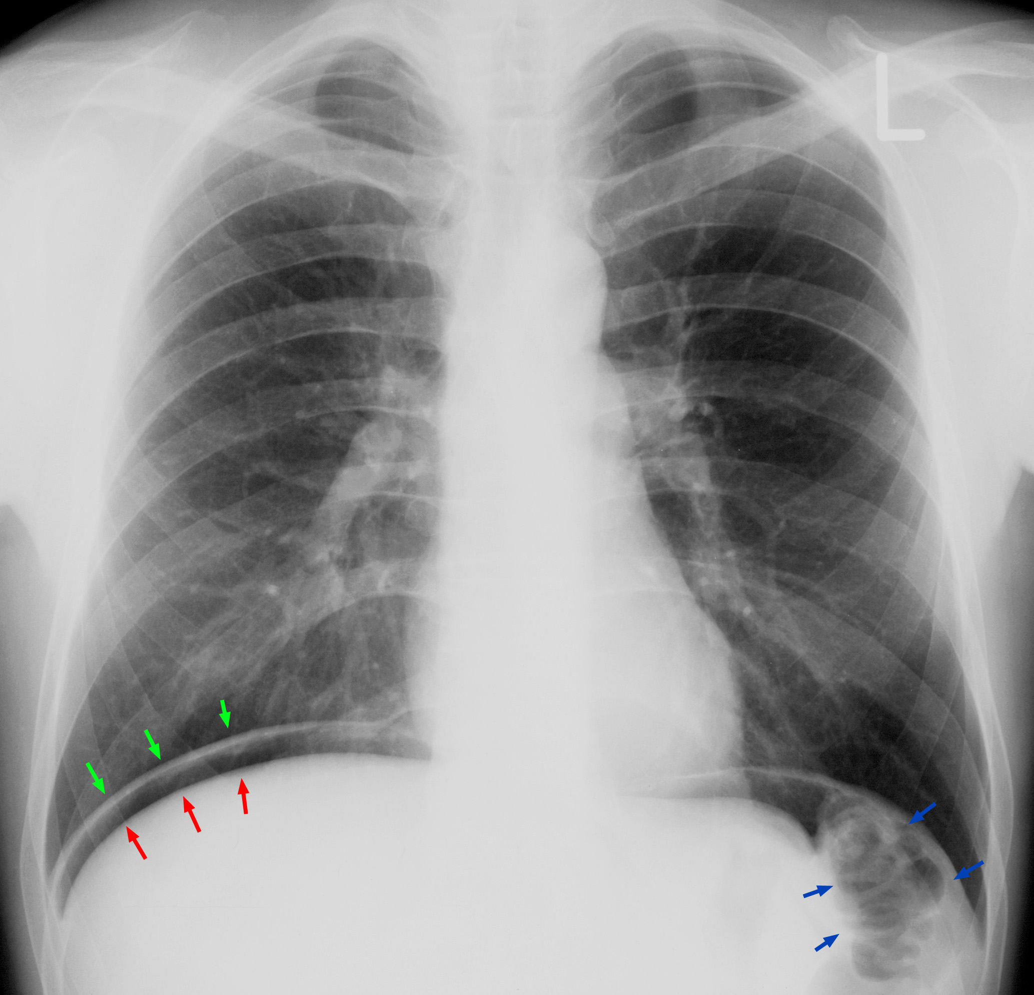
- Free air: free intraperitoneal air on an upright chest film or CT means perforation until proven otherwise (Figure 1). A plain film shows free air only once perforation or infarction has already happened.
Bowel Obstruction
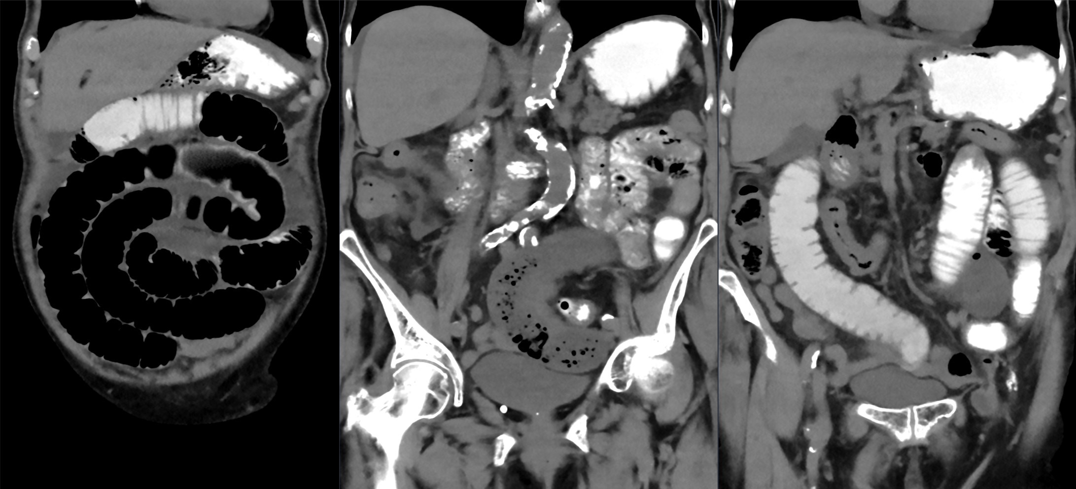
- Pattern: colicky pain, vomiting, distension and constipation. Peritoneal signs, shock or a CT pattern of a closed loop or ischemia: any of these means the loop may be strangulated.
- Contraindications to non-operative care: peritonitis, strangulation or bowel ischemia. Any one of them makes a trial of non-operative management unsafe.
Acute Cholangitis
- Charcot triad: right upper quadrant pain, fever (often with chills) and jaundice. The triad is highly specific but insensitive. Its sensitivity is low in published series, so a missing triad does not exclude cholangitis.
- Severe form (Reynolds pentad): the Charcot triad plus hypotension and confusion, or other new organ dysfunction. Grade severity by organ dysfunction. The triad alone will not tell you.
Appendicitis
- Pattern: periumbilical pain that moves to the right lower quadrant, with anorexia, nausea and fever. No single finding carries much weight. Read the whole picture alongside imaging.
Red Flags
- Lactate above 2 mmol/L alongside abdominal pain. WSES 2022 ties that pairing to irreversible intestinal ischemia. Consider early CT angiography.
- Vasopressor need with abdominal pain or distension and multi-organ dysfunction: consider non-occlusive mesenteric ischemia.
- Free air, pneumatosis or portal or mesenteric venous gas on imaging.
- Hypotension, altered consciousness or falling SpO2 with fever and jaundice: severe cholangitis.
Assessment & Diagnostics
| Check | Why | Act If |
|---|---|---|
| Vital signs, perfusion and mental status | Shock from sepsis, bleeding or ischemia changes the plan | Hypotension or poor perfusion: start fluids and reassess after each bolus (ALS Management) |
| Lactate, serial | Marks hypoperfusion; a rising value supports ischemia or sepsis | Above 2 mmol/L with pain out of proportion: push for early CT angiography. Not a stand-alone test |
| CBC, creatinine, electrolytes, coagulation | Leukocytosis is common in ischemia; creatinine guides contrast and drug dosing; coagulation guides heparin and surgery | Leukocytosis with pain: image. Raised amylase does not confirm pancreatitis, since it is raised in about half of ischemia cases |
| Blood cultures | Identify the organism when sepsis is suspected | Collect before antibiotics when this does not delay them |
| CT angiography or contrast CT | The diagnostic test for ischemia; CT is the main test for perforation and obstruction | Suspected ischemia: CTA without delay (a strong WSES 2022 recommendation). Oral contrast is not indicated for ischemia |
| Plain abdominal or upright chest film | Free air or air-fluid levels; low sensitivity for ischemia | A normal film does not exclude obstruction, ischemia or perforation. Free air: surgical review |
| Point-of-care ultrasound | First-line for appendicitis in adults and children when an experienced operator performs it | Non-diagnostic and still suspicious: move to CT or surgical review per local pathway |
| 12-lead ECG | Excludes inferior or posterior MI mimicking epigastric pain | ST elevation: activate the STEMI pathway. See STEMI Recognition |
| Pregnancy test (urine or serum hCG) | Any woman of childbearing age with abdominal pain; ectopic pregnancy and other pregnancy causes mimic the acute abdomen | Positive or unknown: obstetric evaluation. Pain with bleeding: treat as ectopic until excluded (see Ectopic Pregnancy) |
| Pulsatile mass or bedside aortic check | Ruptured or leaking abdominal aortic aneurysm | Pulsatile mass with hypotension, or back or flank pain: treat as hemorrhage and an aortic emergency |
Obstruction work-up: when the diagnosis is uncertain, CT is the test you want, and it predicts strangulation with high accuracy. Plain films do little here. Water-soluble contrast is an adjunct for adhesive obstruction (see ED & ICU Management).
Differential & Mimics
- Acute pancreatitis: epigastric pain with raised amylase. Amylase rises in about half of mesenteric ischemia cases too, so it cannot tell the two apart. Lean on lipase, CT and the clinical course.
- Inferior or posterior MI: epigastric pain, nausea and vomiting. The 12-lead ECG tells them apart.
- Functional ileus: distension and vomiting with no mechanical cause. CT sorts mechanical obstruction from ileus. The obstruction pathway belongs to the mechanical form only.
- Ruptured or leaking abdominal aortic aneurysm: hypotension, a pulsatile mass, back or flank pain, usually in an older man. Treat it as hemorrhage and an aortic emergency. Do not hold transport for imaging.
- Ectopic pregnancy: pelvic or abdominal pain with bleeding, in any woman of childbearing age. Test hCG early. See Ectopic Pregnancy.
- Aortic dissection: sudden tearing pain that can reach the abdomen, with pulse or blood pressure differences. See Aortic Dissection.
- Diabetic ketoacidosis: abdominal pain, vomiting and deep breathing alongside hyperglycemia. Check glucose and ketones. See Diabetic Ketoacidosis.
- Cholecystitis: right upper quadrant pain, fever and Murphy sign.
- GI bleeding: melena, hematemesis or hypotension with epigastric pain. See GI Bleed.
- Diverticulitis, nephrolithiasis, and testicular or ovarian torsion: common causes of acute abdominal or pelvic pain. The exam and imaging have to separate them.
- Lower lobe pneumonia or pleural disease: upper abdominal pain with cough and fever. A chest exam and film settle it.
BLS Management
- Scene and history: time of pain onset, vomiting, last bowel movement, last oral intake, prior surgery or hernia, pregnancy, anticoagulants and blood thinners.
- Nothing by mouth: give no food, fluids or oral medication. Let the patient take a position of comfort; if vomiting, turn the patient to protect the airway.
- Oxygen and monitoring: give oxygen to the target set by protocol. Check vital signs, mental status and pain at the interval set by protocol, and after any change.
- Vomiting and suction: keep suction ready. Aspiration is the main hazard during vomiting.
- Notify ALS early for hypotension, confusion, rigid abdomen, pain out of proportion to the exam, vomiting with distension, or pregnancy with pain.
- Transport priority: surgical emergencies with shock or peritonitis go as a priority to the nearest appropriate receiving center per local protocol. Report the time of symptom onset and any fluids or medications given.
ALS Management
Doses vary by protocol; follow local protocols and medical direction.
- IV or IO access (Paramedic/RN/CCT): establish access per protocol. See IV Insertion or IO Access
- Fluid for hypoperfusion (Paramedic/RN/CCT): for hypoperfusion or septic shock, give at least 30 mL/kg IV crystalloid within the first 3 hours per SSC 2026, reassessing after each bolus. Use actual body weight, or adjusted or ideal weight when BMI is above 30. In suspected ischemia, start resuscitation immediately. Lactated Ringer's or normal saline per protocol; see Lactated Ringer's
- Antibiotics (Paramedic/RN/CCT, per protocol): for septic shock or high likelihood of sepsis, give antibiotics within 1 hour; for possible infection, within 3 hours per SSC 2026. Whether to give pre-hospital antibiotics when hospital delay is long is a protocol and medical-direction decision. For mild community-acquired intra-abdominal infection, WSES 2017 lists ceftriaxone 2 g IV every 24 hr with metronidazole for anaerobic cover as an option. See Ceftriaxone and Metronidazole for dosing. Severe infection may need piperacillin-tazobactam per local protocol; see Piperacillin-Tazobactam. Check for penicillin or cephalosporin allergy before choosing an agent. Antibiotic administration by EMS is protocol dependent. IV metronidazole intervals differ between sources: WSES 2017 uses 500 mg every 6 hr, and the US label for metronidazole injection uses 7.5 mg/kg every 6 hr after a 15 mg/kg load. Follow local protocol.
- Analgesia (Paramedic/RN/CCT): fentanyl 1 mcg/kg IV slowly (usual 25–100 mcg, maximum initial 100 mcg), titrated to effect. Hold or reduce in hypotension. Analgesia does not change the need for imaging or surgical review. See Fentanyl
- Antiemetic (Paramedic/RN/CCT): ondansetron 4 mg IV or IM for nausea or vomiting per protocol. It may mask progressive ileus or gastric distension after abdominal surgery (per FDA label), so reassess the abdomen after each dose. See Ondansetron
- Metoclopramide: do not give when obstruction or perforation is possible. See Metoclopramide
- Vasopressor (Paramedic/RN/CCT, per protocol): if hypotension persists after fluid, start a vasopressor per protocol toward a MAP target set by protocol and medical direction (65 mm Hg is the usual starting target). See Norepinephrine
- Nasogastric tube (ED/ICU; Paramedic per protocol): decompress the stomach for obstruction or suspected ischemia. The WSES 2022 guideline recommends NG decompression with fluid resuscitation once ischemia is diagnosed.
ED & ICU Management
- Imaging and surgical review: suspected ischemia gets CT angiography without delay. Perforation and obstruction get CT. Any peritonitis or ischemia gets an early surgical consult. Mesenteric ischemia does best in a dedicated center with a multidisciplinary team (a strong WSES 2022 recommendation).
- Ischemia anticoagulation: full-dose unfractionated heparin should be started before surgery in acute mesenteric ischemia, per WSES 2022. Before you start, confirm there is no active bleeding. An uncontrolled active bleed is a labeled heparin contraindication. Check platelets, the baseline CBC and coagulation, and dose per hospital protocol. Therapeutic low-molecular-weight heparin is a good alternative when no surgical intervention is planned. See Heparin
- Revascularization and surgery: overt peritonitis means prompt laparoscopy or laparotomy. For arterial occlusion, endovascular revascularization comes first when the expertise is available. Severe abdominal sepsis calls for damage control surgery with temporary closure.
- Mesenteric venous thrombosis: continuous unfractionated heparin is effective. It is the first line when there is no peritonitis.
- Non-occlusive ischemia: fix the cause and keep cardiac output up. Cut back vasopressors where you can. Noradrenaline with dobutamine may be preferred over vasopressin to protect the bowel microcirculation, depending on cardiac output and vascular resistance.
- Obstruction: NG decompression, plus fluid and electrolyte correction. Give water-soluble contrast by mouth or tube, then check a plain film at 24 hours. Contrast in the colon means the obstruction is resolving. A trial of non-operative management of up to 72 hours is considered safe, unless there are signs of peritonitis, strangulation or ischemia.
- Peritonitis: control the source as soon as the patient is resuscitated. Earlier and more complete is better. In complicated infection, give antibiotics for 3–5 days after adequate source control.
- Cholangitis: antibiotics plus biliary drainage (endoscopic or percutaneous). The drainage is the source control step. Tokyo Guidelines 2018 timing: Grade I (mild), antibiotics with drainage if there is no response. Grade II (moderate), early biliary drainage, which the TG18 analysis associated with lower 30-day mortality. Grade III (severe), urgent drainage once initial organ support has started. The route follows local endoscopy or interventional radiology access.
- Appendicitis: laparoscopic appendectomy is the preferred route. For complicated appendicitis, early appendectomy is best. Antibiotics alone are an option in selected uncomplicated cases. The recurrence risk reaches up to 39% at 5 years.
Transport Considerations
- Destination: send them to a center with general surgery, CT angiography, interventional radiology and an ICU. For suspected mesenteric ischemia, the WSES 2022 guideline supports a dedicated center. WSES 2022 cites an estimate that each 6 hours of delay in diagnosis (delay in CTA) doubles mortality. Delayed diagnosis and revascularization are associated with higher mortality, so the destination and transfer time matter.
- Monitor in transit: SpO2, EtCO2 if intubated, blood pressure trend, mental status and urine output where available. Repeat lactate as ordered.
- Keep the NG tube working: keep it on suction or vented per protocol. Check the tube before and during flight.
- Altitude: as ambient pressure falls, gas in the bowel expands (Boyle's law), so distension and pain can worsen in flight. Reassess abdomen and NG output at each altitude change. See Flight Transport Physiology
- Handoff: onset time, vomiting and last oral intake, fluid volume given, analgesia and antiemetic times, anticoagulant use, lactate, imaging status and the surgical team's contact.
- Equipment: suction, NG tube and drainage bag, IV or IO access, infusion pump for fluids, and a secured airway kit for the patient who vomits and cannot protect the airway.
Special Populations
- Pediatrics: for appendicitis, point-of-care ultrasound is the first-line test in children, same as in adults, when an experienced operator performs it. In children, a CRP of 10 mg/L or more and a white count of 16,000/µL (16 x 10^9/L) or more are strong predictors of appendicitis. Non-operative antibiotics are feasible in selected children. Failure is more likely when an appendicolith is present. Dose by weight. See Weight-Based Dosing. Adhesive obstruction carries a higher lifetime risk in younger patients.
- Bilious vomiting in an infant: malrotation with midgut volvulus until proven otherwise. It is a surgical emergency that needs immediate surgical and imaging evaluation. Do not wait for a diagnosis before transfer.
- Intussusception: colicky pain, currant-jelly stool and lethargy. Follow the surgical and radiology pathway.
- Pyloric stenosis: forceful non-bilious vomiting in an infant.
- Scope: this pediatric list is not exhaustive. Lean on the pediatric pages and local pediatric protocols. Pediatric drug doses are by weight (see Weight-Based Dosing).
- Pregnancy: for suspected appendicitis, graded compression ultrasound is the preferred first test. If ultrasound is inconclusive, use MRI when it is available. The WSES 2020 guideline suggests against non-operative treatment during pregnancy. Laparoscopic appendectomy is safe in pregnancy. Small bowel obstruction is rare in pregnancy, but it carries a risk of fetal loss. Involve obstetrics early. No specific adjustment is established for the drugs on this page, so check with the obstetric team.
- Older Adults: from about age 75, mesenteric ischemia is more common than appendicitis. An 80-year-old's incidence is roughly tenfold that of a 60-year-old. Lower the threshold for CTA.
- Renal Impairment: CT angiography should still be done even when acute kidney injury is present. The WSES 2022 guideline says delayed diagnosis is more harmful than contrast exposure.
- Obesity: crystalloid volume follows SSC 2026. Use adjusted or ideal body weight when BMI is above 30.
- Patients on Anticoagulants: record the agent and last dose before surgery or heparin. See Anticoagulant Reversal
Pitfalls & Safety Checks
- Error: calling pain out of proportion anxiety, constipation or a functional problem. Assume ischemia and move to CT angiography.
- Error: using a normal lactate or a normal plain film to exclude ischemia. Neither one is enough. Use CT angiography while suspicion persists.
- Error: starting oral intake, oral contrast or food before the surgical team has decided. Oral contrast is not indicated for suspected ischemia.
- Error: holding fluid resuscitation while you wait for imaging. Start it immediately in suspected ischemia and in hypoperfusion. Reassess after each bolus.
- Error: missing sepsis in a patient with a benign-looking abdomen. For septic shock or high likelihood of sepsis, antibiotics within 1 hour per SSC 2026.
- Error: anchoring on the Charcot triad. The triad is insensitive. A patient without jaundice can still have severe cholangitis.
- Error: reaching for a prokinetic or an antiemetic to settle a distended patient before the diagnosis is clear. Metoclopramide is contraindicated when obstruction or perforation is possible. Ondansetron may mask progressive ileus or gastric distension.
Metoclopramide and obstruction: the E3 Metoclopramide page lists GI obstruction, perforation and bleeding as contraindications. Until the diagnosis is settled, do not give it.
Clinical Pearls
Mucosa first: ischemia begins in the mucosa. The pain arrives before the exam changes. Believe the pain first.
Plain films lag: free air or infarction shows up on a plain film only after the damage is done. CT angiography is the test that acts early.
The 24-hour film: in adhesive obstruction, contrast in the colon on the 24-hour film means the obstruction is resolving. Contrast still stuck in the small bowel means surgery is more likely.
CEN and CCRN discriminator: pain out of proportion to the exam, with a lactate above 2 mmol/L, calls for CT angiography now. Not another repeat exam. A normal lactate and normal labs do not change that decision.
Charcot triad: pain, fever and jaundice are specific but insensitive for cholangitis. Severe disease is not excluded by a missing triad.
References
- Bala M, Catena F, Kashuk J, et al. (2022). Acute mesenteric ischemia: updated guidelines of the World Society of Emergency Surgery. World J Emerg Surg, 17, 54. https://doi.org/10.1186/s13017-022-00443-x
- Prescott HC, Antonelli M, Alhazzani W, et al. (2026). Surviving Sepsis Campaign: International Guidelines for Management of Sepsis and Septic Shock 2026. Crit Care Med, 54(4), 725–812. https://doi.org/10.1097/CCM.0000000000007075
- Sartelli M, Chichom-Mefire A, Labricciosa FM, et al. (2017). The management of intra-abdominal infections from a global perspective: 2017 WSES guidelines for management of intra-abdominal infections. World J Emerg Surg, 12, 29. https://doi.org/10.1186/s13017-017-0141-6
- ten Broek RPG, Krielen P, Di Saverio S, et al. (2018). Bologna guidelines for diagnosis and management of adhesive small bowel obstruction (ASBO): 2017 update of the evidence-based guidelines from the World Society of Emergency Surgery ASBO working group. World J Emerg Surg, 13, 24. https://doi.org/10.1186/s13017-018-0185-2
- Di Saverio S, Podda M, De Simone B, et al. (2020). Diagnosis and treatment of acute appendicitis: 2020 update of the WSES Jerusalem guidelines. World J Emerg Surg, 15, 27. https://doi.org/10.1186/s13017-020-00306-3
- Podda M, Ceresoli M, De Simone B, et al. (2026). Diagnosis and treatment of acute appendicitis: 2025 edition of the WSES Jerusalem guidelines. JAMA Surgery, pp. 283–295. https://doi.org/10.1001/jamasurg.2025.6218
- Kiriyama S, Kozaka K, Takada T, et al. (2018). Tokyo Guidelines 2018: diagnostic criteria and severity grading of acute cholangitis (with videos). J Hepatobiliary Pancreat Sci, 25(1), 17–30. https://doi.org/10.1002/jhbp.512
Medical Disclaimer
- For Educational Purposes Only: This content is intended for educational reference and should not be used for clinical decision-making.
- Not a Substitute for Professional Judgment: Always consult your local protocols, institutional guidelines, and supervising physicians.
- Verify Before Acting: Users are responsible for verifying information through authoritative sources before any clinical application.
AI Assistance Notice
AI was used to assist in organizing, paraphrasing, and formatting this information.