

At a Glance
- Represents
- Four rhythms with no effective output: coarse and fine VF, pulseless VT, asystole and PEA. Recognition only; management is in Bedside Management.
- Normal (Adult)
- Pulse present with an organized rhythm: sinus 60–100 bpm, QRS ≤110 ms. Arrest rhythms have no pulse.
- Measure It
- Pulse check first. Then QRS width and organization in at least two leads, during a pause in CPR.
- Abnormal → Think
- VF or pulseless VT: shock. Asystole or PEA: CPR, epinephrine and a reversible cause. Artifact: pause and recheck.
- Red Flags
- VF and pulseless VT: no output. Shock as soon as the rhythm is confirmed.
- Bedside Action
- Shock VF or pulseless VT; epinephrine after the second shock. Epinephrine as soon as feasible for PEA or asystole. Do not shock either.
Physiology & What It Represents
The heart is not pumping effectively, yet the monitor can still show electrical activity with no palpable pulse. Four rhythms fall into this group. They differ in one thing: how much organized electrical activity is left.
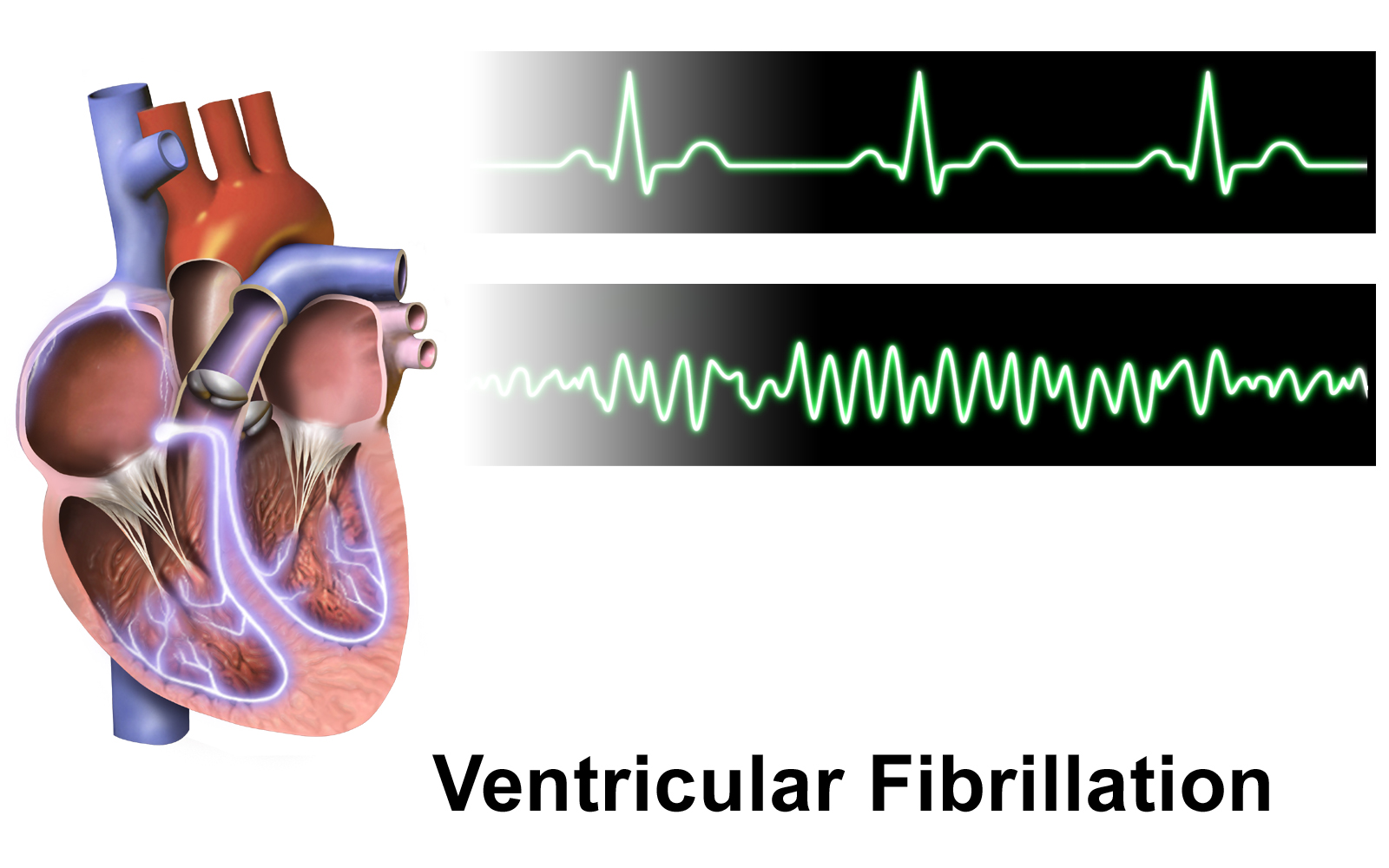
Ventricular fibrillation (VF) is chaos with no organized QRS, ST or T waves. The myocardium quivers instead of contracting. Pulseless ventricular tachycardia (pulseless VT) looks fast and organized on the monitor, with wide complexes, yet it produces no effective output. Both are shockable. Defibrillation can end them.
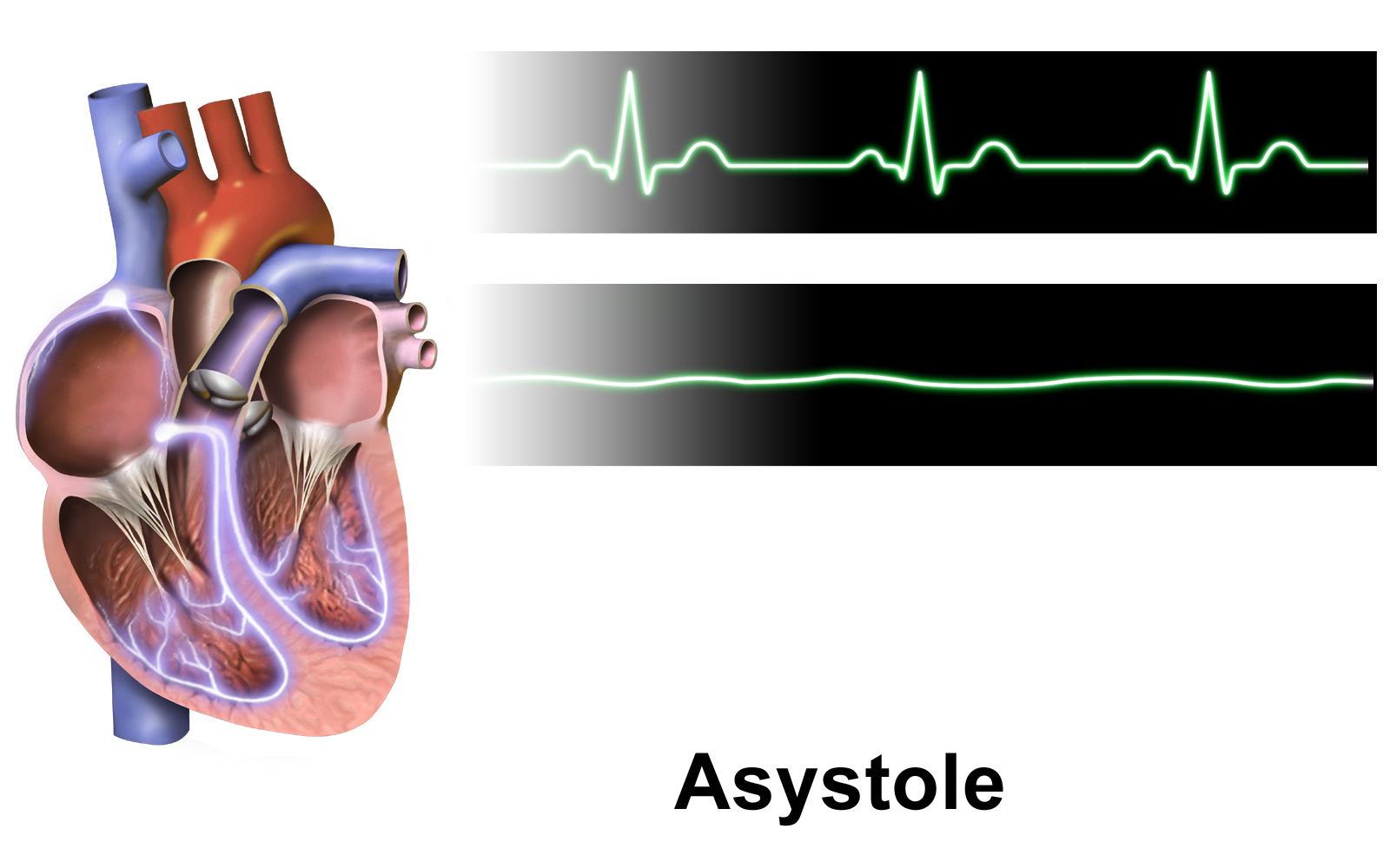
Asystole is no ventricular electrical activity at all. Pulseless electrical activity (PEA) is organized activity on the monitor with no palpable pulse. Neither one responds to defibrillation. Shock is not the tool for either.
In a non-shockable rhythm the conduction hierarchy has failed, and the escape rhythm is slow or gone. Treat with CPR, epinephrine and a search for the reversible cause. A ventricular escape rhythm at 20–40 bpm is the last organized activity before asystole.
Recognition Approach
| Rhythm | Rate (bpm) | Regularity | P Waves | PR | QRS | First-Line Action |
|---|---|---|---|---|---|---|
| Coarse VF | None organized | Chaotic | None | — | None identifiable | CPR; defibrillate now |
| Fine VF | None organized | Chaotic, low amplitude | None | — | None identifiable | CPR; defibrillate if confirmed as VF |
| Pulseless VT | >100 | Regular or near-regular | May dissociate | — | Wide (≥120 ms) | CPR; defibrillate now |
| Asystole | None | — | None | — | None | CPR; epinephrine ASAP; do not shock |
| PEA | Variable | Variable | Variable | Variable | Narrow or wide | CPR; epinephrine ASAP; treat reversible causes |
- Pulse: Confirm there is no pulse before reading the monitor. An organized rhythm with a pulse is not an arrest rhythm.
- Pause: Stop compressions briefly during the scheduled rhythm check, not during compressions.
- Leads: Look at at least two leads. Check gain and electrode contact before calling a flat line asystole.
- Shockable?: Decide whether the rhythm is VF or pulseless VT (shockable) or asystole or PEA (non-shockable).
- QRS width: For PEA, measure the QRS in the widest lead. Narrow is under 120 ms; wide is 120 ms or more.
- Artifact: Ask whether the waveform changes when compressions stop. If it does, it is artifact, not VF.
Normal Conduction Values
| Parameter | Normal | Note |
|---|---|---|
| Sinus rate | 60–100 bpm | Pre-arrest reference; arrest rhythms have no sinus rate |
| QRS | ≤110 ms | 110–119 ms is incomplete BBB or nonspecific IVCD; ≥120 ms is wide |
| PR | 120–200 ms (0.12–0.20 s; 3–5 small boxes) | Not applicable to VF, pulseless VT or asystole; used to exclude AV conduction |
| Escape rates | Junctional 40–60; ventricular 20–40 bpm | Slow escape before arrest is a warning of progressive failure |
Coarse vs Fine Ventricular Fibrillation
Learn VF first, because it is shockable. Coarse and fine VF differ in one feature only: how tall the chaotic waveform is.
ECG Criteria
- Coarse VF: large, irregular undulations that are easy to see in one lead
- Fine VF: small, low-amplitude undulations that can look like asystole
- Both: no identifiable QRS, ST or T complexes and no organized rate
Causes
- Acute myocardial ischemia or infarction
- Electrolyte derangement, including hypokalemia and hyperkalemia
- Hypoxia, acidosis, hypothermia and hypovolemia (H's in the reversible-cause list)
Clinical Significance
- VF is always pulseless. Once the rhythm is confirmed, start CPR and defibrillate without delay.
- Coarse VF is more likely to convert with the first shock. Fine VF after a long arrest may need more CPR before it converts. That is ECG teaching: the AHA algorithm does not grade coarse and fine VF.
Pulseless Ventricular Tachycardia (Pulseless VT)
Pulseless VT is a fast, wide-complex rhythm with no pulse. The electrical rhythm matches VT with a pulse. What differs is perfusion. Treat it the same way you treat VF.
ECG Criteria
- Rate: above 100 bpm, with three or more consecutive ventricular beats
- QRS: wide, 120 ms or more, with no related P waves; AV dissociation may be visible
- Morphology: monomorphic (uniform) or polymorphic (changing from beat to beat)
Causes
- Ischemia, scar from prior infarction, and cardiomyopathy
- Electrolyte derangement and QT-prolonging drugs (polymorphic pulseless VT with a long QT is torsades)
- Toxins and structural heart disease
Clinical Significance
- Same algorithm as VF: CPR and defibrillation. Pulseless VT is managed exactly like VF.
- Torsades is polymorphic pulseless VT with a long QT. Defibrillate per the algorithm, and stop the QT-prolonging drugs. For recurrences, AHA 2025 says magnesium may be considered (Part 9, Section 16; COR 2b, LOE C-LD). Dosing is on Magnesium Sulfate. Routine magnesium in adult cardiac arrest is not recommended (Part 9, Section 11; COR 3: No Benefit, LOE B-R).
- This page does not cover VT with a pulse. For monomorphic and polymorphic VT with a pulse, and for torsades, see Ventricular Rhythms.
Confirming Asystole
Asystole is no ventricular electrical activity. Confirm it before you call it, because fine VF gets a shock and asystole does not.
ECG Criteria
- Flat line: no QRS, P wave or deflection after gain and lead connections are checked
- Two leads: confirm in at least two leads before calling asystole
Causes
- Terminal progression from a slow escape rhythm or from PEA
- Reversible causes from the H's and T's list
Clinical Significance
- No shock for asystole. Give CPR and epinephrine as soon as feasible, and go after the reversible cause.
- The algorithm checks the rhythm every 2 minutes. If it turns shockable at a check, move to the shock step.
PEA: Organized Rhythm Without a Pulse
Organized activity on the monitor, no palpable pulse: that is PEA. It is non-shockable, so the patient needs CPR, epinephrine and a search for the cause.
ECG Criteria
- Organized complexes: recognizable QRS complexes, with or without preceding P waves
- Narrow or wide QRS: measure the width; a wide QRS raises hyperkalemia or sodium-channel blockade (see QRS Complex)
- No pulse: PEA is confirmed by pulse check, not by the monitor alone
Causes
- Reversible causes from the H's and T's list (see Clinical Pearls)
Clinical Significance
- Epinephrine goes in as soon as feasible, then every 3–5 min while CPR continues.
- Hunt for the cause while CPR runs. The rhythm alone will not tell you which cause it is.
CPR Artifact & Rhythm Checks
Compressions and ventilation move the electrodes, and the waveform they leave can pass for VF. Read the rhythm in a pause, never mid-compression.
ECG Criteria
- Compression artifact: regular, high-frequency deflections that stop when compressions stop
- Capnography: waveform capnography is a clue to CPR quality; if ETCO₂ is low or decreasing, reassess CPR quality per the AHA 2025 algorithm
Causes
- Chest compressions and ventilation
- Electrode movement or poor skin contact
- Pacing spikes from a pacemaker or transcutaneous pacing
Clinical Significance
- Pause briefly for the check, then get CPR going again. The AHA algorithm runs a 2-minute CPR cycle between checks.
- Keep compression interruptions short at every check, per the AHA 2025 algorithm.
Differential Diagnosis & Mimics
| Feature | VF | Pulseless VT | Asystole | PEA |
|---|---|---|---|---|
| Organized QRS | No | Yes, wide | No | Yes, narrow or wide |
| Rate | None organized | >100 bpm | None | Variable |
| Shockable | Yes | Yes | No | No |
| First step | CPR, defibrillate | CPR, defibrillate | CPR, epinephrine ASAP | CPR, epinephrine ASAP |
Artifact & Lead Misplacement
- Compressions: they can build a rhythm that looks like VF, until they stop
- Loose or low-gain leads: VF can look like asystole, and asystole can look like noise
- Tremor and movement: shivering, patient motion and vehicle vibration can mimic VF
- Pacing spikes: a transcutaneous or transvenous spike can pass for activity
Bedside Management & Escalation
Start with CPR. Then make the shock or no-shock call, give epinephrine, and hunt for the reversible cause. The doses below are adult and pediatric starting points from the AHA 2025 adult and pediatric cardiac arrest algorithms (Part 9 and Part 8) and the E3 medication pages.
Stability Table
| Situation | BLS (EMT) | ALS (AEMT / Paramedic / RN) | Critical Care / Transport |
|---|---|---|---|
| Pulseless VF or pulseless VT | CPR; AED shock now; resume CPR immediately | CPR; defibrillate; IV/IO access; epinephrine after the second shock, then every 3–5 min; amiodarone or lidocaine after the third shock | As ALS, plus advanced airway with waveform capnography and a search for the cause |
| Pulseless PEA or asystole | CPR; early ALS; do not shock | CPR; epinephrine as soon as feasible, then every 3–5 min; treat the reversible cause | As ALS; advanced airway; treat the identified cause |
| After ROSC | Monitor pulse and breathing; oxygen; prepare for transport | Adults: target SpO₂ 90%–98% and MAP ≥65 mm Hg. Children: SpO₂ 94%–99% (AHA 2025 Part 8, Section 11.3); 12-lead ECG | Post-cardiac arrest care per local protocol |
Therapy Table
| Therapy | Adult | Pediatric | Key Notes |
|---|---|---|---|
| Defibrillation (VF, pulseless VT) | Biphasic: manufacturer dose (for example, initial 120–200 J); if unknown, use the maximum available (AHA 2025 algorithm; Part 9, Section 5, rec 6). Monophasic: 360 J | 2 J/kg, then 4 J/kg; later shocks at least 4 J/kg, max 10 J/kg or the adult dose (AHA 2025 PALS algorithm) | Unsynchronized. Resume CPR for 2 minutes after each shock. Second and subsequent doses should be equivalent, and higher doses may be considered (AHA 2025 algorithm; Part 9, Section 5, rec 5) |
| Epinephrine (all four rhythms) | 1 mg IV/IO every 3–5 min (AHA 2025 Part 9, Section 10, rec 2; COR 2a, LOE B-R); no fixed maximum in the algorithm | 0.01 mg/kg IV/IO (0.1 mL/kg of 0.1 mg/mL), max 1 mg per dose, every 3–5 min | High-alert Shockable rhythm: first dose after the second shock (algorithm Box 6). Part 9, Section 10, rec 4 (COR 2a, LOE B-NR) says after initial defibrillation attempts have failed. PEA or asystole: as soon as feasible (rec 3, COR 2a, LOE B-NR). Pediatric shockable rhythm: the PALS algorithm also gives the first dose after the second shock; Part 8, Section 7, rec 2 (COR 2b, LOE C-LD) allows earlier only when rapid defibrillation is not possible. See Epinephrine 1:10,000 |
| Amiodarone (shock-refractory VF or pulseless VT) | 300 mg IV/IO bolus, then 150 mg IV/IO | 5 mg/kg IV/IO bolus (max 300 mg); may repeat up to 3 doses, with subsequent doses max 150 mg (AHA 2025 PALS algorithm). Differs from FDA label: the label states that safety and effectiveness in pediatric patients are not established and that use in pediatric patients is not recommended; this dose is from the PALS algorithm. | High-alert Give after the third shock (algorithm Box 8; Part 9, Section 11, rec 1, COR 2b, LOE B-R, for VF or pulseless VT unresponsive to defibrillation). The 150 mg second dose follows if VF or pulseless VT persists. Lidocaine is the alternative. See Amiodarone |
| Lidocaine (alternative to amiodarone) | 1–1.5 mg/kg IV/IO first dose, then 0.5–0.75 mg/kg | 1 mg/kg IV/IO (AHA 2025 PALS algorithm; no pediatric maximum stated) | High-alert Use amiodarone or lidocaine, not both by default. See Lidocaine |
Any dose not listed here needs online medical direction.
Pediatric & Special Populations
Pediatrics
- Rhythm mix: In children, VF or pulseless VT as the first rhythm is less common than in adults (AHA 2025 Part 8, Section 8 synopsis, Energy Doses for Defibrillation). Expect a nonshockable first rhythm more often. See Pediatric Cardiac Arrest.
- Doses: Work from the weight-based doses on Weight-Based Dosing. The pediatric values sit in the therapy table above.
- Normal rates by age: Pediatric rates change with age. See Pediatric Vital Signs.
- Pads and attenuator: Reach for a pediatric attenuator or pads when you have them. The pediatric defibrillation and arrest pathway is on Pediatric Cardiac Arrest.
- Bradycardia with poor perfusion: In children this is a pre-arrest state. Progressive respiratory failure or shock drives the bradycardia, and then comes PEA or asystole. Follow the pediatric bradycardia and post-ROSC pathway on Pediatric Cardiac Arrest.
Other Populations
- Pregnancy: When the fundal height is at or above the umbilicus, start manual left lateral uterine displacement with compressions (AHA 2025 Part 10, Section 19.3, rec 4; COR 1, LOE C-LD). Put IV or IO access in the upper extremities (rec 7; COR 2a, LOE C-EO). Calcium is reasonable if the patient is on IV magnesium sulfate (rec 8; COR 2a, LOE C-EO). Resuscitative delivery is indicated when ROSC is not achieved with standard resuscitation and the fundal height is at or above the umbilicus (rec 1; COR 1, LOE B-NR). Preparation starts at recognition, and the goal is delivery complete by 5 minutes (rec 2; COR 1, LOE B-NR).
- Hypothermia: Cold is a reversible cause on the H's and T's list. Warm the patient per local protocol. In severe hypothermia the shock and drug limits change, and rewarming becomes part of the resuscitation. Follow local hypothermia protocols.
- Hyperkalemia: Wide-complex PEA or known renal failure puts hyperkalemia on the list. Consider calcium per protocol. See Electrolyte Abnormalities on the ECG.
- Opioid or toxin arrest: Treat the cause per protocol. In opioid-associated arrest, ventilation comes first. Naloxone follows per protocol.
- Specialist adjuncts: Point-of-care ultrasound and extracorporeal CPR exist only at some centers. This page does not cover them.
- Termination of resuscitation: When to stop depends on protocol. This page does not cover it.
Monitoring & Transport Considerations
- Lead selection: Lead II is a common monitoring lead for arrest rhythms. Before you accept asystole, confirm it in at least two leads.
- Waveform capnography: Run continuous capnography with the advanced airway. A low or falling ETCO₂ is your cue to reassess CPR quality, per the AHA 2025 algorithm.
- Rhythm checks: Every 2 minutes, pause for the check, then get CPR going again.
- Compression interruptions: Keep pauses short during movement and transfer. The algorithm depends on compressions that do not stop.
- Artifact in transport: Vehicle vibration and movement mimic VF. Stop compressions only for the rhythm check, and check the leads before any shock.
- After ROSC: Adults: aim for SpO₂ 90%–98% (AHA 2025 Part 11; COR 2a, LOE B-R) and a MAP of ≥65 mm Hg (COR 1, LOE B-R). Children: SpO₂ 94%–99% (AHA 2025 Part 8, Section 11.3; COR 2b, LOE C-LD). Get a 12-lead ECG as soon as feasible to look for ischemia or arrhythmia (COR 1, LOE B-NR).
Step-by-Step Approach & Pitfalls
- Confirm the arrest: Check for no pulse, start CPR, attach the monitor and defibrillator, and give oxygen with bag-mask ventilation.
- Read the rhythm: Pause and look at at least two leads. Decide whether it is shockable (VF or pulseless VT) or non-shockable (asystole or PEA).
- Shockable: Defibrillate, then resume CPR for 2 minutes with IV or IO access.
- Epinephrine: Give 1 mg every 3–5 minutes. For a shockable rhythm, the first dose follows the second shock; for PEA or asystole, give it as soon as feasible.
- Refractory VF or pulseless VT: After the third shock, give amiodarone 300 mg then 150 mg, or lidocaine 1–1.5 mg/kg then 0.5–0.75 mg/kg.
- Recheck every 2 minutes: If the rhythm becomes shockable, go to the shock step. If it is still non-shockable, continue CPR and epinephrine.
- Find the cause: Work through the H's and T's during every cycle, and place an advanced airway with waveform capnography.
- After ROSC: Adults: target MAP ≥65 mm Hg and SpO₂ 90%–98%. Children: SpO₂ 94%–99%. Obtain a 12-lead ECG.
Common Pitfalls
- Shocking asystole or PEA, or calling fine VF asystole without checking a second lead
- Giving epinephrine before the first shock for VF or pulseless VT, when the algorithm gives it after the second
- Reading the rhythm during compressions, when artifact can mimic VF
- Long rhythm-check pauses that interrupt CPR
- Stopping the search for a reversible cause after the first cycle
ECG Examples
Clinical Pearls
References
- American Heart Association. Part 9: Adult Advanced Life Support: 2025 American Heart Association Guidelines for Cardiopulmonary Resuscitation and Emergency Cardiovascular Care. Circulation. 2025;152(suppl 2):S538-S577. https://doi.org/10.1161/cir.0000000000001376 Full text: https://cpr.heart.org/en/resuscitation-science/cpr-and-ecc-guidelines/adult-advanced-life-support
- American Heart Association and American Academy of Pediatrics. Part 8: Pediatric Advanced Life Support: 2025 American Heart Association and American Academy of Pediatrics Guidelines for Cardiopulmonary Resuscitation and Emergency Cardiovascular Care. Circulation. 2025;152(suppl 2):S479-S537. https://doi.org/10.1161/cir.0000000000001368 Full text: https://cpr.heart.org/en/resuscitation-science/cpr-and-ecc-guidelines/pediatric-advanced-life-support
- American Heart Association. Part 10: Adult and Pediatric Special Circumstances of Resuscitation: 2025 American Heart Association Guidelines for Cardiopulmonary Resuscitation and Emergency Cardiovascular Care. Circulation. 2025;152(suppl 2):S578-S672. https://doi.org/10.1161/CIR.0000000000001380 Full text: https://cpr.heart.org/en/resuscitation-science/cpr-and-ecc-guidelines/adult-and-pediatric-special-circumstances-of-resuscitation
- American Heart Association. Part 11: Post-Cardiac Arrest Care: 2025 American Heart Association Guidelines for Cardiopulmonary Resuscitation and Emergency Cardiovascular Care. Circulation. 2025;152(suppl 2):S673-S718. https://doi.org/10.1161/CIR.0000000000001375 Full text: https://cpr.heart.org/en/resuscitation-science/cpr-and-ecc-guidelines/post-cardiac-arrest-care
- American Heart Association. Advanced Cardiovascular Life Support algorithm posters: Adult Cardiac Arrest Algorithm (with Circular Algorithm and Post-Cardiac Arrest Care), Adult Bradycardia and Tachyarrhythmia With a Pulse, and Cardiac Arrest in Pregnancy, JN-2788, 12/25. 2025. PDF distributed by the Irish Heart Foundation; poster footer reads international.heart.org JN-2788 12/25. https://irishheart.ie/wp-content/uploads/2026/02/JN-2788_ACLS-Posters-International-English.pdf
- Al-Khatib SM, Stevenson WG, Ackerman MJ, et al. 2017 AHA/ACC/HRS Guideline for Management of Patients With Ventricular Arrhythmias and the Prevention of Sudden Cardiac Death. Circulation. 2018;138(13):e272-e391. https://doi.org/10.1161/cir.0000000000000549
- Amiodarone hydrochloride injection, USP (Fresenius Kabi USA). US Food and Drug Administration prescribing information, DailyMed (accessed 2026). Indicated for recurrent ventricular fibrillation and hemodynamically unstable ventricular tachycardia. https://dailymed.nlm.nih.gov/dailymed/drugInfo.cfm?setid=9772ab48-2ef1-4b9f-9a27-794f3e7e5c7a
Medical Disclaimer
- For Educational Purposes Only: This content is intended for educational reference and should not be used for clinical decision-making.
- Not a Substitute for Professional Judgment: Always consult your local protocols, institutional guidelines, and supervising physicians.
- Verify Before Acting: Users are responsible for verifying information through authoritative sources before any clinical application.