

At a Glance
- Covers
- Junctional escape (40–60 bpm); accelerated junctional (60–100 bpm); junctional tachycardia (>100 bpm) and postoperative JET
- Recognize It
- Regular, narrow QRS
- P waves absent, inverted in II, III, aVF, or buried
- Rate names the rhythm: 40–60, 60–100 or >100 bpm
- Most Dangerous
- Digoxin toxicity: a classic toxic rhythm that can progress to VT, VF or AV block. Check K⁺ and a digoxin level, and give digoxin immune Fab when life-threatening.
- Unstable Patient
- Symptomatic escape: atropine 1 mg IV/IO, then pacing or an epinephrine or dopamine infusion. Treat the cause. Narrow-complex tachycardia causing the instability: synchronized cardioversion (Part 9, COR 1, B-NR).
- Stable Patient
- Observe a perfusing escape; get a 12-lead ECG, digoxin level, potassium and magnesium. Adenosine suits regular narrow-complex tachycardia (2025 AHA Part 9, COR 1, B-R), not a junctional focus (standard teaching).
- Do Not
- Give more digoxin or an AV nodal blocker to a patient with junctional tachycardia on digoxin. Do not suppress an escape that is keeping the patient perfusing.
Physiology & What It Represents
The AV junction is the AV node and the proximal His bundle. In a healthy heart, the sinus node runs the show. The junction is the backup. It fires only when the sinus rate drops or sinus impulses fail to get through. When the junction takes over, the atria are activated backward (retrograde) through the AV node. The ventricles still follow the normal His–Purkinje system, so the QRS is usually narrow, and the P wave is absent, inverted or out of step with it.
Three mechanisms make junctional rhythms. The first is an escape: the junction's own automaticity takes over at 40–60 bpm when the sinus node slows or fails. Enhanced automaticity in the junction gives you accelerated junctional rhythm (60–100 bpm) and junctional tachycardia (above 100 bpm). Digoxin toxicity adds triggered activity. Ischemia, surgical injury and electrolyte shifts can also raise junctional automaticity.
The P wave pattern is a consequence of retrograde activation. In II, III and aVF, the sinus P is normally upright, and the retrograde P is inverted there. Where does it land? That depends on the AV delay. It can fall just before the QRS, hide inside it, or follow it.
Rate decides who is in charge. The sinus node runs at 60–100 bpm, the junction at 40–60 bpm and the ventricle at 20–40 bpm. So a junctional escape means the sinus node has slowed, blocked or been suppressed. A junctional tachycardia means the junction has become the faster pacemaker.
Recognition Approach
Start with the rate. It names the rhythm. Then check the P waves and the QRS. The table below has one row per junctional rhythm.
| Rhythm | Rate (bpm) | Regularity | P Waves | PR | QRS | First-Line Action |
|---|---|---|---|---|---|---|
| Junctional escape | 40–60 | Regular | Absent, inverted in II, III, aVF, or buried | Short or not measurable | Narrow | Observe if perfusing; atropine if symptomatic |
| Accelerated junctional | 60–100 | Regular | Retrograde, absent or buried | Short or not measurable | Narrow | Find the cause; no drug if stable |
| Junctional tachycardia | >100 | Regular | Retrograde, absent or buried | Short or not measurable | Narrow | Treat the cause; check digoxin and K⁺ |
- Rate: Measure the R–R interval over several beats and place the rate at 40–60, 60–100 or above 100 bpm.
- Regularity: Junctional rhythms tick along evenly. An irregular narrow rhythm puts atrial fibrillation or multifocal atrial tachycardia on the list.
- P waves: Inspect II, III and aVF for absent, inverted or post-QRS P waves. Check V1 if the baseline is noisy.
- PR or RP: A P before the QRS means the PR is under 120 ms. A P after the QRS means a short RP interval.
- QRS width: Confirm a narrow QRS. Until proven otherwise, a wide escape is usually infranodal or ventricular.
- Patient stability: Check perfusion, blood pressure, mental status and chest pain before deciding on treatment.
| Parameter | Normal | Junctional Note |
|---|---|---|
| PR interval | 120–200 ms (0.12–0.20 s; 3–5 small boxes) | Under 120 ms when a P precedes the QRS |
| QRS | under 120 ms (110–119 ms is a borderline range) | Narrow unless a bundle branch block is present |
| Escape rates | Junctional 40–60 bpm; ventricular 20–40 bpm | Junctional sits between sinus and ventricular |
Junctional Escape Rhythm
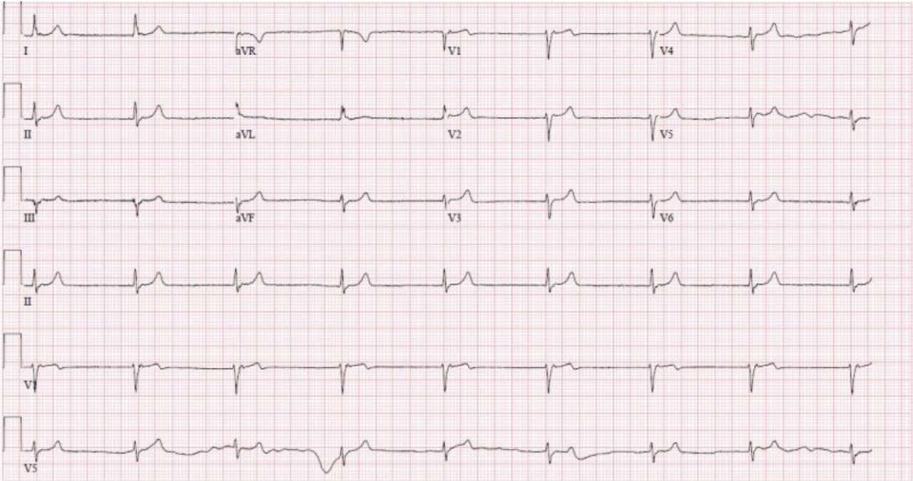
A junctional escape is a regular rhythm at 40–60 bpm. It takes over when the sinus node is slow, blocked or suppressed. It is a protective rhythm. Many patients with it have no symptoms.
ECG Criteria
- Rate: 40–60 bpm and regular
- P waves: Absent, inverted in II, III and aVF, or buried in the QRS
- PR: Under 120 ms when a P precedes the QRS; a P after the QRS has a short RP interval
- QRS: Narrow (under 120 ms) unless a bundle branch block is present
Causes
- Sinus node dysfunction: sinus bradycardia, sinus pause or sick sinus syndrome; see ECG Rate
- Drugs: beta-blockers, calcium channel blockers, digoxin and other sinus-slowing or AV-nodal drugs
- Ischemia: An inferior MI can slow the sinus node, and the junction then gets its chance to escape
- Hyperkalemia: Severe hyperkalemia can slow the sinus node and AV conduction, and a junctional escape can follow. It usually comes with other hyperkalemic changes, such as peaked T waves and a wide QRS. See T Wave
Clinical Significance
Sometimes the escape is the first sign of sinus node failure. Sometimes it is the first sign of drug or potassium toxicity. Check the medication list, the potassium and the 12-lead ECG. A stable escape that keeps the patient perfusing is a rhythm to watch, not one to suppress.
Accelerated Junctional Rhythm
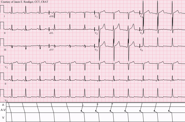
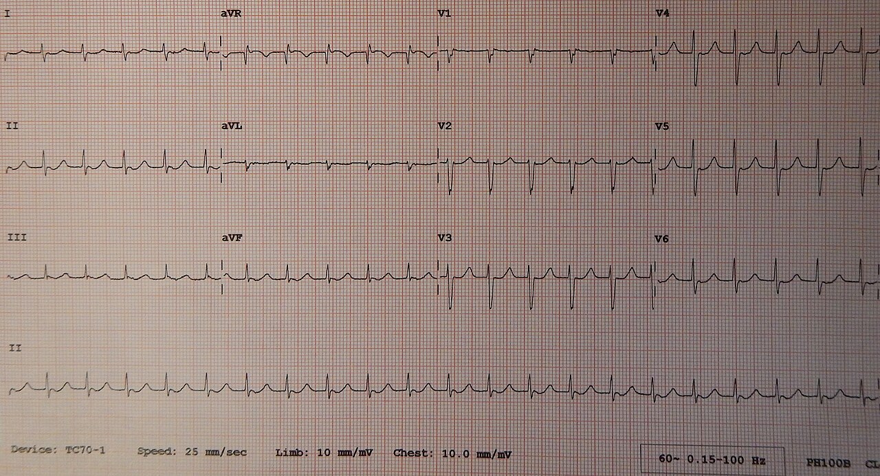
This one is a regular narrow-complex rhythm at 60–100 bpm. It runs faster than an escape, so the junction now competes with the sinus node. It is often transient. When the patient is stable, it is frequently benign.
ECG Criteria
- Rate: 60–100 bpm, regular
- P waves: Retrograde, absent or buried; sinus P waves may appear as fusion beats when the two rhythms compete
- QRS: Narrow unless a bundle branch block is present
Causes
- Digoxin: One of the classic toxic rhythms. Check the level and potassium.
- Inferior MI: AV-nodal ischemia or reperfusion can speed up the junction. Check the ST segments in II, III, aVF and V4R.
- Post-cardiac surgery: This shows up most after valve and congenital repairs. See the junctional tachycardia section.
- Electrolytes: Abnormal potassium or magnesium can change junctional automaticity. Confirm with a lab value.
Clinical Significance
Patient stable, and the rate holding in this band? Your first job is to find the cause: a digoxin level, potassium, magnesium, a 12-lead ECG, and a review of recent surgery or ischemia. Antiarrhythmic drugs are not needed for a stable rhythm.
On digoxin, a stable accelerated junctional rhythm with mild toxicity gets supportive care and observation. Stop the digoxin. Correct potassium and magnesium, and give potassium cautiously. Call the poison center or toxicology. Digoxin immune Fab is for life-threatening toxicity, so a stable rhythm alone does not earn it.
Premature Junctional Complexes
A premature junctional complex (PJC) is an early narrow QRS from the junction. It lands before the next expected sinus beat. Its P wave is absent, inverted in II, III and aVF, or follows the QRS with a short RP interval. Isolated PJCs are common and usually benign in a patient with a normal heart. Frequent PJCs, or a run of junctional beats, get the same check as an accelerated junctional rhythm: digoxin, potassium, magnesium and a 12-lead ECG.
Junctional Tachycardia and JET
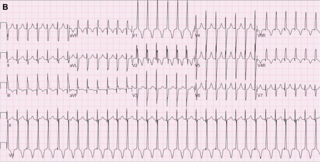
Junctional tachycardia is a regular narrow-complex rhythm above 100 bpm. Its P waves are absent or retrograde. Junctional ectopic tachycardia (JET) is the postoperative form. It follows cardiac surgery, most often in children with congenital heart disease.
ECG Criteria
- Rate: above 100 bpm, regular; can exceed 200 bpm in infants
- P waves: Absent, retrograde or dissociated from the QRS; AV dissociation is common in JET
- QRS: Narrow unless a bundle branch block is present
Causes
| Category | Examples |
|---|---|
| Drug toxicity | Digoxin toxicity, most often in older adults with reduced renal clearance |
| Postoperative | JET after valve or congenital cardiac surgery, from injury to the AV node or His bundle |
| Inflammation or ischemia | Myocarditis, acute ischemia of the AV node |
| Electrolytes | Potassium or magnesium disturbances |
Clinical Significance
Junctional tachycardia from digoxin toxicity or an electrolyte disturbance gets one fix: correct the cause. Congenital JET in neonates and infants is rare and life-threatening, and it is often refractory to conventional antiarrhythmics. A 2025 systematic review found the ivabradine evidence limited to case reports, case series and one cohort. Drug choice and dosing for postoperative JET are center-specific. Decide them with the cardiac team.
Differential Diagnosis & Mimics
Three rhythms get confused at the bedside. Junctional escape, sinus bradycardia and third-degree AV block with a narrow escape all look alike at a glance.
| Feature | Junctional Escape | Sinus Bradycardia | Third-Degree Block |
|---|---|---|---|
| P-wave morphology | Absent, inverted in II, III, aVF, or buried | Upright in I, II and aVF; one P before each QRS | Upright sinus P waves, unrelated to the QRS |
| PR | Short or not measurable | 120–200 ms | Variable; no fixed relation |
| Atrial and ventricular rates | Atrial rate is the sinus rate or none | Same rate, linked by a normal PR | Atrial rate faster than the ventricular rate |
| Response to atropine | Often improves the rate | Usually improves the rate | Variable; a wide escape responds poorly |
Tachycardia Mimics
Separate junctional tachycardia from the other regular narrow-complex rhythms:
- AVNRT: The most common cause of regular narrow-complex SVT. Onset and offset are typically abrupt, often at 150–250 bpm, with buried P waves or a pseudo-R′ in V1. Adenosine often terminates it (2015 ACC/AHA/HRS SVT guideline, reference 8; 2025 AHA Guidelines for CPR and ECC, Part 9, recommends adenosine for acute treatment of regular narrow-complex tachycardia, COR 1, LOE B-R)
- Sinus or atrial tachycardia: Onset and offset are gradual. Every QRS has a P wave in front of it.
- Junctional tachycardia: It warms up gradually. Look for AV dissociation or retrograde P waves, in a digoxin or postoperative setting. Adenosine can briefly block the AV node so the underlying rhythm shows itself. Not in suspected digoxin toxicity (2025 AHA Guidelines for CPR and ECC, Part 9, wide-complex synopsis)
Artifact & Lead Misplacement
- Limb lead reversal: An inverted P and QRS in lead I suggests reversed limb leads. Repeat the lead placement before you call a junctional rhythm from inverted P waves.
- Tremor or movement: Baseline noise can hide P waves. Check a longer strip in lead II or V1.
- Pacing spikes: A pacemaker spike can pass for a P wave. Check whether every spike captures.
- Transport vibration: repeat the strip when the vehicle is stopped
Bedside Management & Escalation
Start with stability. A stable junctional escape gets observed, and its cause gets treated. A symptomatic escape gets atropine, then pacing or an infusion. Doses vary by protocol; follow local protocols and medical direction.
Stability Approach
| Situation | BLS (EMT) | ALS (AEMT / Paramedic / RN) | Critical Care / Transport |
|---|---|---|---|
| Stable junctional escape (40–60 bpm, perfusing) | Monitor the rhythm and pulse; oxygen if SpO₂ <94%; notify ALS | Observe; obtain a 12-lead ECG; check potassium and medications; no rate-raising drug unless symptoms develop | Continuous monitoring; pads placed if the patient is at risk; find the cause |
| Symptomatic junctional escape (hypotension, altered mental status, chest pain, heart failure) | Oxygen; pads on; call ALS; do not delay transport | Atropine 1 mg IV/IO; then transcutaneous pacing or an epinephrine or dopamine infusion while preparing transvenous pacing | Pacing ready; expert consultation; treat the cause |
| Junctional tachycardia with suspected digoxin toxicity | Notify ALS; do not give digoxin | Stop digoxin; draw K⁺, Mg²⁺ and a digoxin level; digoxin immune Fab if life-threatening; hold AV nodal blockers | Defibrillator at bedside; continuous monitoring; toxicology or poison center consultation |
| Unstable junctional tachycardia (hypotension or shock) | Oxygen; pads on; call ALS | Treat the cause. If digoxin toxicity is suspected, give digoxin immune Fab for life-threatening toxicity and involve toxicology. If pulseless VT or VF develops, follow the ACLS arrest pathway; the 2023 AHA focused update says standard defibrillation guidance applies with digoxin Fab added, while animal data raise concern that defibrillation can precipitate new dysrhythmias in digoxin poisoning. Part 9 recommends synchronized cardioversion for hemodynamically unstable narrow-complex tachycardia when the tachycardia is causing the instability rather than being a manifestation of it, such as sepsis (2025 AHA Guidelines for CPR and ECC, Part 9, COR 1, LOE B-NR). | Expert consultation; prepare for electrical instability; treat the cause |
Therapy Table
| Therapy | Adult | Pediatric | Key Notes |
|---|---|---|---|
| Atropine (atropine) | 1 mg IV/IO; repeat every 3–5 min; max 3 mg total | 0.02 mg/kg IV/IO; minimum dose 0.1 mg; maximum 0.5 mg per single dose; may repeat once. Pediatric use is limited to bradycardia from increased vagal tone or atrioventricular block not caused by hypoxia (2025 AHA Guidelines for CPR and ECC, Part 8, COR 1, LOE C-LD); epinephrine is the default drug (see Pediatrics). The no-minimum 0.02 mg/kg dose in Part 8 is for intubation premedication, not for bradycardia. | Works at the SA and AV nodes, so a narrow junctional escape often responds. A wide escape usually does not. Pace if there is no response. 2025 AHA Guidelines for CPR and ECC, Part 9 (Initial Management of Bradycardia, COR 2a, LOE B-NR): atropine is reasonable to increase heart rate in acute bradycardia with hemodynamic compromise. |
| Epinephrine infusion (epinephrine) High-alert | 2–10 mcg/min IV infusion, titrated to heart rate and perfusion | Bradycardia with poor perfusion: 0.01 mg/kg IV/IO (0.1 mL/kg of 1:10,000; 0.1 mg/mL), maximum 1 mg per dose; repeat every 3–5 min. Infusion: 0.05–0.3 mcg/kg/min IV/IO, weight-based, titrate to perfusion and age-based BP (NASEMSO v3.0 Shock, p. 109; reference 7). Specialist use; titrate to perfusion. The NASEMSO range is a state model value, not the AHA PALS drug table (see flags). | Adult dose per the 2025 AHA ACLS bradycardia algorithm. Bridge to transvenous pacing. Titrate to the lowest effective rate. |
| Dopamine infusion (dopamine) High-alert | 5–20 mcg/kg/min IV infusion, maximum 20 mcg/kg/min | Not established in children; specialist use only. NASEMSO v3.0 Bradycardia (p. 36; reference 7) states that dopamine is not indicated for pediatric patients. | Alternative to epinephrine for symptomatic bradycardia (2025 AHA Guidelines for CPR and ECC, Part 9, COR 2b, LOE C-LD: if bradycardia is unresponsive to atropine, IV epinephrine or dopamine, or pacing, may be effective). Off-label for this indication; watch for tachyarrhythmia. |
| Digoxin immune Fab (digoxin immune Fab) | Life-threatening toxicity without arrest, adults: titrated 1–2 vials IV (reference 10), reassess heart rate, rhythm and K⁺ at the bedside, and repeat until toxicity resolves. Cardiac arrest or imminent arrest, or acute ingestion of unknown amount when critically ill: 10 vials IV, repeat 10 if no response (total 10–20). AHA 2025 Part 10 states the ideal empiric arrest dose is unknown; a 2023 European consensus (reference 10) uses 5 vials, repeated once after 30 min. Differs from FDA label: the label calculates the full neutralizing dose, with 20 vials for acute ingestion of unknown amount (10 then 10 more) and 6 vials for chronic toxicity with unknown level. Full-neutralization label formulas, acute ingestion, amount known: vials = body load (mg) ÷ 0.5, where body load = mg ingested × 0.8 for digoxin tablets and the full mg ingested for capsules or digitoxin; round up to whole vials. Label Table 1: 25 tablets of 0.25 mg = 10 vials. Level known: vials = digoxin (ng/mL) × weight (kg) ÷ 100, rounded up | No pediatric titrated vial dose is published: AHA 2025 Part 10 gives none, and the 1–2 vial titrated regimen (reference 10) covers adults only. Call the poison center (1-800-222-1222) for a child who may need Fab. Use the label full-neutralization doses below. Differs from FDA label: the label calculates the full neutralizing dose, and the adult titrated regimen is not applied to children. Full-neutralization label doses: chronic toxicity, level unknown: 1 vial under 20 kg; 6 vials at 20 kg or more. Acute ingestion: 10 then 10 more; monitor volume in children under 20 kg. Level formula applies at any age | Digoxin-Fab should be given for life-threatening digoxin or digitoxin poisoning (2025 AHA Guidelines for CPR and ECC, Part 10, COR 1, LOE B-NR). Give for life-threatening or potentially life-threatening toxicity (DigiFab label): ingestion of 10 mg or more in healthy adults, or 4 mg (or more than 0.1 mg/kg) in healthy children, or an ingestion expected to give steady-state levels of 10 ng/mL or more; chronic steady-state levels above 6 ng/mL in adults or 4 ng/mL in children; severe ventricular arrhythmias, progressive bradycardia, or second- or third-degree heart block not responding to atropine; K⁺ above 5.5 mEq/L in adults or 6 mEq/L in children with rapidly progressive signs and symptoms of toxicity. Potassium can fall quickly after treatment; replace it cautiously when needed. Standard (total) digoxin levels are misleading after Fab, which binds digoxin; the label says free digoxin monitoring may be appropriate for recurrent toxicity in renal failure. A 2023 European consensus (reference 10) lists K⁺ above 6.5 mmol/L among signs of life-threatening toxicity that need immediate Fab. |
Follow local protocols and online medical direction.
Pediatric & Special Populations
Pediatrics
- Not a normal variant to dismiss: In a child or infant, think congenital heart disease, recent cardiac surgery or digoxin exposure first. Healthy children and young adults with high vagal tone can also show a transient junctional rhythm, during sleep for example. Judge it by symptoms and context
- Age-based rates: Compare the heart rate with the age-specific ranges on Vital Signs
- Congenital JET: In neonates and infants it is rare, life-threatening and often refractory. Refer early to pediatric cardiology
- Digoxin exposure: In a healthy child, an ingestion of 4 mg or more, or more than 0.1 mg/kg, meets the DigiFab label criterion for Fab. That is a potentially fatal ingestion. See digoxin immune Fab
- Bradycardia with poor perfusion (PALS): Treat hypoxia and ventilation first. If the heart rate stays under 60/min with cardiopulmonary compromise despite effective ventilation with oxygen, start CPR (2025 AHA Guidelines for CPR and ECC, Part 8, COR 1, LOE C-EO). Give epinephrine 0.01 mg/kg IV/IO (0.1 mL/kg of 1:10,000; maximum 1 mg) once bradycardia persists after other factors such as hypoxia are corrected (COR 1, LOE C-EO), repeated every 3–5 min. Epinephrine is the default drug. Atropine is reserved for bradycardia from increased vagal tone or atrioventricular block that hypoxia does not cause (COR 1, LOE C-LD)
- Pacing: Emergency transcutaneous pacing may be considered for infants and children with bradycardia and cardiopulmonary compromise from complete heart block or sinus node dysfunction that does not respond to CPR (2025 AHA Guidelines for CPR and ECC, Part 8, COR 2b, LOE C-EO). The data on pacing for refractory bradycardia in children are limited, as AHA notes. Decide with medical direction.
- Dosing: Use Weight-Based Dosing for atropine and infusions; the pediatric doses are in the therapy table
Other Populations
- Older adults on digoxin: Reduced renal clearance raises the risk of toxicity. Check the level, potassium and creatinine together
- Heart transplant (denervated heart): The heart may not respond to atropine (NASEMSO v3.0, reference 7). Move to an adrenergic agonist or transcutaneous pacing. See the atropine page
- Pacemaker or ICD patients: Pacing spikes and paced beats can pass for a junctional rhythm. Check capture and the device settings
- Postoperative patients with epicardial wires: Wire pacing and a junctional rhythm can show up together. Check the wire mode before you change therapy
Monitoring & Transport Considerations
- Monitoring lead: Use lead II to see P waves; check II, III and aVF when the P-wave origin is unclear
- 12-lead ECG: Obtain one when the rhythm changes, and serially in an inferior MI; add V4R for right ventricular involvement
- Digoxin: Record the time of the last dose and draw the level 6 hours or more after it; keep potassium and magnesium on the transport record
- Pacing readiness: Place anterior–posterior pads before moving an unstable slow patient; check capture by the pulse and the SpO₂ waveform
- Artifact: Vibration and movement can fake P waves or bury them. Repeat the strip when the vehicle is stopped
- Handoff: Report any digoxin ingestion, the time of the last dose, the rate and the response to atropine
Step-by-Step Approach & Pitfalls
- Measure the rate over several R–R intervals and place it at 40–60, 60–100 or above 100 bpm.
- Check regularity; a regular narrow rhythm at these rates is a junctional candidate.
- Inspect II, III and aVF for absent, inverted or post-QRS P waves.
- Measure the PR or RP to separate junctional from sinus origin.
- Confirm a narrow QRS; a wide one needs a ventricular or aberrancy analysis.
- Decide on stability from perfusion, blood pressure, mental status and chest pain.
- Find the cause: digoxin, potassium, magnesium, inferior MI, recent surgery or sinus node disease.
Common Pitfalls
- Calling a sinus bradycardia junctional. Upright P waves in II say otherwise
- Suppressing a stable escape that keeps the patient perfusing
- Giving digoxin, or another AV nodal blocker, to a patient who is already toxic
- Reading a digoxin level drawn less than 6 hours after the last dose
- Treating an unstable tachycardia without looking for digoxin toxicity, potassium or other causes
- Missing the potassium drop after digoxin immune Fab, or redosing from a post-Fab total digoxin level
ECG Examples
Clinical Pearls
References
- Wigginton JG, Agarwal S, Bartos JA, et al. Part 9: Adult Advanced Life Support: 2025 American Heart Association Guidelines for Cardiopulmonary Resuscitation and Emergency Cardiovascular Care. Circulation. 2025;152(16 suppl 2). https://doi.org/10.1161/CIR.0000000000001376 Guidelines online: https://cpr.heart.org/en/resuscitation-science/cpr-and-ecc-guidelines/adult-advanced-life-support
- Lasa JJ, Dhillon GS, Duff JP, et al. Part 8: Pediatric Advanced Life Support: 2025 American Heart Association and American Academy of Pediatrics Guidelines for Cardiopulmonary Resuscitation and Emergency Cardiovascular Care. Circulation. 2025;152(16 suppl 2). https://doi.org/10.1161/CIR.0000000000001368 Guidelines online: https://cpr.heart.org/en/resuscitation-science/cpr-and-ecc-guidelines/pediatric-advanced-life-support
- Lavonas EJ, Akpunonu PD, Arens AM, et al. 2023 American Heart Association Focused Update on the Management of Patients With Cardiac Arrest or Life-Threatening Toxicity Due to Poisoning. Circulation. 2023;148(16):e149–e184. https://doi.org/10.1161/CIR.0000000000001161
- Kusumoto FM, Schoenfeld MH, Barrett C, et al. 2018 ACC/AHA/HRS Guideline on the Evaluation and Management of Patients With Bradycardia and Cardiac Conduction Delay. Circulation. 2019;140(8). https://doi.org/10.1161/CIR.0000000000000628
- Bokhari SFH, Mushtaq MM, Mushtaq M, et al. Ivabradine in the treatment of congenital junctional ectopic tachycardia: A systematic review. World J Cardiol. 2025;17(4):104465. https://doi.org/10.4330/wjc.v17.i4.104465
- BTG International. DigiFab (digoxin immune fab [ovine]) injection, powder, lyophilized, for solution: prescribing information. DailyMed SPL version 11, effective September 29, 2026. National Library of Medicine. https://dailymed.nlm.nih.gov/dailymed/drugInfo.cfm?setid=c05ee6a5-c98b-45f4-83fd-40781639d653
- National Association of State EMS Officials. NASEMSO National Model EMS Clinical Guidelines, Version 3.0 (March 2022): Bradycardia (pp. 35–36) and Shock (pp. 108–109). https://nasemso.org/docs.ashx?id=1444322
- Page RL, Joglar JA, Caldwell MA, et al. 2015 ACC/AHA/HRS Guideline for the Management of Adult Patients With Supraventricular Tachycardia. Circulation. 2016;133(14). https://doi.org/10.1161/CIR.0000000000000311
- 2025 American Heart Association Guidelines for Cardiopulmonary Resuscitation and Emergency Cardiovascular Care, Part 10: Adult and Pediatric Special Circumstances of Resuscitation. Digoxin and Related Cardiac Glycosides: recommendations for life-threatening poisoning (COR 1, LOE B-NR). https://cpr.heart.org/en/resuscitation-science/cpr-and-ecc-guidelines/adult-and-pediatric-special-circumstances-of-resuscitation
- Andrews P, Anseeuw K, Kotecha D, Lapostolle F, Thanacoody R. Diagnosis and practical management of digoxin toxicity: a narrative review and consensus. Eur J Emerg Med. 2023;30(6):395-401. doi:10.1097/MEJ.0000000000001065. https://pmc.ncbi.nlm.nih.gov/articles/PMC10599802/
Medical Disclaimer
- For Educational Purposes Only: This content is intended for educational reference and should not be used for clinical decision-making.
- Not a Substitute for Professional Judgment: Always consult your local protocols, institutional guidelines, and supervising physicians.
- Verify Before Acting: Users are responsible for verifying information through authoritative sources before any clinical application.