

At a Glance
- Pattern
- Right ventricular infarction: RV free-wall ischemia, usually from a proximal right coronary artery occlusion, that comes with inferior STEMI.
- Normal (Adult)
- V4R ST under 1 mm (1 mm or more is positive, per AFP 1999); isoelectric PR segments in II, III and aVF.
- Confirm It
- Record V4R on every inferior STEMI (Figure 1 for placement); compare J points with baseline; check the PR segments and rhythm.
- Abnormal → Think
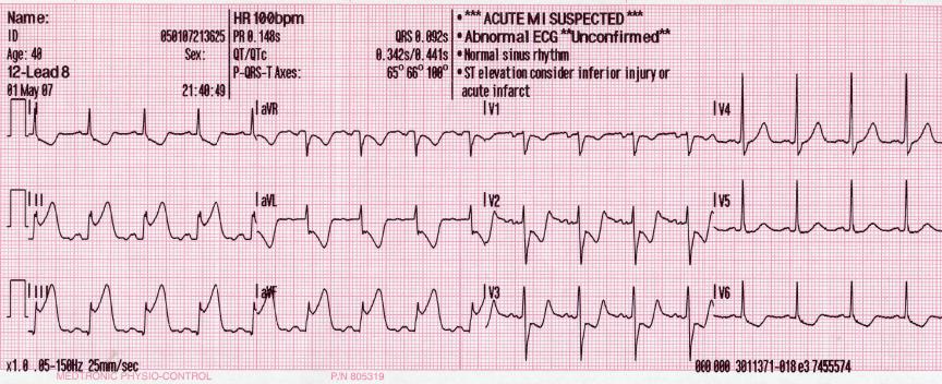
- STE in V4R with inferior STE: RV involvement
- STD in V2–V4 with inferior STE: RV ischemia
- Hypotension with high JVP: preload-dependent RV
- Red Flags
- Inferior STE with V4R elevation and hypotension; sinus bradycardia or AV block; any nitrate order when the blood pressure is falling.
- Bedside Action
- Treat per the ACS protocol; withhold nitrates and other preload-reducing drugs when RV involvement is suspected; fluid in boluses with reassessment, stopping when there is no response; pace for block that persists.
Physiology & What It Represents
The right ventricle is a thin-walled pump that works at low pressure. Its walls are thinner, and its systolic pressure and wall stress sit lower. It burns less oxygen than the left ventricle. Blood keeps reaching it through systole as well as diastole. That extra supply is why it handles ischemia better than the left does.
Most RV infarctions trace back to a proximal right coronary artery (RCA) occlusion, upstream of the branches that feed the RV free wall. That one blockage often reaches the sinoatrial node, the atria and the AV node too. Result: bradycardia or AV block can show up alongside RV injury.
When the RV fails, it delivers less blood to the left heart. Left ventricular filling falls, and systemic pressure follows it down. The RV is preload-dependent. It needs filling. Restricting fluid, the approach for a failing left ventricle, is the wrong move here.
Normal Values & How to Measure
| Parameter | Adult Normal | Pediatric Note | Where / How to Measure |
|---|---|---|---|
| V4R ST segment (V3R also used) | Under 1 mm; 1 mm or more is positive (threshold in AFP 1999). Protocols vary; follow local rules. | Adult criteria; not verified for children | J point of V4R (and V3R) against baseline |
| Inferior ST (II, III, aVF) | Isoelectric; ST elevation of 1 mm or more in 2 contiguous leads is STEMI | Adult criteria | J point in II, III and aVF against baseline |
| ST segment, V2–V4 | Isoelectric; depression here is an RV ischemia sign | Adult criteria | J point in V2–V4 |
| PR segment, II, III, aVF | Isoelectric; displacement suggests atrial injury | Adult criteria | End of the P wave to QRS onset |
- Set the baseline: use the TP or PR segment, and keep the same reference on every tracing.
- Place V4R in the fifth right intercostal space at the midclavicular line, the mirror image of V4 (Figure 1). Mark the position on the chart.
- Measure the J point in II, III and aVF against the baseline.
- Measure the J point in V4R (and V3R), and compare with the inferior leads.
- Check V2–V4 for ST depression and the PR segments in II, III and aVF.
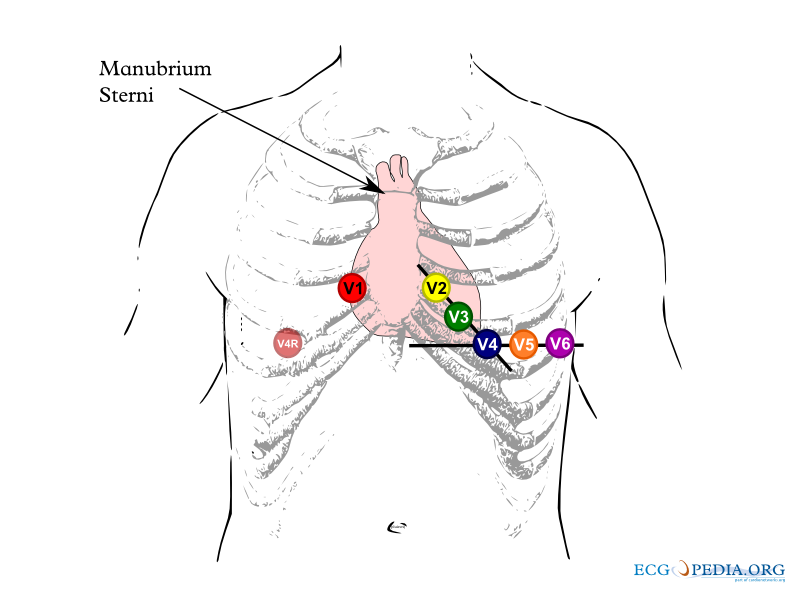
Right Ventricular Involvement in Inferior STEMI
Right ventricular infarction is injury to the RV free wall during an inferior STEMI. How common is it? One study of 200 consecutive inferior MI patients found ST elevation in V4R in 107 (54%).
ECG Criteria
- V4R: ST elevation of 1 mm or more in V4R is the AFP 1999 threshold, and protocols vary. In the 200-patient cohort, it had 88% sensitivity, 78% specificity and 83% accuracy for RV infarction.
- V5R–V6R: per AFP 1999, ST elevation greater than 1 mm in V4R through V6R confirms RV ischemia. Record V4R first.
- Inferior leads: ST elevation in II, III and aVF.
- Precordial leads: ST depression in V2–V4.
- Atrial injury: PR segment displacement in II, III and aVF. See the PR Segment page.
Causes
- Proximal RCA occlusion: the main cause, upstream of the RV branches.
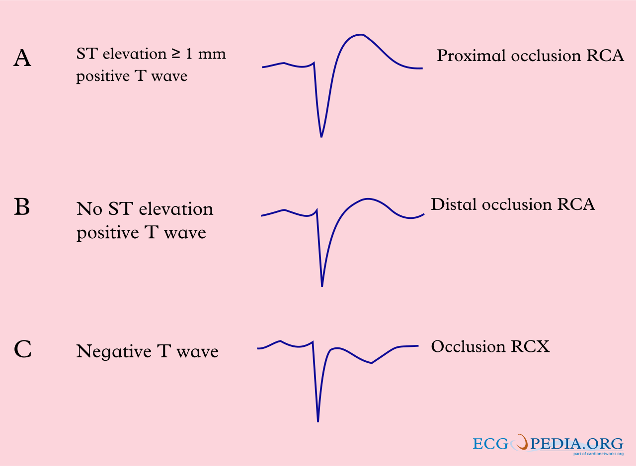
Clinical Significance
- Outcome: with V4R elevation, in-hospital mortality was 31% against 6% without it. Major complications were 64% against 28%. The finding independently predicted both.
- Older adults: RV injury raises the risk of death. That risk is higher in the elderly.
- Screening: the authors recommended right-sided ECG assessment in every acute inferior MI.
Look-Alikes
- Inferior STEMI with a normal V4R: the left-ventricle-only pattern, covered in the Differential section below.
- Posterior MI shows ST depression in V1–V3, not right-sided elevation. See the STEMI Equivalents page.
Bradycardia and AV Block in Inferior STEMI
Proximal RCA occlusion can cut the blood supply to the sinoatrial and AV nodes. The RV free wall is not the only thing at risk.
ECG Criteria
- Sinus bradycardia in an inferior STEMI.
- AV node block and atrial infarction (PR displacement in II, III and aVF).
- Atrial fibrillation also appears among the associated findings.
Causes
- Proximal RCA occlusion that feeds the SA and AV nodes as well as the RV.
Clinical Significance
- Atropine can fail: if it does not correct symptomatic bradycardia or AV block, pacing may be needed.
- Temporary pacing: AFP 1999 advises early AV sequential pacing to keep the atrial contribution. It is usually short term. Permanent pacing may be needed later.
- AF or flutter with hypotension: losing the atrial kick can drop output. Unstable rapid AF needs synchronized cardioversion per the AHA ACLS tachycardia algorithm and local protocol.
- Infranodal block: atropine is less reliable when the escape rhythm is wide-QRS. See the Conduction Blocks page.
Look-Alikes
- Bradycardia from a non-ischemic cause (see Heart Rate).
Preload Dependence and Hypotension
An infarcted RV is preload-dependent. When it fails, left ventricular filling drops and systemic pressure falls. So a drug that lowers preload can push the blood pressure lower still.
Hemodynamic Signs
- Hypotension with an elevated jugular venous pressure.
- Kussmaul sign: jugular pressure rises paradoxically on inspiration.
- Contrast with LV failure: without RV involvement, extensive left ventricular infarction gives low output with congestion. It needs parsimonious fluid. RV involvement can need fluid loading, with monitoring for RV overdistension, as AFP 1999 advises.
Causes
- RV ischemia: the proximal RCA occlusion takes away RV contractility.
Clinical Significance
- Nitrates: nitroglycerin lowers preload. It can cause severe hypotension, especially in volume-depleted or already hypotensive patients. The Nitrostat label contraindicates it in circulatory failure or shock (section 4.5) and warns of severe hypotension (section 5.2).
- Other preload-reducing drugs: morphine (venodilation), diuretics, ACE inhibitors, beta-blockers, and sedative or induction agents can also drop output in RV infarction. Use them only with great caution until volume status and BP are established.
- Preload-dependent output: the IV nitroglycerin label (Baxter, Contraindications) contraindicates use in pericardial tamponade, restrictive cardiomyopathy and constrictive pericarditis. Cardiac output in those conditions depends on venous return.
- Contested guidance: AHA and ESC guidance has historically advised avoiding nitrates in RV infarction. A 2023 systematic review and meta-analysis (Wilkinson-Stokes, PubMed 36180168, 5 studies, n=1,113) found no significant difference in adverse events, with very low certainty. The 2023 ESC ACS guideline (section 4.2.2.2) says nitrates should not be given to patients with hypotension, marked bradycardia or tachycardia, or RV infarction. Medical direction decides.
Differential Diagnosis & Mimics
| Feature | RV Infarction | Inferior LV Infarct, No RV Involvement |
|---|---|---|
| V4R | ST elevation (88% sensitive in the 200-patient study) | No ST elevation in V4R |
| V2–V4 | ST depression | Not part of the RV pattern |
| Hemodynamics | Hypotension with elevated JVP | Low output with pulmonary congestion |
| Fluid | Volume loading may be needed | Parsimonious fluid |
Artifact & Lead Misplacement
- V4R position: it has to be the mirror image of V4 on the right chest (Figure 1). Mark the spot so serial tracings line up.
- Motion or shivering can distort the ST segment. Repeat the tracing once the patient is still.
- Transport vibration adds noise to the tracing. Repeat it after the vehicle stops.
Bedside Management & Escalation
| Situation | BLS (EMT) | ALS (AEMT / Paramedic / RN) | Critical Care / Transport |
|---|---|---|---|
| Stable, inferior STEMI | 12-lead with V4R where allowed; oxygen per local protocol (many EMS protocols use SpO₂ below 94%); early ALS | 12-lead with V4R; IV access; ACS treatment per protocol; notify the receiving PCI center | Emergent reperfusion pathway per protocol; serial ECGs; avoid preload-reducing drugs when RV involvement is present |
| Unstable (SBP <90 mmHg or shock) | Call ALS; oxygen per local protocol (many EMS protocols use SpO₂ below 94%) | Stop nitrates and other preload-reducing drugs; IV fluid in boluses with reassessment after each, up to about 2 L total with JVP and lung checks, the AFP 1999 ceiling; stop fluid when there is no response; then dobutamine, with norepinephrine if still hypotensive, per protocol | Titrate inotrope and vasopressor to perfusion; pacing ready; reperfusion or mechanical support per protocol |
| Symptomatic bradycardia or AV block | Oxygen per local protocol; AED and pads placed; early ALS | Atropine per AHA 2025; transcutaneous pacing if atropine does not work | Pacing, AV sequential if possible; dopamine or epinephrine infusion while transvenous pacing is arranged (see the dosage table) |
| Therapy | Adult | Pediatric | Key Notes |
|---|---|---|---|
| Nitroglycerin (withhold) | Withhold when RV involvement is suspected or SBP is below 90 mmHg; medical direction only | Not established; IV use limited to pediatric cardiology and PICU | Contraindicated with PDE-5 inhibitors or sGC stimulators (riociguat), in circulatory failure or shock, in severe anemia, and with increased intracranial pressure (Nitrostat label, sections 4.1–4.5). |
| Isotonic crystalloid (balanced or 0.9% NaCl) | Small IV/IO boluses with reassessment (BP, JVP, ECG) after each; bolus size per medical direction; total about 1–2 L (AFP 1999 ceiling of 2 L, with RA pressure or JVP monitoring). Not the sepsis 30 mL/kg target | 10 mL/kg IV/IO aliquots with reassessment after each; pediatric cardiology input. No RV-infarct volume was verified for children; use medical direction. | Stop when BP does not respond, JVP keeps rising, or congestion appears. RV-infarct lungs are often clear, so do not wait for crackles. Then add an inotrope. |
| Epinephrine infusion | 2–10 mcg/min IV infusion, titrated to heart rate and BP (bradycardia unresponsive to atropine) | Infusion dosing per PICU protocol; see the Epinephrine page | High-alert Bridge to pacing. See the Epinephrine page. |
| Atropine | 1 mg IV/IO; repeat every 3–5 min; max 3 mg total | 0.02 mg/kg IV/IO; min 0.1 mg, max 0.5 mg per dose; may repeat once | Less reliable in infranodal block: pace early. See the Atropine page. |
| Dopamine | 5–20 mcg/kg/min IV infusion, titrated to HR and BP; max 20 mcg/kg/min | Start 2–5 mcg/kg/min IV/IO; usual range 2–20 mcg/kg/min (PICU protocol only) | High-alert Correct hypovolemia first. See the Dopamine page. |
| Norepinephrine | 0.05–0.5 mcg/kg/min IV infusion; titrate to MAP 65 mmHg; no fixed max | 0.05–2 mcg/kg/min IV/IO infusion, titrated to perfusion; usual max 2 mcg/kg/min (PICU protocol only) | High-alert Add an inotrope for low output. See the Norepinephrine page. |
| Dobutamine | Start 0.5–1 mcg/kg/min IV infusion (RV-infarction teaching in AFP 1999 starts at 2–5 mcg/kg/min; titrate to perfusion); usual range 2–20 mcg/kg/min; max 40 mcg/kg/min (rarely needed) | 2–20 mcg/kg/min IV/IO infusion; usual max 20 mcg/kg/min | High-alert If BP is low, run it with norepinephrine. See the Dobutamine page. |
Follow local protocols and medical direction.
Pediatric & Special Populations
Pediatrics
- Rarity: RV infarction in children is uncommon. No pediatric RV-infarction ECG criteria were verified for this page. Involve pediatric cardiology and follow PALS 2025 and local protocols.
- Bradycardia: find the age-based heart rate and blood pressure norms on the pediatric vital signs page. Atropine dosing by weight is in Bedside Management.
- Fluid: weight-based dosing is on the weight-based dosing page. Fluid volumes are in Bedside Management.
Other Populations
- Pregnancy: RV infarction is rare. Involve obstetrics and cardiology. Avoid supine positioning, which can drop the BP.
- Older adults: RV involvement was linked to higher in-hospital mortality in the Zehender cohort. Age-specific data were not verified for this page.
- PDE-5 inhibitor users: nitrates are contraindicated with PDE-5 inhibitors and sGC stimulators such as riociguat (Nitrostat label, section 4.1). See the Nitroglycerin page for timing.
- Shock: the Nitrostat label contraindicates nitrates in circulatory failure or shock (section 4.5).
Monitoring & Transport Considerations
| Issue | What to Do |
|---|---|
| Right-sided leads | Record V4R on every inferior STEMI and repeat it if the first reading is unclear. |
| Blood pressure | Check before any preload-reducing drug and after each bolus. An SBP below 90 mmHg means stop nitrates (see the Nitroglycerin page). |
| Lungs and SpO₂ | Reassess after every bolus. Stop fluid for a falling BP, a rising JVP or congestion; RV-infarct lungs are often clear. |
| Ventilation | Positive-pressure ventilation, high PEEP and sedation or induction agents can drop preload and BP. Use the lowest effective PEEP, have a vasopressor ready, and watch for sudden hypotension. |
| Rhythm | Watch for sinus bradycardia and AV block. Keep pads on and pace per protocol. AF or flutter with hypotension needs prompt synchronized cardioversion per protocol. |
| Serial 12-leads | Repeat per protocol while ischemia persists; compare V4R with the first tracing each time. |
| Transport | Keep pacing and access ready for an unstable patient. Pad position follows the transcutaneous pacing page and protocol. |
Step-by-Step Approach & Pitfalls
- Get a 12-lead ECG and add V4R (Figure 1) to every inferior STEMI.
- Measure the J point in II, III and aVF against baseline, then in V4R.
- Check V2–V4 for ST depression and the PR segments in II, III and aVF.
- Read the rhythm for sinus bradycardia, AV block and atrial fibrillation.
- Check BP and JVP before any nitrate, morphine, diuretic or other preload-reducing drug.
- Give fluid in boluses and reassess after each. Stop when BP does not respond or the JVP keeps rising, and add an inotrope.
- Call for reperfusion early: primary PCI is definitive treatment. Shock triggers immediate revascularization or mechanical-support review per protocol.
- Notify the receiving team with the V4R result, per protocol.
Common Pitfalls
- Using III greater than II to judge the RCA: that sign had 17% specificity for a dominant RCA culprit.
- Skipping V4R: RV involvement gets missed when right-sided leads are never recorded.
- Reflex nitrates for chest pain: check V4R and BP first.
- Treating RV hypotension like LV failure: restricting fluid is the wrong response.
- Stopping at crackles only: RV-infarct lungs are often clear. Overload shows up as a rising JVP and no BP response.
- Expecting atropine to fix every block: atropine may fail, so pace early.
ECG Examples
Clinical Pearls
References
- Rao SV, O’Donoghue ML, Ruel M, et al. 2025 ACC/AHA/ACEP/NAEMSP/SCAI guideline for the management of patients with acute coronary syndromes. J Am Coll Cardiol. 2025;85:2135–2237. https://doi.org/10.1016/j.jacc.2024.11.009
- Thygesen K, Alpert JS, Jaffe AS, et al. Fourth universal definition of myocardial infarction (2018). Circulation. 2018;138(20):e618–e651. https://doi.org/10.1161/CIR.0000000000000617
- Byrne RA, Rossello X, Coughlan JJ, et al. 2023 ESC guidelines for the management of acute coronary syndromes. Eur Heart J. 2023;44:3720–3826. https://doi.org/10.1093/eurheartj/ehad191
- Wigginton JG, Agarwal S, Bartos JA, et al. Part 9: Adult advanced life support: 2025 American Heart Association guidelines for cardiopulmonary resuscitation and emergency cardiovascular care. Circulation. 2025;152:S538–S577. https://doi.org/10.1161/CIR.0000000000001376
- Zehender M, Kasper W, Kauder E, et al. Right ventricular infarction as an independent predictor of prognosis after acute inferior myocardial infarction. N Engl J Med. 1993;328(14):981–988 (landmark study). https://doi.org/10.1056/NEJM199304083281401
- Zhan ZQ, Wang W, Dang SY, et al. Electrocardiographic characteristics in angiographically documented occlusion of the dominant left circumflex artery with acute inferior myocardial infarction: limitations of ST elevation III/II ratio and ST deviation in lateral limb leads. J Electrocardiol. 2009;42(5):432–439. https://doi.org/10.1016/j.jelectrocard.2009.03.015
- Wilkinson-Stokes M, Betson J, Sawyer S. Adverse events from nitrate administration during right ventricular myocardial infarction: a systematic review and meta-analysis. Emerg Med J. 2023;40(2):108–113. https://doi.org/10.1136/emermed-2021-212294
- Horan LG, Flowers NC. Right ventricular infarction: specific requirements of management. Am Fam Physician. 1999;60(6):1727–1734. https://www.aafp.org/pubs/afp/issues/1999/1015/p1727.html
- Viatris Specialty LLC. Nitrostat (nitroglycerin sublingual tablets) prescribing information. Revised 2/2023. DailyMed, U.S. National Library of Medicine. https://dailymed.nlm.nih.gov/dailymed/drugInfo.cfm?setid=9a52da75-d0af-4237-96c0-0b12adfa8636
- Baxter Healthcare Corporation. Nitroglycerin in 5% dextrose injection prescribing information. Current DailyMed version published April 6, 2026. DailyMed, U.S. National Library of Medicine. https://dailymed.nlm.nih.gov/dailymed/drugInfo.cfm?setid=457a026c-ebce-4701-9deb-2d7652759a99
Medical Disclaimer
- For Educational Purposes Only: This content is intended for educational reference and should not be used for clinical decision-making.
- Not a Substitute for Professional Judgment: Always consult your local protocols, institutional guidelines, and supervising physicians.
- Verify Before Acting: Users are responsible for verifying information through authoritative sources before any clinical application.