

At a Glance
- Pattern
- ST, QRS and T-wave patterns that imitate acute coronary occlusion but come from another cause: pericarditis, early repolarization, LVH strain, LV aneurysm and takotsubo.
- Normal (Adult)
- Isoelectric J point in every lead group, with no ST elevation that meets the STEMI thresholds for sex and age.
- Confirm It
- Measure the J point against the TP baseline in each lead. Check V1–V3 and aVR, calculate LVH voltage, and compare with any prior ECG.
- Abnormal → Think
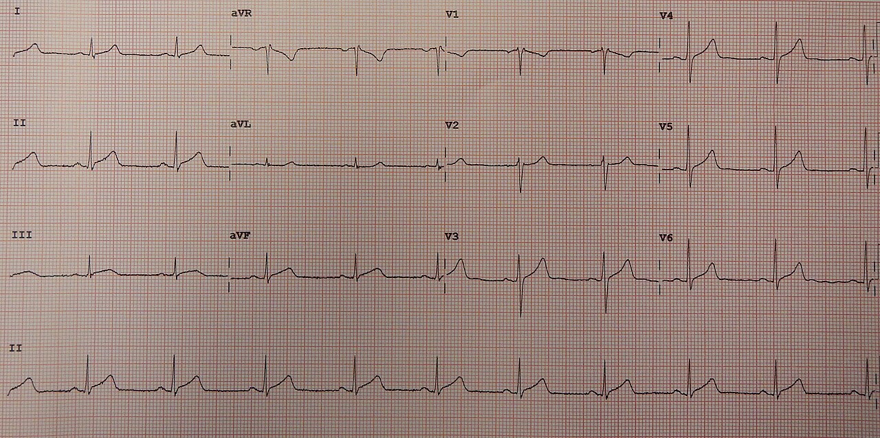
- Diffuse STE, no reciprocal depression: pericarditis or early repolarization
- Deep S with STE in V1–V3: LVH
- Persistent STE with Q or QS waves: LV aneurysm
- Red Flags
- Regional STE with reciprocal depression; ST change that is new compared with a prior ECG; takotsubo still needs angiography to exclude occlusion.
- Bedside Action
- Treat a regional or new pattern per the reperfusion protocol. For a diffuse pattern, use history, troponin, serial ECGs and echo to separate the mimics.
Physiology & What It Represents
The ST segment and T wave record ventricular recovery. They shift for reasons other than coronary occlusion. An acute occlusion hurts one coronary territory, so the injury shows up as regional ST elevation (STE), often with reciprocal ST depression (STD) on the opposite wall. Most mimics do not have that geometry.
Five mechanisms produce the mimics on this page. Each one leaves a different fingerprint.
- Epicardial inflammation (pericarditis): the inflamed pericardium overlies the whole ventricle. That makes the ST shift diffuse, not confined to one coronary territory.
- Early J-point elevation (early repolarization): a notch or slur at the end of the QRS lifts the J point. It shows up most often in the inferior and lateral leads.
- Increased muscle mass (LVH): thicker walls raise QRS voltage. They also shift repolarization, so the leads that face the thick wall show secondary ST and T changes.
- Old scar (LV aneurysm): a thinned, dyskinetic segment can hold ST elevation. It stays on serial ECGs after the infarction.
- Stress-related wall motion (takotsubo): a transient left ventricular dysfunction. It often extends past one coronary territory, and that can change the ST segment and T waves.
Normal Values & How to Measure
Measure at the J point, where the QRS ends, against the TP baseline. Every threshold depends on that spot. Amplitudes are in mm (1 mm = 0.1 mV). Read the values below and decide: mimic, or occlusion signal?
| Parameter | Adult Threshold | Pediatric Note | How to Measure |
|---|---|---|---|
| Normal ST segment | Isoelectric at the J point | Adult values; use pediatric references | Every lead, against the TP baseline |
| Early repolarization | J point ≥0.1 mV (≥1 mm) in at least 2 contiguous inferior and/or lateral leads; STE not required | Common in adolescents and young adults; adult thresholds are not validated in children | Peak of the end-QRS notch or onset of the slur |
| STE (ischemic) | ≥1 mm in 2 contiguous leads; V2–V3 ≥2.5 mm men under 40, ≥2 mm men 40 and over, ≥1.5 mm women | Adult criteria | J point; not valid with LVH or LBBB |
| STD (ischemic) | Horizontal or down-sloping ≥0.5 mm at the J point in 2 or more contiguous leads | Adult criteria | J point, every lead |
| LVH voltage | Sokolow-Lyon S(V1) + R(V5 or V6) ≥35 mm; Cornell R(aVL) + S(V3) >28 mm men, >20 mm women | Age-specific; use pediatric references | Standard gain (10 mm/mV) |
| PR segment (pericarditis) | PR depression, most marked in II and V5–V6, with PR elevation in aVR | Confirm with pediatric cardiology | Baseline between P wave and QRS |
- Find the J point: locate where the QRS ends in each lead. An early repolarization notch or slur sits at this point.
- Set the baseline: use the TP segment. Note any PR depression, because it is a separate pericarditis clue.
- Check V1–V3 separately: compare with the sex and age thresholds before calling STE in V2–V3 ischemic.
- Look for reciprocal depression: check the opposite wall, and add posterior leads (V7–V9) when V1–V3 depression suggests posterior injury.
- Calculate voltage first: work out Sokolow-Lyon and Cornell before interpreting STE in V1–V3, because LVH changes the expected pattern.
- Compare with prior ECGs: a change from the patient's baseline matters more than any single value.
- Repeat the ECG when unsure: obtain serial 12-leads every 15–30 min during ongoing chest pain.
Acute Pericarditis
Acute pericarditis is inflammation of the pericardium. The ECG shows it as diffuse ST elevation across many leads. That elevation is usually concave, and it does not stay inside one coronary territory.
ECG Criteria
- ST segment: diffuse STE across most leads, not a single coronary territory
- Reciprocal change: no reciprocal STD that matches one territory
- PR segment: PR depression, most marked in II and V5–V6, with PR elevation in aVR (see PR Segment)
- Course: ST and T changes evolve over days to weeks; serial ECGs show the course
Causes
- Viral or idiopathic: the usual primary presentations
- After myocardial infarction or cardiac surgery: the pattern can show up after an infarct or a procedure
- Systemic or inflammatory: autoimmune disease and uremia
Clinical Significance
- Diagnosis: the ECG pattern, a compatible history and imaging support pericarditis. The ECG alone does not rule out MI.
- Treatment: aim anti-inflammatory therapy at the cause, per the 2025 ESC and ACC statements. Drug choice and doses sit outside this page.
- Escalation: a suspected effusion with hemodynamic compromise is a separate emergency for the receiving team.
Look-Alikes
- Early repolarization: a J-point notch with a stable pattern. The comparison table below sorts it out.
- Myocarditis also produces diffuse STE. The 2026 Universal Definition lists it alongside pericarditis.
Early Repolarization
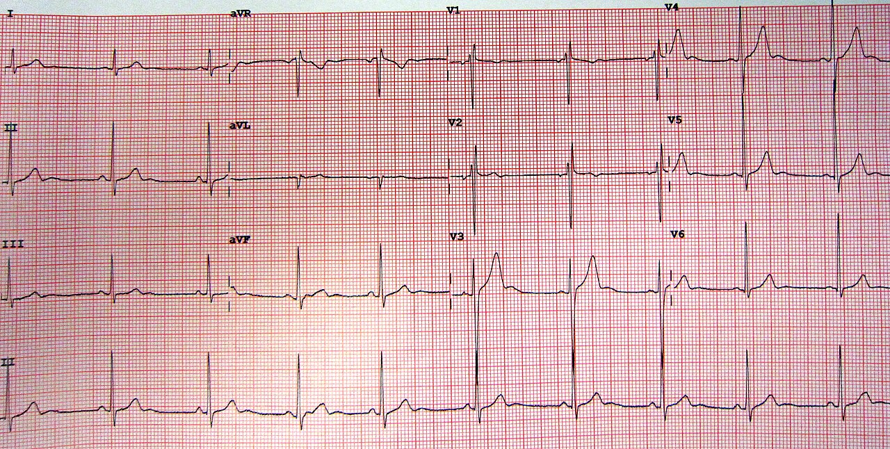
Think of it as a J point that rides up. The notch or slur at the QRS end is the giveaway, most often in the inferior and lateral leads. It is a common repolarization variant. How worried you should be depends on three things: how high the J point sits, which leads show it, and where the patient is.
ECG Criteria
- J point: ≥0.1 mV (≥1 mm) at the peak of an end-QRS notch or the onset of a slur. ST elevation is not required.
- Leads: at least 2 contiguous inferior and/or lateral leads (V1–V3 excluded)
- Shape: concave ST segment that follows the notch or slur
Causes
- Normal variant: plenty of healthy people carry this pattern. Classify it as a finding. On its own, it is not a diagnosis.
Clinical Significance
- Risk by amplitude: amplitude matters. In a 30-year community cohort, inferior J-point elevation of at least 0.1 mV carried a higher cardiac-death risk. Go above 0.2 mV and the risk of cardiac and arrhythmic death climbs markedly.
- Acute setting: early repolarization can look like a subtle anterior STEMI. The derivation study found that STEMI had lower R-wave amplitude, more ST elevation and a longer QTc. A separate validation set separated the two groups with an equation (1.196 × J+60 ms ST elevation in V3 [mm] + 0.059 × QTc [ms] − 0.326 × R wave in V4 [mm]). Above 23.4, it favors STEMI. Two urban centers derived it, retrospectively. Call it a research adjunct, never a bedside rule. Do not use it to clear a pattern that meets the clinical concern.
- Follow-up: new or bigger than the prior ECG? Get cardiology review per local practice.
Look-Alikes
- Pericarditis: diffuse STE with PR depression. The comparison table below shows the difference.
- Subtle anterior STEMI: compare with prior ECGs and serial tracings.
Left Ventricular Hypertrophy (LVH) and Strain
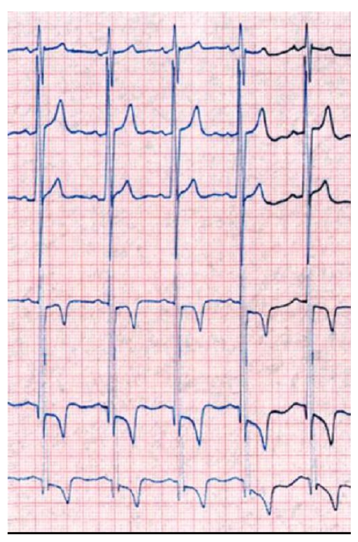
A thick left ventricle shows up through the leads that face it. Voltage goes up. Repolarization shifts with it, so the ST and T waves change in response. That strain pattern can pass for ischemia.
ECG Criteria
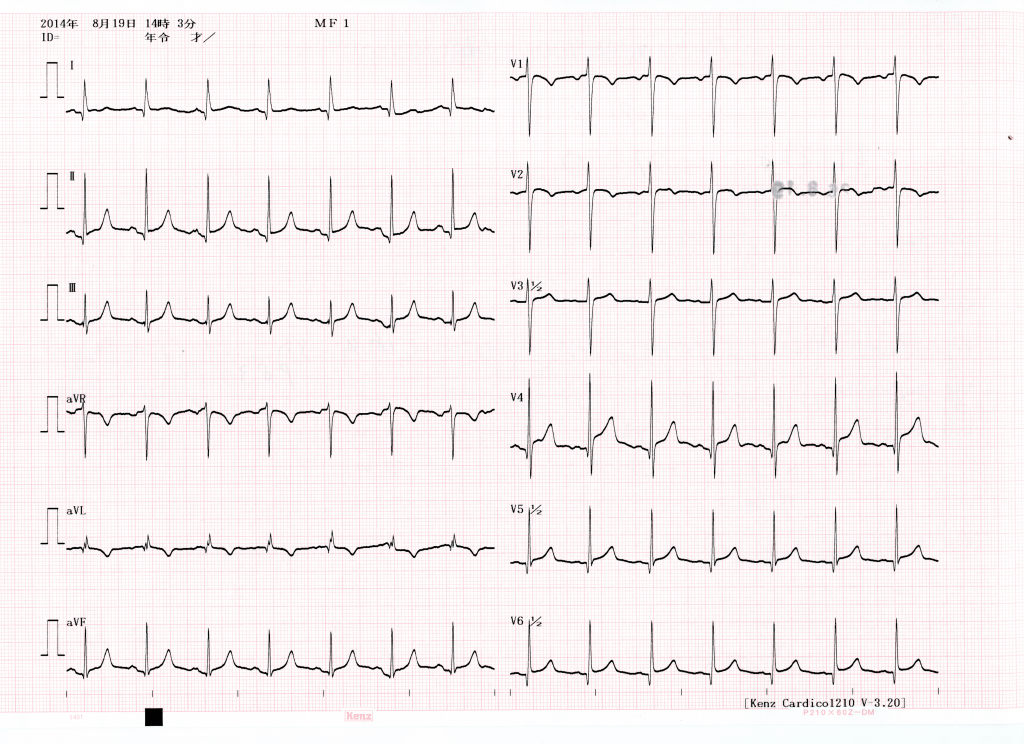
- Sokolow-Lyon: S in V1 plus R in V5 or V6 ≥35 mm
- Cornell: R in aVL plus S in V3 >28 mm in men, >20 mm in women
- Strain: asymmetric ST depression with T-wave inversion in the lateral leads (I, aVL, V5–V6), opposite the dominant QRS direction
- Anterior leads: STE in V1–V3 can follow a deep S wave. The STEMI thresholds do not apply with LVH.
Causes
- Pressure load: long-standing hypertension and aortic valve disease
- Myocardial disease: hypertrophic cardiomyopathy, and infiltrative disease such as Fabry disease, which can show voltage with strain (see Figure 3)
Clinical Significance
- Baseline: got an old ECG showing LVH? Then the pattern is chronic. Lay today's tracing next to it.
- Acute chest pain: strain does not clear the patient. New STE or STD in a new distribution still means serial ECGs and the reperfusion pathway.
- Imaging: the echo is where wall thickness and chamber size get measured.
Left Ventricular Aneurysm
After a large myocardial infarction, a thinned and dyskinetic segment can persist as an aneurysm. Over the scar, the ECG shows ST elevation that stays, plus Q or QS waves.
ECG Criteria
- ST segment: persistent STE that is usually convex in the leads over the scar and does not resolve on serial ECGs
- Q waves: Q or QS waves in the same leads, reflecting the old infarct (see Q Wave)
- Comparison: a stable pattern that matches a prior ECG favors an old scar
Causes
- Prior infarction: an earlier myocardial infarction, most often in the anterior wall
Clinical Significance
- Confirmation: non-invasive imaging identifies a left ventricular aneurysm. The ECG suggests one. It does not confirm one.
- Acute event or old scar: STE that is new, or that has changed from a prior ECG, gets managed as an acute event. That holds until the history and the comparison settle it.
- Handoff: tell the receiving team about a known aneurysm, with the date of the infarct.
Look-Alikes
- Acute anterior STEMI: a new pattern without Q waves. Compare with the prior ECG.
- Pericarditis: diffuse STE without Q waves. See Acute Pericarditis.
Takotsubo Syndrome
Takotsubo syndrome is a transient left ventricular dysfunction, often with apical ballooning. A stress trigger usually sets it off, either emotional or physical, or a neurologic event. It mimics acute coronary syndrome.
ECG Criteria
- New ECG changes: ST elevation, ST depression, T-wave inversion and QTc prolongation. Rare cases have no ECG change.
- Wall motion: usually extends beyond a single epicardial territory. Apical, midventricular, basal and focal patterns occur.
- Biomarkers: troponin is moderately elevated in most cases; creatine kinase is typically only slightly raised; natriuretic peptide is often significantly elevated.
Causes
- Emotional or physical trigger: common, but not required
- Neurologic events: subarachnoid hemorrhage, stroke or transient ischemic attack, and seizures
- Catecholamine excess: pheochromocytoma
Clinical Significance
- Coronary disease: significant coronary disease does not rule takotsubo out. Angiography with left ventriculography is the gold standard to exclude or confirm it.
- Complications: heart failure, pulmonary edema, stroke, cardiogenic shock and cardiac arrest can be the first problem you see.
- QTc and torsades: QTc prolongation is common at presentation. It can progress to torsades de pointes. Check the QTc on serial ECGs, and avoid QT-prolonging drugs.
- Imaging first: in unstable patients, echocardiography for LV outflow-tract obstruction and angiography are used to rule out acute MI.
- Vasoactive drugs: LV outflow-tract obstruction occurs in about 20% of patients. In that setting, and in cardiogenic shock, medical direction decides the inotrope and catecholamine choice. Levosimendan has been suggested as an alternative inotrope.
- Who: postmenopausal women are predominantly affected. Cases have also been described in children, including a premature neonate.
Look-Alikes
- Acute STEMI: a regional wall-motion pattern that matches one artery. Takotsubo often crosses territories.
- Neurogenic T-wave changes follow an intracranial event. See the T Wave page.
Differential Diagnosis & Mimics
The 2026 Fifth Universal Definition of MI puts diffuse STE with no reciprocal change in pericarditis and early repolarization. That pattern is the tell. A territorial occlusion does not look like it. Each table pits a mimic against the pattern it most resembles.
| Feature | Acute STEMI | Pericarditis | Early Repolarization |
|---|---|---|---|
| Distribution | Regional, one coronary territory | Diffuse, across most leads | Diffuse or global; inferior and lateral J points |
| Reciprocal depression | Usually present | Absent as a territorial pattern; PR elevation in aVR is expected | Absent |
| QRS-end notch or slur | Not part of the pattern | Not part of the pattern | Present; defines the pattern |
| PR segment | Not the main feature | PR depression, PR elevation in aVR | Not the main feature |
| Over time | Evolves with the occlusion | Changes over days to weeks | Stable; compare with prior ECG |
Takotsubo, LV aneurysm and acute STEMI share a regional wall-motion or ST story. Put them side by side:
| Feature | Takotsubo | Acute STEMI | LV Aneurysm |
|---|---|---|---|
| Wall motion | Often crosses coronary territories | Regional, matches the culprit | Fixed segment over an old scar |
| ST pattern | STE, STD, T inversion, QTc prolongation | Regional STE with reciprocal change | Persistent STE, usually convex, with Q or QS |
| Trigger | Emotional, physical or neurologic | Coronary occlusion | Prior myocardial infarction |
| Confirmation | Echo wall motion; angiography excludes culprit disease | Angiography shows occlusion | Imaging shows aneurysm; prior ECG shows Q waves |
Artifact & Lead Misplacement
- Limb lead reversal: swapped arm leads scramble the lead I, II and aVR patterns. That can fake an axis shift or an inferior change (see Cardiac Axis).
- Precordial placement: leads sitting high or low move the R and S waves around. Hypertrophy or a bundle branch block can appear out of nowhere (see QRS Complex).
- Tremor and transport vibration: shaking can draw ST artifact and wide, bizarre complexes. Confirm on a second lead before acting.
Other Mimics
- Hyperkalemia: the trace can imitate STE or T-wave change. See Electrolyte Abnormalities on the ECG.
- Brugada pattern, hypothermia (Osborn J waves) and right bundle branch block: each of these can fake an infarct with an ST-elevation or J-point pattern. Look at the morphology, the context and the prior tracing.
Bedside Management & Escalation
Start with one question: is this an occlusion? A regional or new pattern takes the reperfusion pathway per local protocol. In the US, the 2025 ACC/AHA/ACEP/NAEMSP/SCAI ACS guideline sits behind that pathway. A diffuse pattern with no reciprocal change earns a mimic only after history, troponin, serial ECGs and imaging agree.
- Pericarditis: the cause and the inflammation get treated per the 2025 ESC and ACC statements. An ECG pattern on its own does not prove occlusion.
- Takotsubo: handle the complications (heart failure, shock, arrhythmia) per protocol, and watch the QTc. In shock or outflow-tract obstruction, the vasoactive choice belongs to medical direction. Angiography still has to exclude or confirm it.
- LV aneurysm: check the prior ECGs. New or changed means it runs as an acute event.
- Early repolarization: a stable pattern that matches the prior ECG is not an acute finding alone. With ischemic symptoms, a new or larger J-point or ST pattern against the prior ECG runs as a possible occlusion until excluded, per protocol and medical direction.
Stack up serial 12-leads, a troponin trend and an echo. When V1–V3 or the inferior leads raise the question, add posterior or right-sided leads (see STEMI Recognition). Follow local protocols and medical direction.
Pediatric & Special Populations
Pediatrics
- Adult thresholds: the STE, STD and LVH values on this page are adult values. For age-specific voltage and repolarization, apply pediatric references (see Vital Signs).
- Takotsubo: it has been described in children, including a premature neonate born at 28 weeks gestation.
- Pericarditis and myocarditis: bring pediatric cardiology in early. The same mimic check applies.
Other Populations
- LBBB or ventricular pacing: use the Sgarbossa criteria. The Smith-modified version scores discordant STE with an ST/S ratio of 25% or more (see Sgarbossa & Smith-Modified Criteria).
- LVH: the STEMI thresholds are unreliable here. Compare with a prior ECG, and lean on serial ECGs and troponin (see STEMI Equivalents).
- Older adults after infarction: a remote infarct raises the chance of an aneurysm. Pull up the prior ECG and the echo.
Monitoring & Transport Considerations
- Serial 12-leads: repeat every 15–30 min during ongoing chest pain. Send the prior ECG along for comparison.
- Single lead: one monitor lead cannot show the full ST pattern. Get a 12-lead before you judge the ST segment.
- Right-sided and posterior leads: V4R and V7–V9 when the pattern suggests right ventricular or posterior involvement, per protocol.
- Artifact: shivering, movement or vibration can distort the trace. Confirm on a second lead.
- Takotsubo: watch for arrhythmia and deterioration. Heart failure, shock and arrest can be how it presents.
- Handoff: send the ECG with the time of onset, the prior ECG and the mimic you suspect.
Step-by-Step Approach & Pitfalls
- Check lead placement and artifact before you interpret any ST change.
- Measure the J point and T wave in every lead against the TP baseline.
- Decide whether the ST change is regional or diffuse, and whether reciprocal depression is present.
- Calculate Sokolow-Lyon and Cornell voltage to see whether LVH changes the picture.
- Look for a notch or slur at the QRS end and measure the J-point elevation.
- Compare with every prior ECG and note the time of onset.
- Name the most likely mimic and the one finding that would change management, then act per protocol.
Common Pitfalls
- Calling diffuse STE pericarditis without checking reciprocal depression and prior ECGs.
- Calling early repolarization benign without measuring the J point.
- Applying STEMI thresholds in LVH or LBBB.
- Accepting a chronic LV aneurysm pattern without comparing a prior ECG.
- Assuming takotsubo rules out coronary disease. Angiography decides.
ECG Examples
Clinical Pearls
References
- Mills NL, Newby LK, Zaman S, et al. Fifth Universal Definition of Myocardial Infarction (2026). Global Heart. 2026;21(1):64. https://doi.org/10.5334/gh.1578
- Schulz-Menger J, Collini V, Gröschel J, et al. 2025 ESC Guidelines for the management of myocarditis and pericarditis. Eur Heart J. 2025;46(40):3952–4041. https://doi.org/10.1093/eurheartj/ehaf192
- Wang TKM, Klein AL, Cremer PC, et al. 2025 Concise Clinical Guidance: An ACC Expert Consensus Statement on the Diagnosis and Management of Pericarditis. J Am Coll Cardiol. 2025;86(25):2691–2719. https://doi.org/10.1016/j.jacc.2025.05.023
- Ghadri JR, Wittstein IS, Prasad A, et al. International Expert Consensus Document on Takotsubo Syndrome (Part I): Clinical Characteristics, Diagnostic Criteria, and Pathophysiology. Eur Heart J. 2018;39(22):2032–2046. https://doi.org/10.1093/eurheartj/ehy076
- Tikkanen JT, Anttonen O, Junttila MJ, et al. Long-term outcome associated with early repolarization on electrocardiography. N Engl J Med. 2009;361(26):2529–2537 (landmark cohort). https://doi.org/10.1056/NEJMoa0907589
- Macfarlane PW, Antzelevitch C, Haissaguerre M, et al. The early repolarization pattern: a consensus paper. J Am Coll Cardiol. 2015;66(4):470–477 (consensus definition). https://doi.org/10.1016/j.jacc.2015.05.033
- Smith SW, Khalil A, Henry TD, et al. Electrocardiographic differentiation of early repolarization from subtle anterior ST-segment elevation myocardial infarction. Ann Emerg Med. 2012;60(1):45–56.e2 (derivation study). https://doi.org/10.1016/j.annemergmed.2012.02.015
- Hancock EW, Deal BJ, Mirvis DM, et al. AHA/ACCF/HRS recommendations for the standardization and interpretation of the electrocardiogram: part V: electrocardiogram changes associated with cardiac chamber hypertrophy. J Am Coll Cardiol. 2009;53(11):992–1002 (landmark standard). https://doi.org/10.1016/j.jacc.2008.12.015
- Rao SV, O'Donoghue ML, Ruel M, et al. 2025 ACC/AHA/ACEP/NAEMSP/SCAI Guideline for the Management of Patients With Acute Coronary Syndromes. J Am Coll Cardiol. 2025;85(22):2135–2237. https://doi.org/10.1016/j.jacc.2024.11.009
- Ghadri JR, Kato K, Cammann VL, et al. International Expert Consensus Document on Takotsubo Syndrome (Part II): Diagnostic Workup, Outcome, and Management. Eur Heart J. 2018;39(22):2047–2062. https://doi.org/10.1093/eurheartj/ehy077
Medical Disclaimer
- For Educational Purposes Only: This content is intended for educational reference and should not be used for clinical decision-making.
- Not a Substitute for Professional Judgment: Always consult your local protocols, institutional guidelines, and supervising physicians.
- Verify Before Acting: Users are responsible for verifying information through authoritative sources before any clinical application.