At a Glance

Why It's Ordered
To classify acid-base disorders, judge ventilation and oxygenation, and check tissue perfusion in ventilated, unstable or critically ill patients. It guides immediate airway, ventilator and resuscitation decisions.
Check First
pH, PaCO₂ and HCO₃⁻ together classify the disorder; PaO₂ and SaO₂ show oxygenation against the FiO₂ used; lactate shows hypoperfusion. Read the FiO₂ on the request.
Critical Values
pH <7.20 or >7.60 · PaCO₂ <20 or >70 mmHg · PaO₂ <40 mmHg · HCO₃⁻ <10 or >40 mmol/L · Lactate ≥4 mmol/L · COHb >25% (typical)
Don't Miss
A normal SpO₂ or PaO₂ does not exclude carbon monoxide poisoning or methemoglobinemia. Measure COHb and MetHb by co-oximetry whenever exposure is possible. A normal pH can also hide two opposing disorders.
Field & Transport
Point-of-care blood gas cartridges report pH, PCO₂, PO₂ and calculated values; lactate and ionized calcium depend on the cartridge. Send the sample promptly, record the FiO₂ and the time oxygen was started, and repeat en route only when the result will change treatment.

Panel Overview

The arterial blood gas (ABG) panel measures pH, PaCO₂, PaO₂ and SaO₂ from one arterial sample. HCO₃⁻ and base excess are calculated, and most analyzers also report lactate. Co-oximetry models add carboxyhemoglobin (COHb) and methemoglobin (MetHb). Six core values (pH, PaCO₂, PaO₂, SaO₂, HCO₃⁻ and base excess) are reported from the gas. Lactate is a seventh value on most analyzers.

The panel is drawn in emergency departments, ICUs, operating rooms and during transfers. Venous blood gas, ionized calcium and anion gap are common add-ons: they do not replace the arterial sample when oxygenation or ventilation is in question.

When to Order:
  • Shock or sepsis: lactate and acid-base status to guide resuscitation
  • Altered mental status or unexplained tachypnea: acidemia, hypercapnia or hypoxemia
  • Respiratory failure or ventilator changes: PaO₂ for oxygen need, PaCO₂ and pH for ventilation
  • DKA, toxic ingestion or poisoning: metabolic acidosis and the cause
  • Suspected carbon monoxide or methemoglobin exposure: co-oximetry
  • Pre-transfer or pre-procedure check: a baseline before transport or intubation

Acid-Base

pH

Normal: 7.35–7.45 (arterial)

Hydrogen ion concentration and overall acid-base status. Acidemia below 7.35, alkalemia above 7.45. Direction only: the cause is found in PaCO₂ and HCO₃⁻.

Learn More

PaCO₂

Normal: 35–45 mmHg (arterial)

Respiratory component of acid-base balance and a direct measure of ventilation. Raised in hypoventilation, low in hyperventilation.

Learn More

HCO₃⁻ (Bicarbonate)

Normal: 22–26 mmol/L (arterial calculated)

Metabolic component of acid-base balance. Low in metabolic acidosis; high in metabolic alkalosis or chronic respiratory acidosis.

Learn More

Base Excess/Deficit

Normal: −2 to +2 mmol/L

Calculated metabolic component independent of PaCO₂. Negative values indicate a metabolic acidosis or deficit; positive values an alkalosis or excess.

Learn More

Oxygenation

PaO₂

Normal: 80–100 mmHg (room air)

Oxygen dissolved in arterial blood; reflects gas exchange. Interpret with the FiO₂ used. Below 60 mmHg is respiratory failure in most adults.

Learn More

SaO₂

Normal: 95–100%

Percentage of hemoglobin saturated with oxygen, measured by co-oximetry. It is not the same as pulse-oximetry SpO₂, and it misses CO and methemoglobin effects.

Learn More

P/F Ratio (PaO₂/FiO₂)

Expected: about 380–480 (room air, FiO₂ 0.21)

PaO₂ divided by fractional inspired oxygen. Lower values mean worse oxygenation. The 2024 global ARDS definition uses 300 or less, with its support criteria.

Learn More

Co-Oximetry & Metabolites

Lactate

Normal: 0.5–1.5 mmol/L; raised above 2

Marker of tissue hypoperfusion. Raised above 2 mmol/L (1.5–2.0 sits above the textbook 0.5–1.5 range but below the sepsis flag; many lab ranges run to 2.2); 4 mmol/L or more means shock. Trend it during resuscitation.

Learn More

Carboxyhemoglobin (COHb)

Normal: <3% (nonsmoker); up to 10% (smoker)

Hemoglobin bound to carbon monoxide. Pulse oximetry cannot separate it from oxyhemoglobin. Draw co-oximetry early; severity is judged clinically as well as by value.

Learn More

Methemoglobin (MetHb)

Normal: about 1% (lab-dependent)

Hemoglobin with oxidized iron that cannot carry oxygen. Causes a low SaO₂ with a normal PaO₂. Treat if symptomatic or above 30% (common threshold; protocol-dependent). Methylene blue can make SpO₂ read falsely low.

Learn More

Calculated Values

Anion Gap

Normal: 8–12 mEq/L (without K⁺; analyzer-dependent)

Na⁺ minus (Cl⁻ plus HCO₃⁻), from a chemistry panel. Raised in high-anion-gap acidosis: lactic acidosis, ketoacidosis, toxic alcohols, salicylate, uremia.

Learn More

Common Add-Ons

Venous Blood Gas (VBG)

Normal: pH 7.31–7.41; pCO₂ 41–51 mmHg

Venous pH and HCO₃⁻ track arterial values closely; venous pCO₂ runs about 6 mmHg higher and agrees poorly with PaCO₂ in individuals. Not a substitute for PaO₂.

Learn More

Ionized Calcium (iCa²⁺)

Normal: 1.12–1.32 mmol/L (4.5–5.3 mg/dL)

Physiologically active calcium. Low values cause tetany, seizures and arrhythmia; check it with K⁺ in acidemia, alkalemia and transfusion.

Learn More

ScvO₂ / SvO₂

Normal: SvO₂ 65–75%

Venous saturation from a central line (ScvO₂) or pulmonary artery catheter (SvO₂). A low value warns that delivery is not meeting demand, and a normal one does not prove perfusion.

Learn More

Reference Ranges & Critical Values

ABG reference ranges, arterial and venous, with critical values
TestArterial NormalVenous NormalSI UnitsCriticalNotes
pH
7.35–7.45
7.31–7.41
[H⁺] 35–45 nmol/L (arterial)
<7.20 or >7.60
Venous about 0.04 lower
PaCO₂ / PvCO₂
35–45 mmHg
41–51 mmHg
4.7–6.0 kPa (arterial)
<20 or >70 mmHg
Venous poorly predicts arterial
HCO₃⁻
22–26 mmol/L
About the same as arterial
22–26 mmol/L
<10 or >40 mmol/L
Calculated on arterial samples
Base excess
−2 to +2 mmol/L
Similar to arterial
−2 to +2 mmol/L
—
Lab-dependent for venous
PaO₂
80–100 mmHg (room air)
Not used
10.7–13.3 kPa
<40 mmHg
Falls with age; record FiO₂
SaO₂
95–100%
Not used
0.95–1.00
—
Co-oximetry measured value
Lactate
0.5–1.5 mmol/L
0.5–1.5 mmol/L (venous plasma)
0.5–1.5 mmol/L
≥4 mmol/L
Raised above 2 mmol/L
COHb
<3% (smoker up to 10%)
Not used
<0.03 fraction
>25% (typical; severe)
Pregnancy: lower threshold
MetHb
About 1%
Not used
About 0.01 fraction
Treat if symptomatic or >30% (protocol)
Co-oximetry; SpO₂ reads low after dose
Ionized Ca²⁺
1.12–1.32 mmol/L
Lab-dependent
1.12–1.32 mmol/L
<0.8 or >1.6 mmol/L (typical)
Calibrate to pH
Anion gap
8–12 mEq/L
Not used
8–12 mmol/L
—
Analyzer-dependent; no K⁺
Lab variation: Every lab draws its own line. Intervals and critical lists change with the lab, the analyzer and the assay. Use your lab's reported interval and its critical-call policy. The critical values on this page are typical, not universal.

Systematic Interpretation

  1. Check the sample and the patient: arterial or venous, FiO₂ and device, time oxygen started, temperature, and whether the value was reported as measured or calculated.
  2. Acidemia or alkalemia: Below 7.35 is acidemia. Above 7.45 is alkalemia.
  3. Primary disorder: The primary cause pushes the pH in its own direction. Low HCO₃⁻ with a low pH is metabolic acidosis. High PaCO₂ with a low pH is respiratory acidosis. Flip the pattern and you get alkalosis.
  4. Compensation: Watch the second value. Did it move as expected? When the PaCO₂ lands outside the Winter's range, a second disorder is hiding in the picture.
  5. Anion gap for metabolic acidosis: A raised gap points toward lactate, ketones, toxic alcohols, salicylate or uremia. A normal gap with low HCO₃⁻ points elsewhere, to a non-gap cause such as diarrhea or renal tubular acidosis.
  6. Oxygenation: Never judge the PaO₂ alone. Set it against the FiO₂ you gave (P/F ratio) and against the expected alveolar-arterial gradient for the patient's age.

Compensation and Oxygenation Formulas

Winter's formula (metabolic acidosis)
Expected PaCO₂ = 1.5 × HCO₃⁻ + 8 ± 2 mmHg

The normal compensation band runs from 1.5 × HCO₃⁻ + 6 to + 10 mmHg. Above that band, think concurrent respiratory acidosis. Below it, think concurrent respiratory alkalosis.

Alveolar gas equation and A–a gradient
PAO₂ = FiO₂ × (Patm − 47) − PaCO₂ / 0.8; A–a gradient = PAO₂ − PaO₂

At sea level on room air, the expected A–a gradient is about age ÷ 4 + 4 mmHg. Wider than that? Think shunt, ventilation–perfusion mismatch or diffusion limitation.

P/F ratio
PaO₂ ÷ FiO₂ (for example, 96 mmHg ÷ 0.21 = 457)

The Berlin definition grades ARDS by P/F ratio, with PEEP of at least 5 cmH₂O. Mild: above 200 to 300. Moderate: above 100 to 200. Severe: 100 or less mmHg. The 2024 global definition is different. It uses a P/F of 300 or less, or a SpO₂/FiO₂ of 315 or less when SpO₂ is 97% or less. Support counts too: HFNO at 30 L/min or more, CPAP or NIV at 5 cmH₂O or more, or PEEP of at least 5 cmH₂O when intubated.
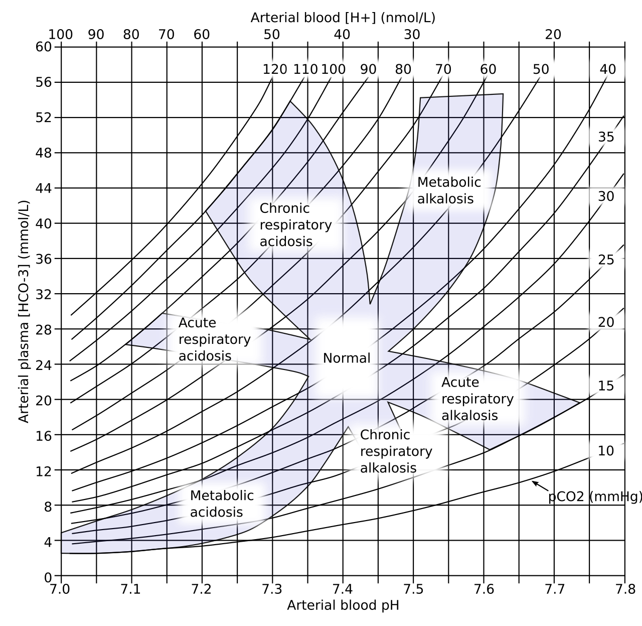
Acid-base nomogram plotting arterial pH against plasma bicarbonate, with PaCO₂ lines and bands for simple disorders
Figure 1: Acid–base nomogram: arterial pH against plasma HCO₃⁻, with PaCO₂ lines and a band for each simple disorder. A result outside every band suggests a mixed disorder. Source: Wikimedia Commons, Huckfinne, public domain
Worked Example: Take a 50-year-old on room air. Arterial pH 7.28, PaCO₂ 30 mmHg, HCO₃⁻ 14 mmol/L, PaO₂ 96 mmHg, Na⁺ 140, Cl⁻ 100 mEq/L. Low pH and low HCO₃⁻ point to metabolic acidosis as the primary disorder. Expected PaCO₂ is 1.5 × 14 + 8 = 29 ± 2 mmHg (27–31). At 30 mmHg, that is appropriate compensation. Anion gap: 140 − (100 + 14) = 26 mEq/L. Raised. The A–a gradient is about 112 − 96 = 16 mmHg. That falls within the expected value for age 50 (about 16.5 mmHg). Your next checks: lactate, ketones and glucose, plus toxicology for salicylate and toxic alcohols.

Patterns & Differential

Check each pattern against pH, PaCO₂, HCO₃⁻, the anion gap and PaO₂. Mixed disorders are common in critical illness. Start with the simple patterns in the table, then look for the second one.

PatternKey FindingsCommon CausesNext Step
Acute respiratory acidosis
pH low, PaCO₂ high, HCO₃⁻ near normal
Opioid or sedative overdose, COPD or asthma exacerbation, neuromuscular weakness, airway obstruction
Support ventilation; find and treat the cause; naloxone if opioid toxicity
Chronic respiratory acidosis
PaCO₂ high, HCO₃⁻ high, pH near normal
COPD, obesity hypoventilation, chronic neuromuscular disease
Correct ventilation slowly; a rapid PaCO₂ fall causes alkalemia
High-anion-gap metabolic acidosis
pH low, HCO₃⁻ low, gap raised
Lactic acidosis (sepsis, shock), DKA, toxic alcohols, salicylate, uremia
Lactate, ketones, glucose, toxicology, creatinine
Normal-gap metabolic acidosis
pH low, HCO₃⁻ low, gap normal, chloride high
Diarrhea, renal tubular acidosis, large-volume chloride-rich fluid
Urine electrolytes, stool losses, fluid review
Metabolic alkalosis
pH high, HCO₃⁻ high, PaCO₂ compensating upward
Vomiting or NG suction, diuretics, low K⁺
Volume status, K⁺, chloride, medication review
Respiratory alkalosis
pH high, PaCO₂ low
Hyperventilation from hypoxemia, PE, sepsis, pain, anxiety; early salicylate toxicity
Treat the trigger; check PaO₂ and lactate
Hypoxemia with normal PaCO₂
PaO₂ low, PaCO₂ normal or low
Ventilation–perfusion mismatch, shunt, diffusion limitation
Check P/F ratio, chest imaging, PEEP and FiO₂
Low SaO₂ with normal PaO₂
SaO₂ low, PaO₂ normal
Carboxyhemoglobin (reads as oxyhemoglobin on pulse oximetry), methemoglobin
Co-oximetry for COHb and MetHb; oxygen for CO; methylene blue for MetHb per protocol

Serial Testing & Kinetics

AnalyteRises or ChangesPeaksRepeat IntervalMonitoring Target
PaO₂ / SaO₂
Changes within minutes of an FiO₂, PEEP or ventilator change
—
After each change, per unit protocol
Target set by the ventilator or ARDS protocol
PaCO₂ and pH
Move with minute ventilation; a rate or tidal volume change shifts both
—
After each ventilator change, per unit protocol
Stable or improving pH while PaCO₂ falls gradually
Lactate
Rises with hypoperfusion
—
Repeat when the initial value is above 2 mmol/L, per sepsis protocol
Falling lactate guides resuscitation
MetHb
Falls after methylene blue
—
One hour after the first dose if MetHb stays above 30% or symptoms persist (label repeat dose 1 mg/kg)
Below 30% and symptoms resolved
COHb
Falls with oxygen; the rate depends on FiO₂
—
Repeat only if the result will change management
Clinical recovery; severity is judged clinically

Using the ABG to Adjust the Ventilator

  • Ventilation: Never read PaCO₂ alone. Read it with the pH. A falling pH with a rising PaCO₂ means ventilation is not keeping up. Raise minute ventilation per protocol.
  • Chronic hypercapnia: The HCO₃⁻ runs high. Drop the PaCO₂ fast and the pH climbs (post-hypercapnic alkalemia). Correct it gradually, then recheck the pH.
  • Oxygenation: After every FiO₂ or PEEP change, check the PaO₂ against the FiO₂ (P/F ratio). Titrate to the target in the ventilator or ARDS protocol.
  • Settings: Write the ventilator settings and the FiO₂ on every request. Without them, no value can be read in context.
Full ventilator guidance: The stepwise acid-base method and ventilator management get their own guides, on mechanical ventilation and acid-base interpretation. Both are planned, and neither is linked from this page yet.

Critical Findings & Emergency Actions

Severe acidemia (pH <7.20)

Recognize: pH below 7.20 with falling blood pressure, dysrhythmia, or altered mental status.

  1. Find and treat the cause: sepsis or shock, DKA, toxic ingestion, or respiratory failure (Paramedic/RN/CCT).
  2. Support ventilation: if PaCO₂ is high, improve alveolar ventilation per protocol (RN/CCT).
  3. Bicarbonate is not routine: the Surviving Sepsis Campaign suggests against bicarbonate to improve hemodynamics in hypoperfusion lactic acidemia. It suggests bicarbonate when pH is 7.2 or lower with stage 2 or 3 AKI. See the sodium bicarbonate page; give only per medical direction.
  4. Check K⁺ and ionized calcium and obtain a 12-lead ECG. In DKA, check K⁺ before insulin and replace it per protocol when low; bicarbonate is reserved for severe acidosis (pH below 6.9 in most guidance).

Acute hypercapnic respiratory failure

Recognize: PaCO₂ rising while pH falls, somnolence, COPD exacerbation, or sedative or opioid overdose.

  1. Secure the airway and ventilation: bag-valve-mask assistance or intubation per protocol (Paramedic/RN).
  2. Opioid toxicity: naloxone per protocol, with ventilation support.
  3. COPD exacerbation: noninvasive ventilation is the recommended support for acute hypercapnic failure (ERS/ATS guideline).
  4. Oxygen target: 88–92% for patients at risk of hypercapnic failure until a blood gas is available (BTS oxygen guideline). Repeat the gas after each change.

Refractory hypoxemia

Recognize: PaO₂ falling despite higher FiO₂, low P/F ratio, or SpO₂ that does not respond to oxygen.

  1. Check the device: tube position, circuit leaks, suction, and oxygen supply (RN/CCT).
  2. Recruit and titrate: PEEP and FiO₂ per the ventilator protocol; consider prone positioning per ARDS protocol.
  3. Refer early: ECMO referral when protocol criteria are met (see the ECMO overview).

Lactate ≥4 mmol/L or septic shock

Recognize: lactate of 4 mmol/L or more, or hypotension with a rising lactate.

  1. Fluids: IV crystalloid per the sepsis protocol. The Surviving Sepsis Campaign suggests at least 30 mL/kg within the first 3 hours for sepsis-induced hypoperfusion (conditional, low certainty).
  2. Vasopressors: norepinephrine per protocol for persistent hypotension (Paramedic/RN/CCT).
  3. Serial lactate: guide resuscitation with repeat lactate, not a fixed fluid volume alone.

Suspected carbon monoxide poisoning

Recognize: headache, dizziness, nausea or confusion in several people, or an exposure source. SpO₂ can read normal.

  1. Remove from the source and protect responders (Paramedic/CCT).
  2. Give the highest available oxygen concentration per protocol until COHb is known.
  3. Send blood for co-oximetry and record the time oxygen was started.
  4. Refer for hyperbaric oxygen per receiving-center criteria: loss of consciousness, neurologic or cardiac signs, severe acidosis, pregnancy, or very high COHb.

Methemoglobinemia (symptomatic, or MetHb above 30%)

Recognize: SaO₂ or SpO₂ low with a normal PaO₂ that does not rise with oxygen; chocolate-brown blood; dyspnea or altered mental status.

  1. Oxygen and support per protocol (Paramedic/RN/CCT).
  2. Methylene blue (ProvayBlue): High-alert 1 mg/kg IV over 5–30 minutes. If MetHb stays above 30% or symptoms persist, a repeat dose of 1 mg/kg may be given one hour after the first (label). For moderate or severe renal impairment (eGFR 15–59), give a single dose only (label). The label allows dilution in 50 mL D5W; avoid sodium chloride diluent (label).
  3. If no response: consider alternative treatment if MetHb has not resolved after 2 doses. Response may fail or rebound with aniline or dapsone causes (label).
  4. Before dosing: check for SSRIs, SNRIs, MAOIs and opioids (boxed warning; avoid concomitant use), and for G6PD deficiency (contraindicated).
  5. After dosing: methylene blue can make pulse oximetry read falsely low; use co-oximetry on an arterial sample rather than SpO₂ alone (label).
Pitfall: a normal PaO₂ and a normal SpO₂ do not exclude carbon monoxide or methemoglobin. Only co-oximetry measures them.

Prehospital & Transport Considerations

Point-of-Care Testing

Device or MethodAnalytesSpecimenLimitations
Handheld blood gas analyzer (cartridge)
pH, PCO₂, PO₂ and calculated HCO₃⁻, base excess, SO₂; lactate or iCa on some cartridges
Arterial or venous whole blood
Confirm which analytes the cartridge reports; check expiry and storage temperature; follow medical direction for use
Handheld lactate meter
Lactate
Capillary or venous whole blood
Results differ from laboratory plasma values; use the trend and the protocol cut-off
Pulse CO-oximetry (SpCO, SpMetHb)
COHb and MetHb estimates
Noninvasive, fingertip
Screening only; cannot confirm CO poisoning; motion and poor perfusion reduce accuracy; confirm with blood co-oximetry

Interfacility Transfer & Handoff

  • Record: FiO₂ and device, time of sampling, time oxygen was started, and the last ventilator settings
  • Send: the most recent gas and its trend, with lactate, glucose and potassium if available
  • Pending: co-oximetry, lab-only tests and any repeat gas that is due
  • En route: Repeat the gas only when the result will change treatment. Analyze it promptly and follow the lab's stability limits.
  • Altitude: Cabin pressure lowers PaO₂ in flight. Compare with the pre-flight baseline, not a sea-level target.

Scope Notes

  • EMS: Arterial sampling is not routine in most systems. Use it only where protocol and medical direction allow.
  • ED/ICU: draw, analyze and act on the result within the unit's protocol
  • Transport: confirm the analyzer and cartridge before departure; document the value and the time

Special Populations

Pediatrics

Past the newborn period, the pH, PaCO₂ and HCO₃⁻ ranges sit close to adult values. Neonates and infants use the lab's age-specific range. A capillary sample can screen pH and PCO₂, but it does not give a reliable PaO₂. When an oxygenation decision depends on that number, draw an arterial sample.

Pediatric dosing: methylene blue 1 mg/kg IV over 5–30 minutes. The label gives no separate pediatric dose. It cites two pediatric case series with the same safety profile. Repeat only as in the label. See weight-based dosing and pediatric vital signs.

Pregnancy

Progesterone drives hyperventilation, and PaCO₂ falls to about 27–32 mmHg. The compensating HCO₃⁻ sits around 18–22 mmol/L. Near 40 mmHg in pregnancy? That suggests CO₂ retention. Maternal COHb underestimates fetal exposure to carbon monoxide. Keep a low threshold for hyperbaric consultation.

Chronic CO₂ Retainers

Think about COPD or obesity hypoventilation. These patients may carry a high HCO₃⁻ and a breathing drive that leans on hypoxemia. Keep the 88–92% oxygen target until a blood gas is available (BTS oxygen guideline). Re-check the gas after every oxygen change.

Specimen Collection & Interfering Factors

Arterial Puncture

  • Site: The radial artery is the most common choice. Before a radial puncture, check collateral circulation (modified Allen test or ultrasound) per unit protocol. Brachial and femoral sites are the alternatives.
  • Cautions: infection or cellulitis at the site, absent collateral flow, bleeding disorder, or anticoagulation (relative; per protocol and medical direction).
  • Hemostasis: apply direct pressure for at least 5 minutes, longer when anticoagulated; check distal perfusion afterward.
  • Complications: hematoma, arterial spasm or thrombosis, ischemia, nerve injury and infection; watch for bleeding in anticoagulated patients.
  • Temperature: The lab does not routinely apply a temperature correction. Follow its policy.
  • Specimen: Arterial blood goes in a heparinized gas syringe. Use lyophilized heparin only, because excess heparin dilutes the sample.
  • Handling: Expel air bubbles at once and cap the syringe. Analyze promptly, within the lab's stability limit. Delay lowers PO₂ and raises PCO₂, lactate and potassium.
  • Order of draw: The gas syringe is not part of the CLSI GP41 tube sequence (blood cultures, light blue, red or gold, green, lavender, gray). When it is drawn alongside tubes, follow lab policy.
  • Tube colours: Institution-dependent. An ABG uses a heparinized syringe, not a coloured tube.
Error or InterferentEffect on ResultsPrevention
Air bubbles
PO₂ falsely high, PCO₂ falsely low
Expel bubbles immediately and cap
Venous contamination
PO₂ falsely low, PCO₂ falsely high
Confirm arterial sampling; repeat if the result does not fit the patient
Delayed analysis
PO₂ falls; PCO₂, lactate and potassium rise; pH falls
Analyze promptly; follow laboratory stability time
Excess heparin
Dilution lowers HCO₃⁻ and PCO₂; ionized calcium falsely low
Use lyophilized heparin; fill to the required volume
Very high WBC count
In-vitro oxygen consumption lowers PO₂
Analyze promptly; chill a delayed sample only as laboratory policy directs
Carbon monoxide or methemoglobin
Calculated SO₂ and SpO₂ overstate oxygen content
Use co-oximetry when exposure is possible

Clinical Pearls

Field and transport: Put the FiO₂, device and time on every gas. Without all three, you cannot turn the PaO₂ into a P/F ratio or an A–a gradient.
Nursing: Trend pH and PaCO₂ together after every ventilator change. Falling pH plus rising PaCO₂: ventilation is failing. Rising PaCO₂ with a stable pH may be chronic retention, already compensated. Check the baseline before you decide.
Critical care: Bicarbonate is not routine for lactic acidemia. Fix the cause and the perfusion before anything else. The exceptions: pH 7.2 or lower with stage 2 or 3 AKI.
Winter's formula: Outside 1.5 × HCO₃⁻ + 8 ± 2, the PaCO₂ is telling you about a second disorder. That holds even when the pH looks simple.
Oxygenation: Under the 2024 global ARDS definition, a P/F ratio of 300 or less hits the oxygenation threshold, provided the support criteria also apply. Read it with PEEP and imaging, never alone.
Transfer: Send the last gas with its trend and time. One value with no FiO₂ and no time stamp is hard to act on.
Pitfall: A normal pH can hide a mixed disorder. Opposing disorders cancel. Read PaCO₂, HCO₃⁻ and the anion gap every time.
Pitfall: Methylene blue carries a boxed warning for serotonin syndrome with SSRIs, SNRIs, MAOIs and opioids. Check the medication list before the first dose. Then weigh that risk against untreated methemoglobinemia. Methylene blue can also make pulse oximetry read falsely low.

References

  1. Prescott H, Antonelli M, Alhazzani W, et al. Surviving Sepsis Campaign: international guidelines for management of sepsis and septic shock 2026. Crit Care Med. 2026; published online 23 March 2026. https://doi.org/10.1097/CCM.0000000000007075
  2. Matthay MA, Arabi Y, Arroliga AC, et al. A new global definition of acute respiratory distress syndrome. Am J Respir Crit Care Med. 2024;209(1):37–47. https://doi.org/10.1164/rccm.202303-0558WS
  3. Berend K, de Vries APJ, Gans ROB. Physiological approach to assessment of acid-base disturbances. N Engl J Med. 2014;371(15):1434–1445. https://doi.org/10.1056/NEJMra1003327
  4. Rochwerg B, Brochard L, Elliott MW, et al. Official ERS/ATS clinical practice guidelines: noninvasive ventilation for acute respiratory failure. Eur Respir J. 2017;50(2):1602426. https://doi.org/10.1183/13993003.02426-2016
  5. O'Driscoll BR, Howard LS, Earis J, Mak V; BTS Emergency Oxygen Guideline Group. BTS guideline for oxygen use in adults in healthcare and emergency settings. Thorax. 2017;72(Suppl 1):ii1–ii90. https://doi.org/10.1136/thoraxjnl-2016-209729
  6. ProvayBlue (methylene blue injection) prescribing information. US Food and Drug Administration; revised 02/2024. https://www.accessdata.fda.gov/drugsatfda_docs/label/2024/204630Orig1s023correctedlbl.pdf
  7. Shih RD, Tomaszewski CA, Kaji A, Diercks DB; ACEP Clinical Policies Subcommittee on Carbon Monoxide Poisoning. A critical issue in the management of adult patients presenting to the emergency department with acute carbon monoxide poisoning. Ann Emerg Med. 2025;85(4):e45–e59. https://doi.org/10.1016/j.annemergmed.2024.12.005

Last updated Spotted something wrong? Report an error and include the page URL.

Medical Disclaimer
  • For Educational Purposes Only: This content is intended for educational reference and should not be used for clinical decision-making.
  • Not a Substitute for Professional Judgment: Always consult your local protocols, institutional guidelines, and supervising physicians.
  • Verify Before Acting: Users are responsible for verifying information through authoritative sources before any clinical application.
AI Assistance Notice
AI was used to assist in organizing, paraphrasing, and formatting this information.