

Bedside Snapshot
- Normal (Adult)
- Arterial pH 7.35–7.45; PaCO₂ 35–45 mmHg; HCO₃⁻ 22–26 mEq/L (22–26 mmol/L); anion gap lab-specific; above 12 mmol/L is high-gap by the 2024 DKA consensus
- Critical Values
- pH <7.20 or >7.60; PaCO₂ <20 or >70 mmHg; HCO₃⁻ <10 or >40 mEq/L; typical, lab-specific
- High Suggests
- PaCO₂ high with pH low: respiratory acidosis
- HCO₃⁻ high with pH high: metabolic alkalosis
- Normal pH with both components abnormal: mixed disorder
- Low Suggests
- PaCO₂ low with pH high: respiratory alkalosis
- HCO₃⁻ low with pH low: metabolic acidosis; check the gap
- Gap high with a normal pH: offsetting alkalosis
- Act Now If
- pH <7.20 or >7.60, or PaCO₂ rising as pH falls: support ventilation, monitor and 12-lead ECG, check K⁺, glucose and ionized Ca²⁺; notify receiving/medical control
- Watch Out For
- A normal pH can hide two opposing disorders. Read PaCO₂, HCO₃⁻ and the anion gap every time.
Reference & Critical Ranges
Reference ranges
| Population | Conventional | SI | Notes |
|---|---|---|---|
| Adult: arterial pH | 7.35–7.45 | [H⁺] 35–45 nmol/L | Acidemia below 7.35; alkalemia above 7.45 |
| Adult: PaCO₂ | 35–45 mmHg | 4.7–6.0 kPa | The respiratory component. See the PaCO₂ page |
| Adult: blood gas HCO₃⁻ | 22–26 mEq/L | 22–26 mmol/L | Calculated by the analyzer. Serum total CO₂ runs higher (22–29 mmol/L); see the bicarbonate page |
| Adult: anion gap | Lab-specific | Lab-specific | Na⁺ − (Cl⁻ + HCO₃⁻). The normal gap depends on the analyzer and on whether K⁺ is included. The 2024 DKA consensus calls a gap above 12 mmol/L high-gap acidosis |
| Pregnancy | PaCO₂ about 27–32 mmHg | 3.6–4.3 kPa | Physiologic respiratory alkalosis; pH stays near the upper end of normal |
| Pregnancy: HCO₃⁻ | About 18–21 mEq/L | About 18–21 mmol/L | Lower than the non-pregnant range; a normal-range HCO₃⁻ (22 mEq/L or more) may be a relative metabolic acidosis |
| Older adult | Same as adult | Same as adult | No clinically meaningful difference in pH or PaCO₂ |
| Children | Same as adult | Same as adult | Neonates: use the laboratory's age-specific range; cord and first-day values differ |
Severity & critical values
| Level | Conventional | SI | What it means |
|---|---|---|---|
| Critical (low pH) | pH <7.20 | [H⁺] >63 nmol/L | Severe acidemia: falling contractility and blood pressure, poor vasopressor response, dysrhythmia |
| Critical (high pH) | pH >7.60 | [H⁺] <25 nmol/L | Severe alkalemia: low ionized Ca²⁺ with tetany, seizures, delirium; dysrhythmia |
| Critical (PaCO₂) | <20 or >70 mmHg | <2.7 or >9.3 kPa | Severe hyperventilation (low) or ventilatory failure with narcosis (high). Read with the pH |
| Critical (HCO₃⁻) | <10 or >40 mEq/L | <10 or >40 mmol/L | Severe metabolic acidosis (for example DKA or toxin) or severe metabolic alkalosis |
Conversion: [H⁺] (nmol/L) = 10(9 − pH). A 0.1 drop in pH lifts [H⁺] by about 25%.
Normal: whatever the lab's range says. Analyzers move that line, so a gap sitting just above the limit can still be abnormal. The 2024 DKA consensus calls a gap above 12 mmol/L high-gap acidosis. Kraut and Madias 2010 covers the formula and its limits (see References).
Normal: at albumin 4 g/dL, the corrected gap matches the measured one. Low albumin hides a high gap.
Near zero suggests a pure high-gap acidosis. A positive value suggests a second disorder: a concurrent metabolic alkalosis. A negative value suggests a concurrent normal-gap acidosis. The 12 and 24 are conventional, not this lab's numbers. The result is unreliable without a known baseline bicarbonate.
Reach for it only when metabolic acidosis is the primary disorder. Example: HCO₃⁻ 10 mEq/L gives an expected PaCO₂ of 23 ± 2 mmHg (21–25 mmHg).
Causes of Abnormal Results
Low (acidemia, pH <7.35)
Which value pulled the pH down? A high PaCO₂ means respiratory acidosis. A low HCO₃⁻ means metabolic acidosis. Both together is the most dangerous pattern.
| Mechanism | Common causes | Bedside clue |
|---|---|---|
| Respiratory: drive | Opioids, sedatives, alcohol, head injury, stroke | Slow, shallow breathing; rising EtCO₂; pinpoint pupils |
| Respiratory: lungs and airway | COPD exacerbation, tiring asthma, pneumonia, airway obstruction | Wheeze or silent chest, accessory muscles, fatigue, confusion |
| Respiratory: pump | Neuromuscular weakness, high spinal cord injury, flail chest, obesity hypoventilation | Weak cough, paradoxical breathing, falling vital capacity |
| Metabolic, high gap: lactate | Shock, sepsis, hypoxia, seizures, CO or cyanide, metformin with AKI | Poor perfusion; raised lactate |
| Metabolic, high gap: ketoacids | DKA, including euglycemic DKA on an SGLT2 inhibitor; alcoholic ketoacidosis; starvation | Kussmaul breathing, vomiting, ketones |
| Metabolic, high gap: toxins and kidney | Methanol, ethylene glycol, salicylate; kidney failure | Raised osmolal gap; tinnitus (salicylate); rising creatinine |
| Metabolic, normal gap | Diarrhea, large-volume saline, renal tubular acidosis; the recovery phase of DKA | Hyperchloremia; gap normal |
Mnemonic: GOLDMARK covers the high-gap causes (glycols, oxoproline, L-lactate, D-lactate, methanol, aspirin, renal failure, ketoacidosis). MUDPILES is the older version.
High (alkalemia, pH >7.45)
Two roads to alkalemia: a low PaCO₂ (respiratory alkalosis) or a high HCO₃⁻ (metabolic alkalosis). Vomiting, NG suction and diuretics are the common treatment-related causes of metabolic alkalosis.
| Mechanism | Common causes | Bedside clue |
|---|---|---|
| Respiratory: hypoxemia and lung | Pulmonary embolism, pneumonia, pulmonary edema | Tachypnea; low SpO₂ (PE can have a normal SpO₂) |
| Respiratory: central drive | Sepsis, early salicylate toxicity, pain, anxiety, fever, pregnancy | Tachypnea out of proportion to the illness |
| Respiratory: iatrogenic | Bag-valve-mask or ventilator overventilation | Low EtCO₂ on the monitor |
| Metabolic: acid loss | Vomiting, NG suction | Low Cl⁻ and K⁺ |
| Metabolic: kidney and volume | Loop or thiazide diuretics, mineralocorticoid excess, low K⁺ or Mg²⁺ | Low K⁺; hypertension if mineralocorticoid excess |
| Metabolic: alkali | Sodium bicarbonate, citrate in massive transfusion; a chronic CO₂ retainer ventilated down fast | Alkalemia after treatment or intubation |
Interpretation & Bedside Approach
Step-by-step approach
- Is it real? Confirm the sample type (arterial or venous). Check that the pH, PaCO₂ and HCO₃⁻ agree with the [H⁺] check. If they don't, redraw the gas before you act.
- How sick is the patient? Check mental status, breathing rate and depth, EtCO₂, perfusion, ECG and K⁺. A near-normal pH in a tiring patient can fool you. Such a patient can be minutes from arrest.
- Read the pH first. Below 7.35 is acidemia. Above 7.45 is alkalemia. The primary disorder is the one that moved the pH in that direction.
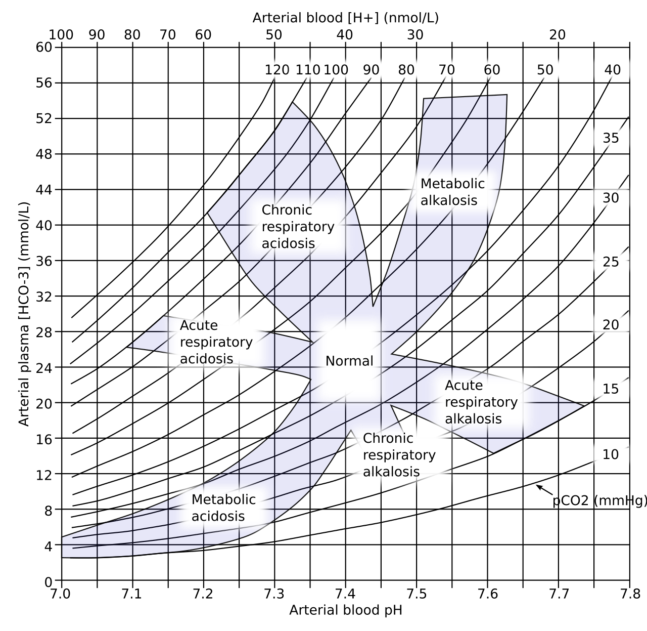
- Find the component that moved the same way. Watch PaCO₂ and the pH together. Same direction means metabolic. Opposite directions mean respiratory. Acute or chronic? Work it out from the history and the trend. The trend outranks any single value.
- Check compensation and derived values. Set the measured value against the expected one (table below). In any metabolic acidosis, work out the albumin-corrected anion gap and the delta gap. Compare the gap with lactate (see the lactate page). An unexplained high gap needs an osmolal gap and a toxic-alcohol check (see the toxic alcohols page). Compensation that is too small or too large points to a second disorder.
- Name it and escalate. Name the criteria you used. For DKA, use the 2024 consensus: glucose ≥11.1 mmol/L (200 mg/dL) or known diabetes, β-hydroxybutyrate ≥3.0 mmol/L, and pH <7.30 and/or HCO₃⁻ <18 mmol/L (severity below). For sepsis, use Sepsis-3 with the 2026 Surviving Sepsis Campaign.
Expected compensation
| Primary disorder | Primary change | Expected compensation |
|---|---|---|
| Metabolic acidosis | ↓ HCO₃⁻ | PaCO₂ = (1.5 × HCO₃⁻) + 8 ± 2 mmHg (Winter's formula). Roughly 1 mmHg of PaCO₂ fall per 1 mmol/L fall in HCO₃⁻ |
| Metabolic alkalosis | ↑ HCO₃⁻ | PaCO₂ rises about 0.6–0.75 mmHg per 1 mEq/L rise; not above about 55 mmHg from compensation alone |
| Respiratory acidosis, acute | ↑ PaCO₂ | HCO₃⁻ rises about 1–2 mEq/L per 10 mmHg rise |
| Respiratory acidosis, chronic | ↑ PaCO₂ | HCO₃⁻ rises about 3–4 mEq/L per 10 mmHg rise |
| Respiratory alkalosis, acute | ↓ PaCO₂ | HCO₃⁻ falls about 1–2 mEq/L per 10 mmHg fall |
| Respiratory alkalosis, chronic | ↓ PaCO₂ | HCO₃⁻ falls about 4–5 mEq/L per 10 mmHg fall |
Compensation pulls the pH back toward normal and stops short of it. It never overshoots.
Patterns
| Pattern | Suggests |
|---|---|
| pH low, PaCO₂ high, HCO₃⁻ normal | Acute respiratory acidosis (opioids, tiring asthma) |
| pH low, HCO₃⁻ low, PaCO₂ within Winter's range | Metabolic acidosis with appropriate compensation |
| pH low, HCO₃⁻ low, PaCO₂ normal or high | Metabolic plus respiratory acidosis: ventilation is failing |
| pH high, PaCO₂ low, HCO₃⁻ low or normal | Respiratory alkalosis; an HCO₃⁻ below the expected value means an added metabolic acidosis (sepsis, salicylate) |
| pH normal, PaCO₂ and HCO₃⁻ both abnormal | Mixed disorder until proven otherwise |
| pH normal, PaCO₂ and HCO₃⁻ normal, anion gap high | Hidden high-gap acidosis offset by a metabolic alkalosis (DKA with vomiting) |
Management Priorities
Emergency: Severe acidemia (pH <7.20)
- Ventilation first if PaCO₂ is high: start with the airway and bag-valve-mask. Naloxone treats opioid toxicity. NIV is for a COPD exacerbation, and intubation follows if NIV fails or is contraindicated (Paramedic/RN/CCT, per local protocol).
- Protect compensation if PaCO₂ is low: Don't oversedate, because that strips the compensation away. If intubation can't be avoided, shorten the apnea and match the pre-intubation minute ventilation or EtCO₂ right away.
- Treat the cause: restore perfusion and control the source in shock or sepsis. For DKA, fluids, insulin and K⁺. Call poison center. For toxic alcohols, fomepizole and early dialysis planning. For salicylate that meets EXTRIP criteria, dialysis planning.
- Watch K⁺ and glucose: Watch the ECG, K⁺ and glucose now and often. Dangerous hyperkalemia gets treated directly: calcium first, then insulin with dextrose, per protocol. Bicarbonate is only an adjunct. Don't sit and wait for the pH to fix the potassium.
Emergency: Severe alkalemia (pH >7.60)
- Monitor: ECG; K⁺, ionized Ca²⁺, Mg²⁺ and phosphate; seizure precautions.
- Respiratory cause: Bring the bag or ventilator rate or tidal volume down toward the patient's baseline PaCO₂. Treat hypoxemia, pain and the cause.
- Metabolic cause: Where possible, stop alkali, citrate or diuretics. Replace K⁺ and Mg²⁺. Give volume and chloride per protocol for vomiting or NG loss.
Medications
Doses vary by protocol; follow local protocols and medical direction.
| Drug | Adult dose | Pediatric dose (max) | Onset / key notes |
|---|---|---|---|
| Sodium bicarbonate: septic shock with severe acidemia | pH ≤7.20 and AKI stage 2–3 (AKIN score): IV infusion per ICU protocol, titrated to pH (titrate to effect: no fixed max) | Not routinely used (consult) | High-alert Surviving Sepsis Campaign suggests it only for severe metabolic acidemia. It suggests against use to improve blood pressure in lactic acidemia. Ventilation must clear the extra CO₂; watch Na⁺ and ionized Ca²⁺ |
| Sodium bicarbonate: TCA or sodium-channel blocker toxicity | 1–2 mEq/kg IV/IO bolus, repeated to narrow the QRS, per protocol | 1–2 mEq/kg IV/IO, per protocol | For QRS widening, dysrhythmia or hypotension. Stop at a pH of about 7.55 (see the sodium bicarbonate page) |
| Sodium bicarbonate: hyperkalemia with ECG changes | 50 mEq IV/IO as an adjunct, per protocol; calcium, insulin and dextrose come first | 1 mEq/kg IV/IO slowly (max 50 mEq) | An adjunct only. It does not replace calcium or insulin, and it shifts K⁺ mainly when acidemia is present. Repeat the gas to guide dosing |
| Sodium bicarbonate: salicylate poisoning | 1–2 mEq/kg IV bolus, then infusion, per toxicology protocol | Per toxicology protocol | Alkalinizes the urine. Monitor serum and urine pH, K⁺ and Na⁺; see the salicylate page |
| Sodium bicarbonate: DKA | Not routine. Consider only when pH is below 7.00 (2024 consensus); insulin and fluids come first | Not routine: consult | Routine use gives no outcome benefit and can cause hypokalemia |
| Insulin: DKA | IV infusion per DKA protocol; hold while K⁺ is below 3.5 mmol/L and replace K⁺ first (titrate to glucose and ketones: no fixed max) | Weight-based DKA protocol; see weight-based dosing | High-alert Hypoglycemia and hypokalemia are the main treatment complications in the 2024 consensus |
| Naloxone: opioid hypoventilation | 0.2–2 mg IV/IO/IM or 2–4 mg IN, every 2–3 min, titrated to breathing (no fixed max). Start low in opioid-dependent patients | 0.1 mg/kg IV/IO, max 2 mg per dose (as on the naloxone page). The naloxone injection label starts at 0.01 mg/kg IV, then 0.1 mg/kg if needed. Titrate to breathing. The lower label start suits neonates and opioid-dependent children, because a full dose can precipitate withdrawal | IV onset within 2 min. Ventilate first; titrate to breathing, not alertness. Effect can wear off before the opioid |
| Sodium chloride 0.9%: chloride-responsive alkalosis | Volume and chloride repletion per protocol (titrate to response) | Per pediatric protocol | Used for vomiting or NG loss with a low Cl⁻; watch K⁺ and volume status |
Cardiac arrest: Skip routine bicarbonate. The AHA doesn't recommend it in cardiac arrest. Save it for a specific indication, such as hyperkalemia or sodium-channel blockade, per protocol.
Definitive care & disposition
- ICU: pH <7.20 or >7.60, rising PaCO₂ despite NIV, severe DKA (pH <7.00 or HCO₃⁻ <10 mmol/L), any bicarbonate or acid infusion.
- Dialysis: salicylate toxicity meeting EXTRIP criteria (see the salicylate page), toxic alcohols (start fomepizole early; dialysis per EXTRIP criteria, see the toxic alcohols page), refractory acidemia, or alkalosis with kidney failure and volume overload. Call nephrology and toxicology early. For an unstable patient, continuous renal replacement is the alternative; see the CRRT page.
- Chronic CO₂ retainers: Go slow. Bring PaCO₂ down over hours, toward the patient's baseline, to avoid posthypercapnic alkalosis.
Doses are typical references. Follow local protocols, medical direction and institutional policy.
Field & Transport Considerations
Point-of-care testing
Cartridge analyzers give you pH and PaCO₂, and they calculate HCO₃⁻ for you. Stay inside the cartridge temperature and hematocrit limits. Trend on one device. Venous gas can work for pH in many patients who aren't in shock. It won't answer PaO₂. Waveform EtCO₂ watches ventilation, not pH. Check it against a gas, because the PaCO₂–EtCO₂ gap widens with shock, PE and lung disease.
Transport cases
- DKA: a high gap with deep, rapid breathing. Never slow it. Make sure the insulin and fluids are running and that glucose and K⁺ are being checked. Severe band (pH <7.00 or HCO₃⁻ <10 mmol/L): ICU-level transport.
- Salicylate: respiratory alkalosis with a high gap. Respiratory depressants strip away the compensation, so avoid them. If EXTRIP criteria are met, redirect to a dialysis center.
- COPD: rising PaCO₂ with falling pH and slow, shallow breathing. NIV comes first where protocol allows. Titrate oxygen to target, since uncontrolled oxygen can worsen hypercapnia.
Before and during interfacility transport
| Setting | Consideration |
|---|---|
| Before departure | Timed pH, PaCO₂, HCO₃⁻, lactate, K⁺ and glucose with the ventilator settings and EtCO₂ at the time of the draw |
| Ventilator change | Match the sending minute ventilation, especially in metabolic acidosis; recheck a gas after changes if a POC analyzer is carried |
| Running infusions | Insulin, bicarbonate and K⁺ change pH and K⁺ en route: confirm drug, concentration and rate |
| Altitude and flight | Hypoxemia at cabin altitude drives hyperventilation in spontaneously breathing patients; keep SpO₂ at target |
| Redirect | Toxic alcohol, salicylate toxicity meeting EXTRIP criteria, or refractory acidemia: go to a center that can start hemodialysis |
Special Populations
| Population | What changes | Clinical implication |
|---|---|---|
| Neonates & infants | Transitional values after birth; the laboratory's neonatal range applies | Interpret cord and first-day gases with neonatal norms, not adult cut-offs |
| Children & adolescents | Adult range for pH, PaCO₂ and HCO₃⁻. DKA is diagnosed and staged with the pediatric criteria | Follow the pediatric DKA protocol; see weight-based dosing |
| Pregnancy | Progesterone drives ventilation: PaCO₂ about 27–32 mmHg; HCO₃⁻ about 18–21 mEq/L; pH in the upper half of normal | A PaCO₂ of 40 mmHg is relatively high in late pregnancy and may signal tiring or ventilatory failure. An HCO₃⁻ of 22 mEq/L or more is relatively high for pregnancy and may signal an added metabolic acidosis |
| Older adults | No clinically meaningful difference in pH or PaCO₂ | Review diuretics and other drugs that change acid–base status |
| Chronic CO₂ retainers | Baseline chronic respiratory acidosis with a raised HCO₃⁻ | Target the patient's baseline PaCO₂, not 40 mmHg; a normal pH may be a sign of failure |
| CKD / dialysis | Chronic metabolic acidosis is common as GFR falls | Compare with the patient's baseline; refractory acidemia in kidney failure needs dialysis |
Interfering Factors & Pitfalls
Collection & handling
| Factor | Effect | Why | Avoid / fix |
|---|---|---|---|
| Air bubbles | pH ↑ (usually slight); PaCO₂ ↓ | CO₂ escapes into the bubble | Expel air at once, cap, mix gently |
| Delay at room temperature | pH ↓; PaCO₂ ↑ | Cells keep producing CO₂ and acid | Analyze promptly; the pH and PaCO₂ pages give the time limits |
| Excess liquid heparin | PaCO₂ and HCO₃⁻ falsely ↓ | Dilution | Use dry-heparin blood-gas syringes; fill to the stated volume |
| Vigorous mixing | Hemolysis; K⁺ falsely ↑ on the same tube | Red-cell damage | Roll and invert gently |
| Line sample with flush | Variable; PaO₂ falsely ↓ | Saline or drug contamination | Discard enough dead space before drawing |
Physiologic / patient factors
| Factor | Effect | Why | Avoid / fix |
|---|---|---|---|
| Painful or anxious puncture | pH ↑; PaCO₂ ↓ | Hyperventilation | Local anesthetic; calm the patient; note the breathing pattern |
| Venous sample read as arterial | pH ↓ (about 0.03); PaCO₂ ↑ | Venous blood carries more CO₂ | Label the sample type; check that PaO₂ and SaO₂ fit |
| Low albumin | Anion gap falsely low (False −) | Albumin is an unmeasured anion | Correct the gap for albumin; see the formula in Reference & Critical Ranges |
Assay & device-specific
| Factor | Effect | Why | Avoid / fix |
|---|---|---|---|
| Calculated HCO₃⁻ | Inherits pH and PaCO₂ errors | Derived by the analyzer, not measured | Compare with serum total CO₂; trust the measured value if discordant |
| Lab-specific anion gap | Different normal limits with and without K⁺ | Methods differ between laboratories | Use the lab's gap formula and reference range |
| POC cartridge outside limits | Unreliable | Temperature or hematocrit outside the operating range | Follow the device limits; send to the lab |
Common misinterpretations
- "A normal pH means no disorder": two opposing disorders can cancel out. Look at PaCO₂, HCO₃⁻ and the gap.
- "PaCO₂ inside Winter's range rules out a second disorder": the formula only tests respiratory compensation. A normal-gap acidosis or an alkalosis can sit alongside it.
- "Compensation brought the pH to normal": compensation never fully corrects the pH, so a normal pH with abnormal components signals a mixed disorder.
- "A normal gap after DKA treatment means DKA is still active": after treatment, a hyperchloremic normal-gap acidosis can pass for persistent DKA. Calling it that can hold up the move to subcutaneous insulin.
Physiology & Test Basics
What it measures
This page teaches a method, not a single analyte. The blood gas gives pH, PaCO₂ and HCO₃⁻. The chemistry panel gives the serum anion gap and albumin. Together they name the primary disorder, check compensation and test for a mixed one. The single values are on the pH, PaCO₂, bicarbonate and base excess pages.
Physiology & regulation
- Buffers: Within seconds, bicarbonate, CO₂, hemoglobin and protein all soak up acid.
- Lungs: they move PaCO₂ within minutes, the fastest fix for pH.
- Kidneys: over days, they reclaim HCO₃⁻ and excrete acid.
- Acute high-gap acidosis: organic acids such as ketoacids or lactic acid usually drive it.
- Chronic acidosis: bicarbonate loss or impaired renal acid excretion often drives it.
How it is measured
- Specimen: arterial (usually radial) or venous blood in a pre-heparinized (dry heparin) blood-gas syringe. Expel air, cap, mix gently, and record the sample type, FiO₂ and temperature.
- Method: the analyzer measures pH and PaCO₂, then works out HCO₃⁻ and base excess. The anion gap runs on serum Na⁺, Cl⁻ and HCO₃⁻ from the chemistry panel, plus albumin from that same panel.
- Fasting and timing: Skip the fasting. Draw before any bicarbonate or ventilator change you want to measure.
Why it matters
One gas shows how dangerous the acid–base problem is right now. Add the gap, and you see whether breathing or metabolism is behind it. That answer decides whether you ventilate, resuscitate, give insulin or call for dialysis.
Clinical Pearls
References
- Kraut, J. A., & Madias, N. E. (2010). Metabolic acidosis: pathophysiology, diagnosis and management. Nature Reviews Nephrology, 6(5), 274–285. https://doi.org/10.1038/nrneph.2010.33
- Figge, J., Jabor, A., Kazda, A., & Fencl, V. (1998). Anion gap and hypoalbuminemia. Critical Care Medicine, 26(11), 1807–1810. https://doi.org/10.1097/00003246-199811000-00019
- Albert, M. S., Dell, R. B., & Winters, R. W. (1967). Quantitative displacement of acid-base equilibrium in metabolic acidosis. Annals of Internal Medicine, 66(2), 312–322. https://doi.org/10.7326/0003-4819-66-2-312
- Singer, M., Deutschman, C. S., Seymour, C. W., et al. (2016). The Third International Consensus Definitions for Sepsis and Septic Shock (Sepsis-3). JAMA, 315(8), 801–810. https://doi.org/10.1001/jama.2016.0287
- Prescott, H. C., Antonelli, M., Alhazzani, W., et al. (2026). Surviving Sepsis Campaign: international guidelines for management of sepsis and septic shock 2026. Critical Care Medicine, 54(4), 725–812. https://doi.org/10.1097/CCM.0000000000007075; PubMed 41869847
- Umpierrez, G. E., Davis, G. M., ElSayed, N. A., et al. (2024). Hyperglycemic crises in adults with diabetes: a consensus report. Diabetes Care, 47(8), 1257–1275. https://doi.org/10.2337/dci24-0032; PMC11272983
- Juurlink, D. N., Gosselin, S., Kielstein, J. T., et al. (2015). Extracorporeal treatment for salicylate poisoning: systematic review and recommendations from the EXTRIP workgroup. Annals of Emergency Medicine, 66(2), 165–181. https://doi.org/10.1016/j.annemergmed.2015.03.031
Medical Disclaimer
- For Educational Purposes Only: This content is intended for educational reference and should not be used for clinical decision-making.
- Not a Substitute for Professional Judgment: Always consult your local protocols, institutional guidelines, and supervising physicians.
- Verify Before Acting: Users are responsible for verifying information through authoritative sources before any clinical application.