

Bedside Snapshot
- Normal (Adult)
- Men 0.7–1.3 mg/dL (62–115 µmol/L)
Women 0.6–1.1 mg/dL (53–97 µmol/L)
Lab-specific; compare with the patient's baseline - Critical Values
- No universal critical value: interpret by trend and clinical context. AKI: rise ≥0.3 mg/dL in 48 h. Stage 3: ≥4 mg/dL or 3× baseline.
- High Suggests
- AKI: shock, sepsis, nephrotoxins
- Obstruction or rhabdomyolysis
- Chronic kidney disease
- Secretion blockers (trimethoprim)
- Low Suggests
- Low muscle mass (older, frail, amputation)
- Pregnancy (normal)
- Cirrhosis or malnutrition
- Dilution after large fluid volumes
- Act Now If
- Rising creatinine with K⁺ ≥6.5 mEq/L or ECG changes, pulmonary edema, severe acidosis, uremic signs or anuria: monitor, 12-lead, IV calcium for ECG changes, bladder scan; urgent nephrology
- Watch Out For
- A normal creatinine can hide a low GFR in older or low-muscle patients, and it lags hours to days behind acute injury. Trend it with urine output.
Reference & Critical Ranges
Reference ranges
| Population | Conventional | SI | Notes |
|---|---|---|---|
| Adult men | 0.7–1.3 mg/dL | 62–115 µmol/L | Higher with more muscle mass |
| Adult women | 0.6–1.1 mg/dL | 53–97 µmol/L | Lower muscle mass on average |
| Older adult | Same as adult | Same as adult | Less muscle: a value inside the range can hide an eGFR below 60 |
| Pregnancy | Upper limit about 0.87 mg/dL | About 77 µmol/L | Runs about 15–25% below nonpregnant values; above 0.87 mg/dL is outside the pregnancy range |
| Neonate (0–2 months) | 0.31–0.92 mg/dL | 27–81 µmol/L | Reflects maternal creatinine at birth, then falls |
| Infant (2 months–1 year) | 0.16–0.39 mg/dL | 14–34 µmol/L | — |
| Child (1–3 years) | 0.17–0.35 mg/dL | 15–31 µmol/L | — |
| Child (3–7 years) | 0.26–0.48 mg/dL | 23–42 µmol/L | Rises with age and muscle mass |
| Child (7–11 years) | 0.32–0.64 mg/dL | 28–57 µmol/L | — |
| Child (11–13 years) | 0.42–0.71 mg/dL | 37–63 µmol/L | — |
| Adolescent (13–18 years) | 0.46–0.81 mg/dL | 41–72 µmol/L | Adult intervals from 18 years |
Severity & critical values
| Level | Conventional | SI | What it means |
|---|---|---|---|
| Low | Below the lab interval | Below the lab interval | Low muscle mass, pregnancy, liver disease or dilution; not proof of good kidney function |
| Normal | 0.6–1.3 mg/dL (sex-specific above) | 53–115 µmol/L | Does not exclude chronic kidney disease (CKD) or early acute kidney injury (AKI); read the eGFR and the trend |
| AKI stage 1 | Rise ≥0.3 mg/dL in 48 h, or 1.5–1.9 × baseline | Rise ≥26.5 µmol/L, or 1.5–1.9 × baseline | KDIGO 2012; or urine output <0.5 mL/kg/hr for 6–12 h |
| AKI stage 2 | 2.0–2.9 × baseline | 2.0–2.9 × baseline | Or urine output <0.5 mL/kg/hr for ≥12 h |
| AKI stage 3 | ≥4 mg/dL or 3× baseline, or start of kidney replacement therapy | ≥353.6 µmol/L or 3× baseline | Or urine output <0.3 mL/kg/hr for ≥24 h or anuria ≥12 h; under 18 years, also eGFR <35 |
| Critical | No universal critical value: interpret by trend and clinical context | — | Typical; lab-specific. Act on K⁺, acid–base status, volume and uremic signs, which drive dialysis decisions |
Conversion: creatinine mg/dL × 88.4 = µmol/L
SCr in mg/dL; κ = 0.7 (female) or 0.9 (male); α = −0.241 (female) or −0.302 (male). Result in mL/min/1.73 m². Normal: ≥90; labs flag <60 as low.
This number is not indexed to body surface area. Many drug labels set renal dose cutoffs in CrCl. Which weight you plug in (actual, ideal or adjusted) is a pharmacy policy call.
Causes of Abnormal Results
High (above the lab interval, or a rise from baseline)
Azotemia means creatinine and blood urea nitrogen (BUN) are both raised. Here's the catch: KDIGO 2012 defines AKI by the change from baseline, not by the range. A value inside the reference range can still be AKI. KDIGO 2024 defines CKD as abnormal kidney structure or function for more than 3 months.
| Mechanism | Common causes | Bedside clue |
|---|---|---|
| Low kidney perfusion (prerenal) | Shock, sepsis, hemorrhage, vomiting, diarrhea, overdiuresis, heart failure (cardiorenal), cirrhosis (hepatorenal) | Hypotension, tachycardia, low urine output; BUN:Cr >20:1; improves within days once perfusion is restored |
| Tubular injury (acute tubular necrosis, ATN) | Prolonged shock or sepsis, nephrotoxins (aminoglycosides, NSAIDs, iodinated contrast in high-risk patients) | Rise continues after perfusion is restored; muddy brown casts; BUN:Cr <10:1 is common |
| Pigment injury | Rhabdomyolysis (crush injury, long lie, seizures, exertional heat illness, stimulants) | Very high creatine kinase (CK), dark urine, high K⁺ and phosphate, low calcium |
| Glomerular or vascular disease | Rapidly progressive glomerulonephritis, vasculitis, thrombotic microangiopathy, malignant hypertension | Blood and protein on urinalysis, red cell casts, edema, severe hypertension, low platelets |
| Interstitial nephritis | Drug reaction (antibiotics, proton pump inhibitors, NSAIDs), infection | New drug in the past weeks; rash or fever in some; white cells in urine |
| Obstruction (postrenal) | Bladder outlet obstruction (prostate, clots, urinary retention from anticholinergics or opioids), obstruction of both ureters or of a single kidney | Distended bladder, large bladder-scan volume, hydronephrosis on ultrasound, anuria |
| Chronic kidney disease | Diabetes, hypertension, glomerular disease, polycystic kidneys | Stable elevation, known low eGFR, albuminuria, anemia |
| Rise without a true GFR change | Trimethoprim, cimetidine, dolutegravir, cobicistat (block tubular secretion); cooked meat; creatine supplements; Jaffe-assay interference | Small rise after a new drug or meal; BUN, urine output and cystatin C unchanged |
Low (below the lab interval)
A low creatinine rarely hurts the patient on its own. The problem is what it does to the eGFR. A low value makes the creatinine-based eGFR read too high, and that can hide AKI or CKD.
| Mechanism | Common causes | Bedside clue |
|---|---|---|
| Low creatinine generation | Older age, frailty, amputation, spinal cord injury, muscle-wasting disease, malnutrition | Low muscle mass; eGFR reads falsely high; cystatin C-based eGFR is more accurate |
| Higher GFR | Pregnancy | Use pregnancy limits; an adult-normal value may already be abnormal |
| Liver disease | Cirrhosis (less creatine made, muscle wasting) | Creatinine understates kidney dysfunction, including hepatorenal AKI |
| Dilution | Massive fluid resuscitation | A low or flat creatinine after large volumes can mask AKI; watch urine output |
Interpretation & Bedside Approach
Step-by-step approach
- Is it real? Same lab, same method, or the comparison isn't fair. Ask about new drugs that block secretion, a recent meat meal, ketoacidosis or N-acetylcysteine (NAC). Repeat an unexpected value before you act on it alone.
- How sick is the patient? The creatinine number doesn't set urgency. K⁺, pH and bicarbonate, volume status (pulmonary edema), urine output and mental status do.
- Acute or chronic? Find a baseline in prior results. No baseline? KDIGO 2012 lets you back-calculate one from an assumed eGFR of 75. The trap: unrecognized CKD then looks like AKI. Trend beats a single value.
- Calculate. Report the absolute and percentage change from baseline. Use the CKD-EPI 2021 eGFR for CKD staging. Use CrCl (Cockcroft-Gault) when a drug label asks for it, and only at steady state.
- Classify. Stage AKI with KDIGO 2012, using creatinine and hourly urine output. Classify CKD with the KDIGO 2024 GFR (G1–G5) and albuminuria (A1–A3) categories, once it has been present for more than 3 months.
- Find the cause. Volume and perfusion checks, a bladder scan, urinalysis with microscopy, urine sodium, CK and renal ultrasound sort the cause: prerenal, intrinsic or postrenal.
- Escalate. Start the KDIGO AKI bundle (Management Priorities). Call nephrology early. Stage 2–3, an unclear cause or any dialysis indication all qualify.
KDIGO 2024 GFR categories
| Category | eGFR (mL/min/1.73 m²) | Meaning |
|---|---|---|
| G1 | ≥90 | Normal or high; CKD only with another marker of kidney damage (e.g., albuminuria) |
| G2 | 60–89 | Mildly decreased |
| G3a | 45–59 | Mildly to moderately decreased |
| G3b | 30–44 | Moderately to severely decreased |
| G4 | 15–29 | Severely decreased |
| G5 | <15 | Kidney failure |
Albuminuria categories (urine albumin-to-creatinine ratio): A1 <30 mg/g, A2 30–300 mg/g, A3 >300 mg/g. KDIGO 2024 wants a validated eGFR equation, not creatinine alone, and it leaves race out of the calculation. When creatinine is the less accurate choice and the result will change management, it recommends eGFRcr-cys, which adds cystatin C.
eGFR or CrCl?
| Measure | Units | Best use |
|---|---|---|
| CKD-EPI 2021 eGFR | mL/min/1.73 m² | CKD detection and staging; KDIGO 2024 also accepts it for most drug dosing |
| Non-indexed eGFR | mL/min | Drug dosing at extremes of body size, especially narrow-therapeutic-range drugs |
| CrCl (Cockcroft-Gault) | mL/min | Drug labels written in CrCl; less accurate with edema, obesity or unstable creatinine |
| eGFRcr-cys or measured GFR | mL/min/1.73 m² | Low or high muscle mass, cirrhosis, cancer, narrow-therapeutic-range or toxic drugs |
Management Priorities
Emergency: AKI with life-threatening complications (K⁺ ≥6.5 mEq/L or ECG changes, pulmonary edema, severe acidosis, uremic signs)
- Monitor and assess: Put on the cardiac monitor, get a 12-lead ECG and IV access. Then check K⁺, the blood gas, a bladder scan and urine output.
- Stabilize the myocardium: Give IV calcium for hyperkalemic ECG changes, and repeat it if the changes persist. Calcium chloride causes tissue necrosis when it leaks from a peripheral line, so give it through a central or IO line where you can. Use calcium with caution in digoxin toxicity: arrhythmia risk.
- Shift K⁺: Insulin with dextrose, plus nebulized albuterol 10–20 mg. That dose runs higher than for bronchospasm and depends on protocol, so see the albuterol page. Check glucose often for several hours. Hypoglycemia is more likely in kidney failure.
- Volume: Fluid only for hypovolemia. In pulmonary edema, give oxygen and ventilatory support, plus a loop diuretic if the patient still makes urine.
- Remove and relieve: Call nephrology urgently for emergent kidney replacement therapy. Catheterize urinary retention.
KDIGO 2012 AKI bundle: all stages
- Stop nephrotoxins where possible: NSAIDs and aminoglycosides are the ones to stop. Consider alternatives to iodinated contrast when they work just as well.
- Perfusion: Restore volume with isotonic crystalloid, not colloid, unless this is hemorrhagic shock. In vasodilatory shock, add vasopressors alongside the fluids.
- Do not use diuretics to prevent or treat AKI. Their only job here is volume overload. Low-dose dopamine is not for kidney protection.
- Monitor: Creatinine, hourly urine output, glucose (avoid hyperglycemia) and the dose of every renally cleared drug. From stage 2, consider ICU care and kidney replacement therapy.
Medications
| Drug | Adult dose | Pediatric dose (max) | Onset / key notes |
|---|---|---|---|
| Calcium gluconate 10% (hyperkalemia with ECG changes) | 3 g (30 mL) IV over 10 min; repeat if ECG changes persist | 60 mg/kg (0.6 mL/kg) IV, max 3 g (protocol-based) | Onset 1–3 min, lasts 30–60 min; does not lower K⁺: start insulin-glucose at once |
| Calcium chloride 10% (arrest or peri-arrest hyperkalemia) | 1 g (10 mL) IV over 5–10 min, IV/IO push in arrest; repeat if ECG changes persist | 20 mg/kg (0.2 mL/kg) IV/IO, max 1 g | 3 times the calcium of gluconate per mL; central line or IO preferred |
| Regular insulin with dextrose (hyperkalemia) | Insulin 10 units IV with dextrose 25 g (some protocols use 5 units in kidney failure) | Insulin 0.1 units/kg IV (max 10 units) with dextrose 0.5 g/kg (D10 5 mL/kg) | High-alert Check glucose often for several hours |
| Sodium bicarbonate 8.4% (hyperkalemia with metabolic acidosis or arrest) | Hyperkalemic arrest: 50 mEq IV/IO; repeat every 5–10 min guided by blood gas | 1 mEq/kg IV/IO slowly (max 50 mEq/dose); use 4.2% (0.5 mEq/mL) in neonates and infants, or dilute 8.4% 1:1 | Adds sodium and volume; not a stand-alone K⁺ treatment |
| Furosemide (volume overload only) | 40 mg IV over 1–2 min, or IV 1–2.5 times the daily oral dose if already on a loop diuretic | 1 mg/kg IV over 1–2 min; may repeat in 1 mg/kg steps no sooner than 2 h apart, cumulative limit 6 mg/kg (protocol-based; many protocols cap a single dose at 40 mg) | Not for preventing or treating AKI; little effect in anuria |
| Isotonic crystalloid (lactated Ringer’s or normal saline; hypovolemia only) | 500 mL IV/IO bolus, reassess, repeat (up to 30 mL/kg) | 10–20 mL/kg IV/IO bolus, reassess after each (up to 40–60 mL/kg in the first hr with intensive care) | Stop at signs of fluid overload; see weight-based dosing below |
Definitive care & disposition
- Emergent kidney replacement therapy: KDIGO 2012 calls for it when fluid, electrolyte or acid–base balance turns life-threatening, such as refractory hyperkalemia, pulmonary edema or acidosis, or uremic pericarditis, encephalopathy or bleeding. No creatinine or BUN number makes that call on its own.
- Consults: Nephrology for stage 2–3 AKI or an unclear cause. Urology for obstruction. ICU for stage 3 AKI or complications.
- Contrast: ACR–NKF 2020 indicates IV normal saline prophylaxis for AKI or an eGFR <30 (not on dialysis). At 30–44 in high-risk cases, it may be considered.
- Follow-up: Recheck kidney function about 3 months after AKI. KDIGO 2012 advises it to catch recovery or new CKD.
Pediatric fluid and drug calculations: weight-based dosing.
Doses are typical references. Follow local protocols, medical direction and institutional policy.
Field & Transport Considerations
Point-of-care testing
Some cartridge blood analyzers report creatinine with electrolytes from whole blood. Some CT departments screen with handheld creatinine meters. Those results can differ from the central lab, because of method, hematocrit and temperature limits. Trend on one platform. Confirm the key decisions on a lab sample.
Before and during interfacility transport
| Setting | Consideration |
|---|---|
| Stroke imaging | Many stroke protocols do not delay CTA and CT perfusion to obtain a creatinine; follow the local stroke protocol |
| Handoff | Latest creatinine, K⁺ and bicarbonate with draw times, the baseline creatinine, urine output since admission, contrast or nephrotoxins given; for dialysis patients, access type and last session |
| En route | Hourly urine output with a catheter if placed; cardiac monitor for K⁺ effects; avoid NSAIDs; confirm renal dose adjustments with the sending team |
| Crush injury or long lie | Creatinine may still be normal; anticipate hyperkalemia and dark urine, and monitor the ECG. Avoid succinylcholine in suspected rhabdomyolysis, crush injury or hyperkalemia; use rocuronium per protocol |
| Destination | Patients meeting dialysis indications need a center with nephrology and kidney replacement therapy |
Scope: POC chemistry use and interpretation follow local protocol (Paramedic/RN/CCT).
Timing & Kinetics
| Parameter | Value | Clinical implication |
|---|---|---|
| Lag after an abrupt GFR fall | Time to a 50% rise: about 4 h with normal baseline, up to 27 h in stage 4 CKD (modeled, 90% fall in clearance) | An early normal value does not exclude AKI; urine output often falls first |
| First-day rise after severe injury | About 1.8–2 mg/dL in 24 h, whatever the baseline (modeled) | In CKD the same injury is a small percentage rise; watch absolute change |
| New steady state | Days after the change in GFR | eGFR and CrCl are unreliable until then |
| Cooked meat or fish meal | Rise of about 0.23 mg/dL, peak 2–4 h | For elective testing, wait at least 12 h after the meal |
| ACE inhibitor or ARB started or increased | Recheck creatinine and K⁺ within 2–4 weeks | Per KDIGO 2024, continue unless creatinine rises >30% within 4 weeks |
| SGLT2 inhibitor started | Early reversible eGFR dip | Per KDIGO 2024, generally not a reason to stop |
| Recovery after AKI | Falls over days once the cause is fixed | Reassess at about 3 months for CKD |
Special Populations
| Population | What changes | Clinical implication |
|---|---|---|
| Neonates & infants | Birth value reflects maternal creatinine, then falls; Jaffe assays are less accurate at low values and with jaundiced or hemolyzed samples | Use age-specific ranges and the trend; KDIGO 2024 favors enzymatic assays in children |
| Children & adolescents | Creatinine rises with age and muscle mass | Estimate GFR with a pediatric equation such as CKiD U25 2021; eGFR <90 is flagged low after age 2; AKI stage 3 includes eGFR <35 under 18 |
| Pregnancy | GFR rises; creatinine falls about 15–25% | Above 0.87 mg/dL is abnormal in pregnancy; >1.1 mg/dL or a doubling is a severe feature of preeclampsia |
| Older adults | Less muscle mass | A normal creatinine can hide an eGFR below 60; dose renally cleared drugs by eGFR or CrCl, not creatinine |
| CKD / dialysis | Chronically high; falls after each dialysis session | Compare with the patient's baseline; percentage criteria detect AKI late in advanced CKD |
| Liver disease | Low creatine synthesis and muscle wasting | Creatinine overestimates kidney function; KDIGO 2024 suggests adding cystatin C |
| Other: muscle and hormones | Amputation, spinal cord injury, bodybuilding, creatine supplements, class III obesity, anabolic or hormone therapy | Non-GFR effects on creatinine; eGFRcr-cys is more accurate. CKD-EPI 2021 calculates eGFR without race |
Interfering Factors & Pitfalls
Collection & handling
| Factor | Effect | Why | Avoid / fix |
|---|---|---|---|
| Unseparated sample (Jaffe assay) | ↑ | Measured creatinine creeps up while serum stays on the cells | Separate serum or plasma within 12 h of collection |
| Hemolysis or icterus | Bias (direction varies) | Spectral interference, worse with Jaffe methods | Redraw; ask for an enzymatic method |
Medications & supplements
| Factor | Effect | Why | Avoid / fix |
|---|---|---|---|
| Trimethoprim, cimetidine, dolutegravir, cobicistat | ↑ | Block tubular creatinine secretion; true GFR unchanged | Check the start date; BUN and cystatin C are unaffected |
| Cephalosporins (e.g., cefoxitin) | False + (Jaffe) | React as non-creatinine chromogens | Ask which method the lab uses; enzymatic assay |
| N-acetylcysteine (NAC), lidocaine metabolites, IV immunoglobulin stabilizers | Bias (enzymatic); NAC causes false − | Interfere with enzymatic reaction steps | Tell the lab; confirm by another method |
| Creatine supplements | ↑ | Extra creatinine generated | Ask about supplements; cystatin C |
Physiologic / biologic
| Factor | Effect | Why | Avoid / fix |
|---|---|---|---|
| Ketoacids (DKA) and marked hyperglycemia | False + (Jaffe) | Acetoacetate and glucose react as non-creatinine chromogens | Repeat with an enzymatic assay or after treatment |
| Cooked meat or fish meal | ↑ | Dietary creatine and creatinine | Wait 12 h for elective tests |
| High or low muscle mass | ↑ / ↓ | Generation follows muscle mass | Use the trend; consider cystatin C |
Assay & device-specific
| Factor | Effect | Why | Avoid / fix |
|---|---|---|---|
| Jaffe vs enzymatic method; POC vs lab | Bias | Different chemistry and interferences | Trend on one method; KDIGO 2024 prefers enzymatic assays |
Common misinterpretations
- Normal creatinine = normal kidneys: Not in older, frail or low-muscle patients. Not in the first hours of AKI either.
- Any rise = kidney injury: Not so. Secretion blockers, a meat meal and assay interference all raise creatinine. None of them changes GFR.
- Stop the ACE inhibitor at any rise: Don't. A rise up to 30% after you start an ACE inhibitor or ARB is acceptable. A larger rise needs a review.
- eGFR works in AKI: Only at steady state. eGFR equations assume one, so while creatinine is rising they overestimate GFR.
Physiology & Test Basics
What it measures
This page is about the filtration marker itself, serum creatinine. eGFR, BUN and the BUN:creatinine ratio each have their own page. Creatinine is what muscle leaves behind as it breaks down creatine. At steady state, the blood level climbs as GFR falls.
Physiology & regulation
- Generation: Muscle makes it from creatine and phosphocreatine, and muscle mass sets the rate. Diet adds to it: cooked meat and creatine supplements both do.
- Filtration: The glomerulus filters it freely. The tubule doesn't reabsorb it.
- Secretion: The proximal tubule adds some creatinine of its own. That's why creatinine clearance reads higher than true GFR. Drugs that block secretion push the level up.
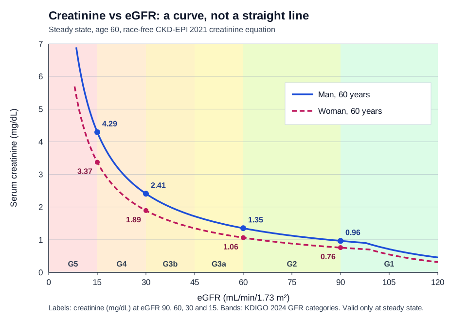
- Curve, not line: At steady state, creatinine and GFR move in opposite directions. Early GFR loss barely moves creatinine. Late loss moves it a lot (Figure 1).
- Not real time: After an abrupt GFR fall, the level takes hours to days to climb toward its new steady state.
How it is measured
- Specimen: Serum (red or gold top) or lithium-heparin plasma (green top). Your lab says which. It's part of the basic and comprehensive metabolic panels and the renal panel. No fasting needed.
- Methods: Jaffe (alkaline picrate, colorimetric) or enzymatic. Both are calibrated to an isotope dilution mass spectrometry (IDMS) reference. KDIGO 2024 prefers enzymatic.
- Turnaround: About 45 minutes at one academic lab. Lithium-heparin plasma stays stable up to 8 hr at room temperature. POC cartridges give you the result at the bedside.
Why it matters
Creatinine defines AKI and stages it. It feeds every eGFR report. It sets the dose of many renally cleared drugs. Read it against the baseline, the urine output and the muscle mass. That's how you tell real kidney injury from a number that only moved on paper.
Clinical Pearls
References
- Kidney Disease: Improving Global Outcomes (KDIGO) Acute Kidney Injury Work Group. (2012). KDIGO clinical practice guideline for acute kidney injury. Kidney International Supplements, 2(1), 1–138. Current guideline; a 2026 AKI/AKD update has completed public review and is in pre-publication (kdigo.org checked 2026-10-14). https://kdigo.org/wp-content/uploads/2016/10/KDIGO-2012-AKI-Guideline-English.pdf
- Kidney Disease: Improving Global Outcomes (KDIGO) CKD Work Group. (2024). KDIGO 2024 clinical practice guideline for the evaluation and management of chronic kidney disease. Kidney International, 105(4S), S117–S314. https://doi.org/10.1016/j.kint.2023.10.018
- Inker, L. A., Eneanya, N. D., Coresh, J., et al. (2021). New creatinine- and cystatin C–based equations to estimate GFR without race. New England Journal of Medicine, 385(19), 1737–1749. https://doi.org/10.1056/NEJMoa2102953
- Waikar, S. S., & Bonventre, J. V. (2009). Creatinine kinetics and the definition of acute kidney injury (landmark modeling study). Journal of the American Society of Nephrology, 20(3), 672–679. https://doi.org/10.1681/ASN.2008070669
- Davenport, M. S., Perazella, M. A., Yee, J., et al. (2020). Use of intravenous iodinated contrast media in patients with kidney disease: Consensus statements from the American College of Radiology and the National Kidney Foundation. Radiology, 294(3), 660–668. https://doi.org/10.1148/radiol.2019192094
- Prabhakaran, S., Gonzalez, N. R., Zachrison, K. S., et al. (2026). 2026 guideline for the early management of patients with acute ischemic stroke: A guideline from the American Heart Association/American Stroke Association. Stroke, 57(8). https://doi.org/10.1161/STR.0000000000000513
- Wiles, K., Bramham, K., Seed, P. T., et al. (2019). Serum creatinine in pregnancy: A systematic review. Kidney International Reports, 4(3), 408–419. https://doi.org/10.1016/j.ekir.2018.10.015
- Oregon Health & Science University Laboratory Services. (2026). CHH-Creatinine (test directory: specimen and age-specific reference ranges). https://www.ohsu.edu/lab-services/chh-creatinine
Medical Disclaimer
- For Educational Purposes Only: This content is intended for educational reference and should not be used for clinical decision-making.
- Not a Substitute for Professional Judgment: Always consult your local protocols, institutional guidelines, and supervising physicians.
- Verify Before Acting: Users are responsible for verifying information through authoritative sources before any clinical application.