

Bedside Snapshot
- Normal (Adult)
- 1.5–2.7 mg/dL (0.6–1.1 mmol/L)
Lab-specific; intervals differ by lab - Critical Values
- <1 mg/dL (<0.4 mmol/L) low; high end is lab-specific
Reflexes lost near ≈12 mg/dL (label): respiratory paralysis hazard - High Suggests
- Kidney failure or oliguria
- IV magnesium overdose or medication error
- Magnesium laxatives, antacids or Epsom salts, especially with kidney disease
- Low Suggests
- GI losses: diarrhea, vomiting, NG suction
- Loop or thiazide diuretics, PPIs, aminoglycosides, cisplatin
- Alcohol use or refeeding after starvation
- Act Now If
- Torsades or sustained polymorphic VT: unsynchronized shock first, then IV magnesium. Lost knee reflexes or slow breathing on magnesium: stop, ventilate, IV calcium
- Watch Out For
- A normal serum level does not exclude deficiency. On IV magnesium, urine under 100 mL in 4 hr means stop and check a level.
Reference & Critical Ranges
Reference ranges
| Population | Conventional | SI | Notes |
|---|---|---|---|
| Adult | 1.5–2.7 mg/dL | 0.6–1.1 mmol/L | Serum. The 50% label cites 1.5–2.5 mEq/L (0.75–1.25 mmol/L; 1.8–3 mg/dL), so labs differ; the lab range governs |
| Children and neonates | — | — | Lab-specific; no verified pediatric interval here. Use the lab's pediatric range |
Severity & critical values
Low (hypomagnesemia)
| Level | Conventional | SI | What it means |
|---|---|---|---|
| Mild | 1.2–1.5 mg/dL | 0.5–0.6 mmol/L | Often no symptoms; oral replacement is usual |
| Moderate | 1.0–1.2 mg/dL | 0.4–0.5 mmol/L | Symptoms usually appear below 0.5 mmol/L (about 1.2 mg/dL): tremor, cramps, weakness |
| Critical | <1 mg/dL | <0.4 mmol/L | Severe: neuromuscular and cardiac complications; IV magnesium (typical; lab-specific) |
High (hypermagnesemia)
| Level | Conventional | SI | What it means |
|---|---|---|---|
| Seizure-control range | 3–9 mg/dL | 1.25–3.75 mmol/L (2.5–7.5 mEq/L) | Anticonvulsant levels in eclampsia (label) |
| Label target | 6 mg/dL | ≈2.5 mmol/L (≈4.9 mEq/L) | Optimal level for seizure control (label) |
| Reflexes weaken | ≈4.9 mg/dL | 2 mmol/L (4 mEq/L) | Deep tendon reflexes begin to decrease (label) |
| Respiratory paralysis hazard (label) | ≈12 mg/dL | ≈5 mmol/L (≈10 mEq/L) | Reflexes may be absent here; respiratory paralysis is a potential hazard; heart block possible at this or lower levels (label) |
| Lab critical call (high) | Lab-specific | Lab-specific | Not verified here; use your lab's critical-call value |
| Potentially fatal (label) | above ≈14.6 mg/dL | above 6 mmol/L (12 mEq/L) | The 50% label says concentrations above 12 mEq/L may be fatal |
Conversion: mg/dL × 0.411 = mmol/L; 1 mmol/L = 2.43 mg/dL = 2 mEq/L.
Causes of Abnormal Results
Low (hypomagnesemia)
| Mechanism | Common causes | Bedside clue |
|---|---|---|
| GI loss | Diarrhea, vomiting, NG suction, malabsorption | Losses from tube or stool; potassium falls with it |
| Renal wasting | Loop and thiazide diuretics, aminoglycosides, amphotericin B, cisplatin and carboplatin, cyclosporine | Magnesium lost in urine; cause found on urine studies |
| Long-term acid suppression | Proton pump inhibitor therapy | Often found on routine labs with no acute signs |
| Alcohol use | Chronic intake with poor nutrition and urinary losses | Often with low potassium |
| Refeeding | Intracellular shift when calories restart after starvation | Fall within 5 days of feeding, by ASPEN 2020 criteria |
| Diabetes | Impaired insulin signaling promotes urinary magnesium loss | Risk rises with insulin resistance |
| Hypokalemia that will not correct | Magnesium deficiency drives renal potassium loss | Potassium stays low despite replacement |
High (hypermagnesemia)
| Mechanism | Common causes | Bedside clue |
|---|---|---|
| Reduced excretion | Kidney failure or oliguria; magnesium leaves the body only through the kidneys | Falling urine output, rising creatinine, lost reflexes |
| Magnesium laxatives, antacids and enemas | Oral or rectal magnesium products; Epsom salt ingestion. Risk is higher with kidney disease or in older adults | Ask about over-the-counter products; check creatinine and urine output |
| Excess dosing | IV magnesium overdose or medication error; continuous infusion in renal impairment | Lost knee jerk, flushing, hypotension, slow breathing |
| Maternal infusion (neonate) | Magnesium given to the mother close to delivery or for prolonged periods (label: beyond 5–7 days harms the fetus) | Hypotonia, hyporeflexia, poor respiratory effort in the newborn |
Interpretation & Bedside Approach
Step-by-step approach
- Is it real? Note whether the sample came during or just after an IV magnesium dose. A level drawn then reflects the infusion, not total body stores. In hypoalbuminemia, total magnesium can mislead. Use an ionized assay where the lab offers one.
- How sick is the patient? Tetany, seizure, arrhythmia or torsades all mean IV therapy on a cardiac monitor. Mild deficits are often treated by mouth.
- Acute or chronic, and why? Look for GI losses, loop or thiazide diuretics, aminoglycosides, cisplatin, long-term proton pump inhibitors, alcohol use and recent refeeding.
- Check potassium and calcium together. Hypokalemia will not stay corrected while magnesium is low. Low magnesium also blunts PTH release and action, plus vitamin D handling. So hypocalcemia will not correct until magnesium is replaced.
- Check kidney function and urine output. Only the kidneys clear magnesium. Falling urine output or rising creatinine changes the plan.
- Escalate or dispose. Torsades or polymorphic VT: treat as a cardiac emergency (see Management). Severe or symptomatic hypomagnesemia: IV replacement per protocol. Magnesium toxicity: stop and ventilate.
Pattern & what it suggests
| Pattern | Suggests |
|---|---|
| Low Mg²⁺ and K⁺ not rising after replacement | Magnesium-driven renal potassium wasting; replace magnesium first or alongside potassium |
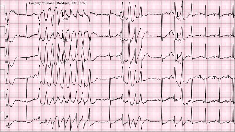
| Low Mg²⁺ with long QT or torsades | Cardiac emergency: unsynchronized shock first, then magnesium as an adjunct |
| Low Ca²⁺ with tetany and low Mg²⁺ | Hypocalcemia that will not correct until magnesium is replaced |
| High Mg²⁺ with falling reflexes or slow breathing | Magnesium toxicity: stop the infusion, ventilate, give IV calcium |
| Falling Mg²⁺ after feeds restart | Possible refeeding syndrome; grade by the ASPEN thresholds below |
Refeeding syndrome: ASPEN 2020 stratification
| Severity | Fall in phosphorus, potassium or magnesium within 5 days of calories |
|---|---|
| Mild | 10–20% decrease in any one or more of the three |
| Moderate | 20–30% decrease |
| Severe | >30% decrease, or organ dysfunction from the drop or from thiamin deficiency |
Management Priorities
Emergency: Torsades de pointes (polymorphic VT with a long QT)
- Shock first: Pulseless torsades or polymorphic VT gets an immediate unsynchronized shock (AHA 2025 class 1). Torsades with a pulse runs the ACLS pathway for VT with a pulse. Unstable? Synchronized cardioversion.
- Magnesium: give IV magnesium sulfate for recurrences with a long QT, per AHA 2025 class 2b (may be considered).
- Remove the cause: stop QT-prolonging drugs, correct potassium and treat bradycardia or pauses.
- Pauses or bradycardia: overdrive pacing or isoproterenol under expert guidance per AHA 2025.
Medications
| Drug and indication | Adult dose | Pediatric dose (max) | Onset / key notes |
|---|---|---|---|
| Magnesium sulfate: torsades | 2 g IV, diluted, over about 15 min (matches the label rate of 150 mg/min); further doses per protocol. Pulseless arrest timing follows ACLS protocol | Weight-based per local protocol/PALS; do not exceed the adult single dose | High-alert Shock first. Infusion generally not faster than 150 mg/min (label; the label's exception is severe eclampsia with seizures) |
| Magnesium sulfate: eclampsia (IV) | 4 g IV at up to 150 mg/min, then 1–2 g/hr; initial total 10–14 g with IM doses (label) | Obstetric use; not applicable | High-alert Max 30–40 g per 24 hr; 20 g per 48 hr with severe renal insufficiency (label). Obstetric protocols may set a different load and rate; follow local protocol |
| Magnesium sulfate: eclampsia (IM, no IV) | 4–5 g IM into each buttock (undiluted 50%), then 4–5 g every 4 hr in alternate buttocks if reflexes and breathing allow (label) | Obstetric use; not applicable | High-alert Continue only while the patellar reflex is present and RR is 16 or more (label) |
| Magnesium sulfate: mild hypomagnesemia | Mild: 1 g (2 mL of 50%) IM every 6 hr for 4 doses (label) | Weight-based per protocol; dilute IM to 20% or less | High-alert Replace potassium and calcium too. Oral replacement for mild deficits per protocol (no oral dose range is sourced here) |
| Magnesium sulfate: severe or symptomatic hypomagnesemia | IV replacement per protocol. Label example: 5 g in 1 L D5W or NS IV over 3 hr. The label also gives an IM regimen for severe deficiency, worded ambiguously, so use protocol | Weight-based per protocol; dilute IM to 20% or less | High-alert Avoid exceeding renal excretory capacity; reduce dose and recheck levels in renal impairment (label) |
| Magnesium sulfate: severe asthma (adjunct, not labeled) | Adults: single 2 g IV over 20 min for those who do not respond to initial treatment and have persistent hypoxemia (per GINA 2025) | Children 2 years and older: 40–50 mg/kg IV, max 2 g, over 10–20 min (per GINA 2025) | High-alert Not for routine use; for severe attacks that fail first-line therapy (per GINA 2025). Nebulized magnesium is not recommended (per GINA 2025) |
| Calcium chloride: magnesium toxicity | The label names no salt: "intravenous calcium, 10 to 20 mL of a 5% solution." The salt, concentration and dose follow local protocol; do not transfer this volume to calcium chloride | Per protocol | Antidote for magnesium toxicity; give with ventilatory support (label). Calcium chloride irritates tissue, so give it through a central or well-secured large vein |
Definitive care & disposition
- Torsades with pauses or bradycardia: expert consultation for pacing or isoproterenol; ICU care.
- Magnesium toxicity with respiratory depression: ICU, airway support, IV calcium; send serum magnesium and creatinine.
- Kidney failure with toxicity: nephrology consult. For severe toxicity, dialysis may be considered, per nephrology and protocol. This page's references do not cover the removal method.
- Hypomagnesemia that persists: review for renal-wasting drugs (cisplatin, aminoglycosides, proton pump inhibitors) and renal tubular disorders.
- Refeeding syndrome: ICU or nutrition-team care; monitor phosphorus, potassium and magnesium through the first days of feeding.
Doses are typical references. Follow local protocols, medical direction and institutional policy.
Field & Transport Considerations
Point-of-care testing
No point-of-care magnesium device appears in the sources reviewed. In the field you decide on the rhythm, the QT interval, knee reflexes, respiratory rate and urine output. The number does not drive the call.
Before and during interfacility transport
- Before each IV dose: check patellar reflexes, respiratory rate and urine output (label: patellar reflex present, about 16 breaths per minute or more).
- Pump and container: run magnesium on a smart pump, and do not connect flexible containers in series (label warning).
- Torsades: send the 12-lead, the QT and the most recent potassium and calcium with the patient.
- Eclampsia without IV access: IM is the labeled route when there is no IV. Keep the neonatal and obstetric teams in the loop on the total maternal dose.
| Setting | Consideration |
|---|---|
| Prehospital | Shock first for pulseless torsades; magnesium per protocol with medical control |
| Interfacility transport | Stop the infusion for lost reflexes or slow breathing; carry calcium and airway equipment |
| Obstetric transport | Watch urine output (label: at least 100 mL every 4 hr); note the time and dose of each bolus |
Special Populations
| Population | What changes | Clinical implication |
|---|---|---|
| Neonates & infants | Maternal infusion reaches the newborn; prolonged maternal use causes fetal hypocalcemia, bone changes and fractures | Tell the neonatal team the total maternal dose; ventilate and give IV calcium for hypotonia or poor breathing |
| Children & adolescents | Reference intervals are lab-specific; IM concentrate must be diluted to 20% or less for children | Weight-based doses per protocol; check the pediatric formulary |
| Pregnancy | Indicated for eclampsia and preeclampsia with severe features; continuous use beyond 5–7 days can cause fetal abnormalities (label) | Limit duration; follow the obstetric protocol. The cited premix label still lists as a contraindication IV magnesium in mothers with toxemia of pregnancy during the 2 hr before delivery. Obstetric practice often continues magnesium through delivery, so follow the obstetric protocol and medical direction |
| Older adults | Lower kidney reserve, so the excretion limit is reached sooner | Check creatinine and urine output before each repeat dose |
| CKD, dialysis & renal impairment | Only the kidney clears magnesium; renal insufficiency can cause intoxication | Severe renal insufficiency: maximum 20 g per 48 hr with frequent levels (label) |
| Neuromuscular disease | Magnesium blocks neuromuscular transmission; excessive block reported with neuromuscular blockers (label) | Expect added weakness; monitor strength and breathing. Myasthenia gravis is a general caution, not a labeled contraindication |
| Heart block or myocardial damage | Label contraindication for parenteral use | Decide with expert guidance; see the contraindication box under Management |
| Digoxin or other cardiac glycosides | Label: extreme caution; IV calcium given for magnesium toxicity can precipitate heart block | Check the rhythm and digoxin level; plan any calcium with cardiology or toxicology |
Interfering Factors & Pitfalls
Collection & handling
| Factor | Effect | Why | Avoid / fix |
|---|---|---|---|
| Sample drawn during or just after IV magnesium | ↑ (reflects the infusion) | Recent dose raises serum level above body stores | Note the infusion time; repeat when the infusion is stable |
| Hypoalbuminemia | Lower total Mg with normal ionized Mg | About 30% of serum magnesium is albumin-bound | Use an ionized assay where the lab offers one |
Medications & supplements
| Factor | Effect | Why | Avoid / fix |
|---|---|---|---|
| Loop and thiazide diuretics | ↓ | Renal magnesium loss | Check magnesium with potassium on therapy |
| Long-term proton pump inhibitors | ↓ | Gut and renal losses | Check magnesium on long-term therapy |
| Aminoglycosides, cisplatin, cyclosporine | ↓, can persist after stopping | Renal wasting | Monitor magnesium during and after therapy |
Physiologic & administration
| Factor | Effect | Why | Avoid / fix |
|---|---|---|---|
| Tissue depletion with normal serum | False − (serum normal) | Intracellular stores can be low while serum stays normal | Judge by symptoms, potassium and calcium; do not rule out deficiency on a normal level |
| Renal impairment | ↑ with dosing | Only the kidney excretes magnesium | Reduce or stop; check creatinine and urine output |
| Co-infusion with bicarbonate, phosphate or calcium | Precipitate, less drug delivered | Magnesium salts precipitate with these (label) | Use a separate line; flush well before calcium |
Common misinterpretations
- A normal serum level does not rule out deficiency. Tissue stores can run low while serum looks normal.
- Magnesium does not stop torsades on its own. Unsynchronized defibrillation comes first, per AHA 2025.
- Magnesium is not routine for every polymorphic VT. Without a long QT, AHA 2025 does not recommend routine use.
Physiology & Test Basics
What it measures
The serum test reads the extracellular pool of this divalent cation. That pool holds about 1% of body magnesium. Most of it sits inside cells, so a normal serum level does not prove normal stores. This page covers one analyte. Magnesium is not part of the standard CMP.
Physiology & regulation
- Second only to potassium as an intracellular cation. It is a cofactor for enzymes and for neuromuscular excitability.
- Most serum magnesium is ionized, the active form. About 30% is albumin-bound (label), and a small share is complexed.
- The kidney is the only route out. Excretion tracks the serum level and the GFR.
- Low magnesium impairs the Na⁺/K⁺-ATPase and lifts ROMK inhibition. Potassium is lost in urine as a result.
- Magnesium works peripherally to dilate vessels. Larger doses lower blood pressure (label).
How it is measured
Serum or plasma goes to the lab, and the tube and additive depend on the analyzer. Your lab's collection manual is the authority. Urine magnesium helps find the cause of a low level. Ionized and red-cell assays exist, but no standard reference range covers them.
Why it matters
Magnesium sets cardiac repolarization. Low levels lengthen the QT and promote torsades. They also make potassium and calcium hard to correct. High levels block neuromuscular transmission and breathing. That is why the knee reflex and respiratory rate are your bedside checks.
Clinical Pearls
References
- Wigginton JG, Agarwal S, Bartos JA, et al. (2025). Part 9: Adult advanced life support: 2025 American Heart Association guidelines for cardiopulmonary resuscitation and emergency cardiovascular care. Circulation, 152(16 suppl 2), S538–S577. https://doi.org/10.1161/CIR.0000000000001376
- Hospira, Inc. (2026). Magnesium sulfate in water for injection: prescribing information (revised August 2026). DailyMed, U.S. National Library of Medicine. https://dailymed.nlm.nih.gov/dailymed/drugInfo.cfm?setid=22ca78b4-f5a3-4144-cf89-5f633acf1e6d
- Hospira, Inc. (2026). Magnesium sulfate injection, USP 50%: prescribing information (revised April 2026). DailyMed, U.S. National Library of Medicine. https://dailymed.nlm.nih.gov/dailymed/drugInfo.cfm?setid=d4b5863a-efb9-4551-a9ad-2b06e8666d87
- da Silva JSV, Seres DS, Sabino K, et al. (2020). ASPEN consensus recommendations for refeeding syndrome. Nutrition in Clinical Practice, 35(2), 178–195. https://doi.org/10.1002/ncp.10474
- Papagiannidou A, Mitropoulou M, Papantzikos K, et al. (2026). Hypomagnesemia: a clinical and nutritional update. Current Nutrition Reports. https://doi.org/10.1007/s13668-026-00745-5
- Diaz V, Long Q, Oladapo OT. (2023). Alternative magnesium sulphate regimens for women with pre-eclampsia and eclampsia. Cochrane Database of Systematic Reviews, CD007388.pub3. doi:10.1002/14651858.CD007388.pub3. https://pmc.ncbi.nlm.nih.gov/articles/PMC10563167/
- Global Initiative for Asthma (2025). Global Strategy for Asthma Management and Prevention: 2025 update (updated November 2025). Magnesium sulfate: Box 12-2 and adult and pediatric sections. https://ginasthma.org/wp-content/uploads/2025/11/GINA-2025-Update-25_11_08-WMS.pdf
- Institute for Safe Medication Practices. ISMP List of High-Alert Medications in Acute Care Settings (2018 posting; PDF retrieved October 2026; magnesium sulfate injection entry). https://www.ismp.org/sites/default/files/attachments/2018-01/highalertmedications(1).pdf
Medical Disclaimer
- For Educational Purposes Only: This content is intended for educational reference and should not be used for clinical decision-making.
- Not a Substitute for Professional Judgment: Always consult your local protocols, institutional guidelines, and supervising physicians.
- Verify Before Acting: Users are responsible for verifying information through authoritative sources before any clinical application.