

At a Glance
- Represents
- Ventricular depolarization and repolarization: QRS onset to T-wave end, corrected for heart rate as the QTc.
- Normal (Adult)
- QTc <450 ms (men), <460 ms (women). Short QTc: ≤340 ms is a screening flag (≤360 ms with history).
- Measure It
- Longest QT in lead II or V5–V6, averaged over several beats. Use Fridericia at HR <60 or >90 bpm.
- Abnormal → Think
- Low K⁺, Mg²⁺ or Ca²⁺, QT-prolonging drugs, bradycardia or pauses, hypothermia, ischemia, raised ICP; congenital LQTS if no cause.
- Red Flags
- QTc ≥500 ms or a rise of ≥60 ms from baseline; polymorphic VT (torsades); syncope with a long QT.
- Bedside Action
- Stop QT drugs; check K⁺ and Mg²⁺; magnesium 2 g IV for recurrent torsades; unsynchronized shock if pulseless; seek expert input for pacing or isoproterenol for pauses.
Physiology & What It Represents
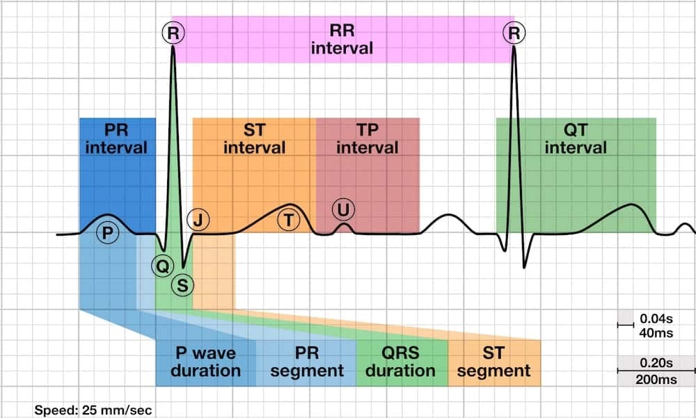
The QT interval is the window from QRS onset (the start of ventricular depolarization) to the end of the T wave (the end of ventricular repolarization). Its length tracks the ventricular action potential, mainly the plateau (phase 2) and repolarization (phase 3). Phase 3 runs largely on potassium current. Low K⁺ or Mg²⁺ stretches the window. So do many drugs and some inherited channel defects.
Repolarization does not happen at the same rate across the ventricular wall. Make it more uneven, and early afterdepolarizations can set off torsades de pointes (TdP): a polymorphic VT that twists around the baseline at about 160–240/min. Most episodes stop on their own. Some degenerate into VF.
The QT shortens as the heart rate rises and lengthens as it falls. A raw QT cannot be compared between beats or patients. Correct for heart rate and you get the QTc, the number you use to judge risk.
- Who is at risk: A long QTc alone does not mean TdP. Risk climbs with a QTc above 500 ms, a rise of 60 ms from baseline, several QT-prolonging drugs, low K⁺ or Mg²⁺, bradycardia or pauses, female sex, older age, and heart failure or other heart disease.
Normal Values & How to Measure
| Parameter | Adult Normal | Pediatric Note | Where / How to Measure |
|---|---|---|---|
| QT interval | Rate-dependent; no fixed normal | Always correct before comparing | Lead II or V5–V6; QRS onset to T-wave end |
| QTc, men | <450 ms (≥450 ms prolonged) | Use age-specific limits | Formula in the table below |
| QTc, women | <460 ms (≥460 ms prolonged) | Use age-specific limits | Formula in the table below |
| Torsades-risk trigger | QTc ≥500 ms, or a rise of ≥60 ms from baseline | Same action threshold; confirm with a pediatric reference | Compare with the prior ECG on the same lead |
Correction Formulas
Measure RR in seconds (RR = 60 / heart rate). Bazett is the default on most machines. Don't assume the machine's default is the best choice. In a 2016 comparison of 6,609 patients, Fridericia and Framingham corrected for heart rate best. Bazett performed worst.
| Formula | Equation | Best Use | Limitation |
|---|---|---|---|
| Bazett | QTc = QT / √RR | Familiar; HR near 60–100 bpm | Overcorrects at high rates and undercorrects at low rates |
| Fridericia | QTc = QT / ∛RR | HR below 60 or above 90–100 bpm | Less often the machine default |
| Framingham | QTc = QT + 154 × (1 − RR) ms, RR in seconds | Rate extremes; performed best in 2016 | Less familiar to most clinicians |
| Hodges | QTc = QT + 1.75 × (HR − 60) ms | Linear; uses heart rate directly | Not among the top performers in the 2016 comparison |
Normal and Abnormal QTc
| Category | QTc, men / women | Clinical Significance |
|---|---|---|
| Normal | <450 ms / <460 ms | Baseline torsades risk |
| Borderline | 450–499 ms / 460–499 ms | Mild increased risk; look for and correct causes |
| Prolonged, high risk | ≥500 ms (both) | Torsades risk rises steeply; act now |
| Rise from baseline | ≥60 ms increase | Act even when the QTc stays under 500 ms |
| Short | ≤340 ms is a screening flag (≤360 ms with supporting history) | Linked to atrial and ventricular fibrillation; diagnosis needs cardiology review; see Short QT Interval |
Measurement Technique
- Choose the lead: Use the lead with the longest, clearest T wave, usually II or V5–V6. Use the same lead on serial ECGs.
- Mark the start: QRS onset, the first deflection of the Q or R wave. In a wide QRS, mark the earliest deflection.
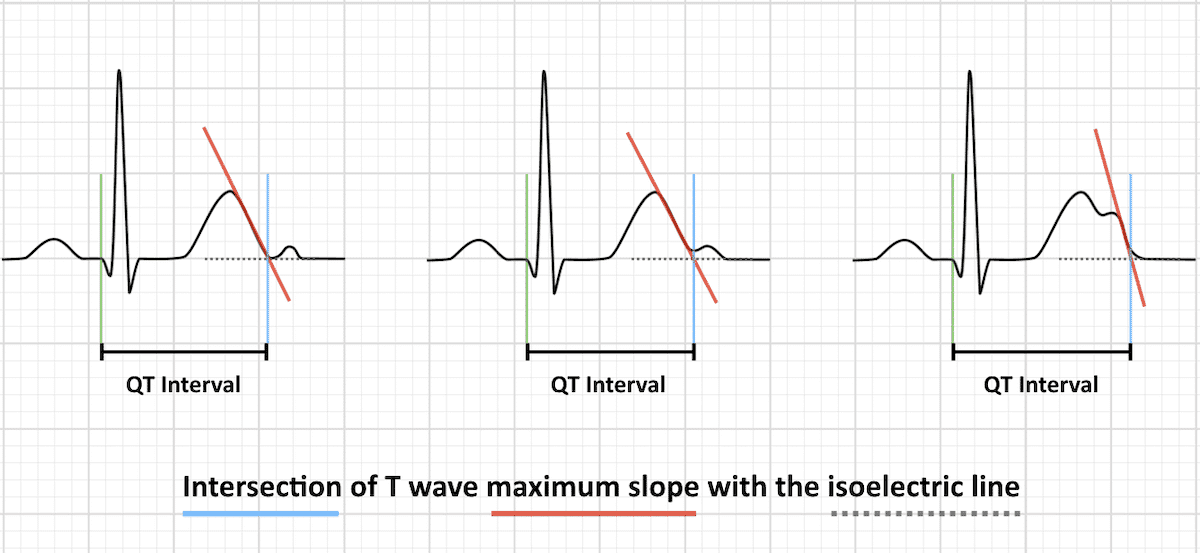
- Mark the end: The T-wave end, using the maximum-slope (tangent) method, where the steepest downslope meets the baseline. Exclude the PR segment.
- Handle U waves: Include a U wave only when it merges with the T wave. When U waves are separate, measure to the nadir between the T and U.
- Average beats: Measure several consecutive beats and average them. In atrial fibrillation or frequent ectopy, average more beats.
- Correct and verify: Use Fridericia or Framingham when the HR is below 60 or above 90–100 bpm, and check any automated QTc by hand.
Acquired Prolonged QT Interval
Acquired QT prolongation means repolarization got slower. Drugs do it. So do electrolyte disturbances, bradycardia or pauses, and some systemic and neurologic conditions. It is usually reversible. That is why you check it first.
ECG Criteria
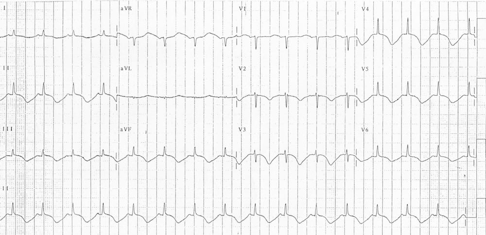
- QTc: ≥450 ms in men or ≥460 ms in women
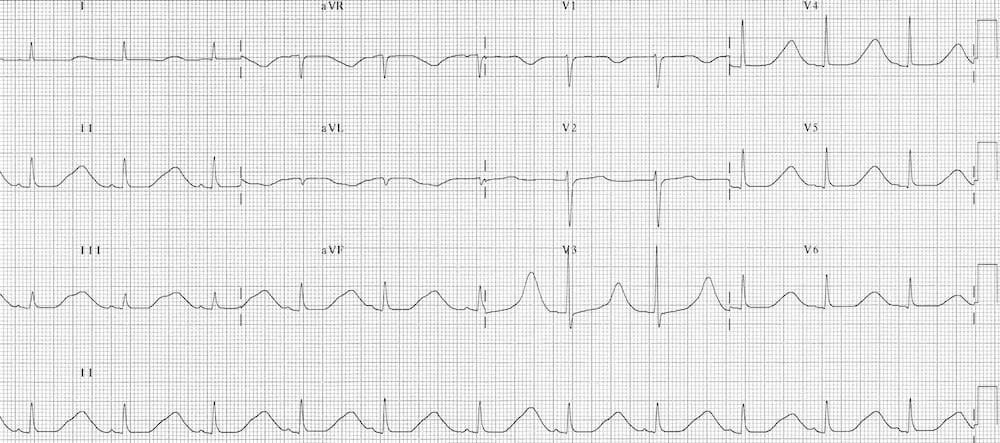
- Hypokalemia: apparent prolongation from fused U waves, with flat T waves and ST depression
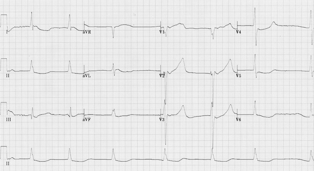
- Hypocalcemia: a long, flat ST segment with a relatively normal T wave
- Trend: a rise of 60 ms from baseline matters even under 500 ms
Causes
| Category | Examples |
|---|---|
| Electrolytes | Low K⁺, low Mg²⁺, low Ca²⁺ |
| Drugs | Antiarrhythmics, antipsychotics, antiemetics, methadone, macrolides and fluoroquinolones (see next section) |
| Bradycardia and pauses | Sinus or AV-block bradycardia; pauses after premature beats |
| Ischemia and MI | Acute ischemia can cause modest prolongation; check the trend, not one value |
| Temperature | Hypothermia: marked prolongation with Osborn (J) waves and shivering artifact |
| Neurologic | Raised intracranial pressure and subarachnoid hemorrhage (cerebral T waves) |
| After cardiac arrest | Common after ROSC; reassess once the patient stabilizes |
| Endocrine and nutritional | Hypothyroidism; anorexia and starvation |
Clinical Significance
- Risk gradient: Congenital LQTS data tie a QTc above 500 ms to a 2- to 3-fold higher TdP risk. Drug-induced case series show a similar rise (2010 AHA statement). Torsades risk climbs with the QTc. No single threshold guarantees it.
- Action threshold: After a QT-prolonging drug, act promptly if the QTc tops 500 ms or rises at least 60 ms from the predrug baseline (2010 AHA statement).
- Reserve: Low K⁺ and Mg²⁺ eat into the heart's repolarization reserve. The same drug then prolongs the QT more.
- Neurogenic changes: Cerebral T-wave changes after subarachnoid hemorrhage come from the neurologic event. They are not primary heart disease.
Drug-Induced QT Prolongation and Torsades Risk
Many drugs block the IKr (hERG) potassium current. That lengthens repolarization. Torsades is the end point you fear. Risk peaks with several QT-prolonging drugs, high doses, rapid IV infusion, and electrolyte or heart-rate risk factors. The 2010 AHA statement on torsades prevention lists them.
Drug Classes and Examples
| Drug Class | Examples | Note |
|---|---|---|
| Antiarrhythmics | Amiodarone, sotalol, dofetilide, ibutilide, procainamide, quinidine | Stacking drugs compounds QT risk |
| Antipsychotics | Haloperidol, ziprasidone, chlorpromazine, prochlorperazine | Ziprasidone is contraindicated with known QT prolongation (label) |
| Antiemetics | Ondansetron, droperidol | Droperidol has a boxed warning; avoid ondansetron in congenital long QT |
| Opioids | Methadone | Boxed warning for life-threatening QT prolongation |
| Antibiotics and antifungals | Azithromycin, erythromycin, levofloxacin, moxifloxacin, fluconazole | Check the list before adding |
| Antidepressants | Citalopram, escitalopram, tricyclics | Dose-related QT effect |
Check drug interactions at CredibleMeds. It sorts QT-prolonging drugs by torsades risk category.
Torsades Risk Factors
- QTc above 500 ms, or a rise of 60 ms from baseline
- More than one QT-prolonging drug, or rapid IV infusion
- Hypokalemia, hypomagnesemia or hypocalcemia; diuretic use
- Bradycardia or pauses; female sex; advanced age; heart failure or other heart disease
Congenital Long QT Syndrome (LQTS)
Congenital long QT syndrome (LQTS) is inherited. Mutations in the genes that build cardiac ion channels lengthen repolarization. That sets up syncope, torsades and sudden cardiac death. The most common forms are LQT1 (KCNQ1), LQT2 (KCNH2) and LQT3 (SCN5A).
ECG Criteria
- Persistently prolonged QTc on serial ECGs with no secondary cause
- Diagnosis rests on the QTc, events, family history and genetic testing; one QTc value is not enough
Triggers by Subtype
| Subtype | Trigger Pattern | Events During Exercise | Events During Rest or Sleep |
|---|---|---|---|
| LQT1 | Exercise | 62% | 3% |
| LQT2 | Emotional stress | 13%* | 29% |
| LQT3 | Rest or sleep | 13%* | 39% |
*Exercise events for LQT2 and LQT3 are pooled. The shares come from the 2001 genotype cohort of 670 symptomatic patients.
Clinical Significance
- Red flags: Syncope with exertion, swimming or emotion. Family history of sudden unexplained death at a young age.
- Therapy: Beta-blockers are the mainstay. In the 2001 cohort, beta-blocker recurrence-free rates were 81% in LQT1 and 59% in LQT2. ICD therapy is considered in high-risk patients (2022 ESC).
- Avoid: QT-prolonging drugs and catecholamine stimulation such as isoproterenol (see Bedside Management)
Short QT Interval (Short QT Syndrome)
A short QT is rarer than prolongation. It still links to atrial fibrillation, ventricular fibrillation and sudden cardiac death. Hypercalcemia and the digoxin effect both shorten it. Digoxin toxicity is a separate, life-threatening emergency.
ECG Criteria
- QTc: ≤340 ms, or ≤360 ms with supporting history, prompts a short-QT workup. Think of it as a screening flag, not a diagnosis. No consensus QT cut-off existed in 2011 (Gollob). Diagnostic scores also weigh family history and genetic findings. Confirm with cardiology.
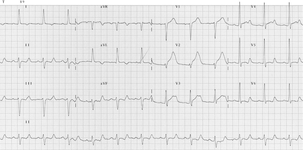
- Hypercalcemia: a shortened ST segment is the most prominent change
- Congenital short QT: very short QTc with tall, peaked T waves
Causes
| Category | Examples |
|---|---|
| Electrolyte | Hypercalcemia from hyperparathyroidism, malignancy, vitamin D toxicity or immobilization |
| Drug effect | Therapeutic digoxin: QT shortening with reverse-tick ST depression |
| Congenital | Short QT syndrome, an inherited potassium-channel disorder |
Clinical Significance
- Outcomes: In a 2011 review of 61 reported cases, 57% presented with sudden cardiac death, aborted arrest, syncope or atrial fibrillation as the index event.
- QTc range: Symptomatic cases sat between 248 and 381 ms, with a mean of 307 ms.
- Family history: Sudden death in relatives, or atrial fibrillation in a young adult, should prompt testing.
U Waves
Think of the U wave as a low bump that follows the T wave and points the same way. Prominent U waves most often point to hypokalemia. They can merge with the T wave and mimic a long QT.
ECG Criteria
- Low-amplitude deflection after the T wave, most visible in V2–V3
- Prominent U waves with flat T waves and ST depression: low K⁺
- T–U fusion: the measured QT can be overestimated; apply the U-wave rules in Normal Values
Clinical Significance
- Action: Prominent U waves mean you check K⁺ and Mg²⁺ now, before any drug decision.
- Mimic: A fused U wave is not a long QT until you check electrolytes and the medication list.
Differential Diagnosis & Mimics
Several conditions can mimic a prolonged QT, and some hide one.
| Feature | Hypokalemia (T–U fusion) | Congenital LQTS | Wide QRS or Paced Rhythm |
|---|---|---|---|
| ECG clue | Prominent U waves, flat T, ST depression | Long QTc with no cause on repeat ECGs | QT includes the QRS; the interval is hard to judge |
| History | Diuretics, vomiting, poor intake | Syncope with exertion or emotion; family history | Bundle branch block or ventricular pacing |
| Next step | K⁺ and Mg²⁺ now; repeat ECG after repletion | Cardiology and genetic evaluation | Compare with the prior ECG; subtract the QRS-width change or use the JT interval (2010 AHA statement) |
Artifact and Lead Misplacement
- Shivering or tremor: Blurs the T-wave end and can fake a long or irregular QT. Repeat the ECG when the patient is still.
- Limb lead reversal: Flips lead polarity and can distort the T wave.
- Precordial misplacement: Shifts V-lead morphology. Check the electrode positions.
- Transport vibration: Adds baseline noise. Repeat the tracing once the vehicle is stopped.
Normal Variants
- Sex: Women run a higher baseline QTc than men. That is why the cut-offs are sex-specific.
- Sinus arrhythmia: The QT changes with each RR interval. Average several beats.
Bedside Management & Escalation
Treatment follows the cause and the rhythm. Your priorities: unsynchronized defibrillation for pulseless torsades, magnesium for torsades, electrolyte correction, and stopping QT-prolonging drugs.
| Situation | BLS (EMT) | ALS (AEMT / Paramedic / RN) | Critical Care / Transport |
|---|---|---|---|
| Unstable with a pulse (hypotension, altered mental status, chest pain; torsades with a pulse) | 12-lead if allowed; oxygen if SpO₂ <94%; AED pads on; notify; early ALS | Unsynchronized shock for sustained polymorphic VT (AHA 2025 Part 9, COR 1); magnesium 2 g IV for recurrent torsades with a long QT (COR 2b); seek expert input for pacing or isoproterenol if pause-dependent | Continuous ECG; repeat QTc after magnesium; pads on; expert input before adding a second QT drug |
| Stable (asymptomatic prolonged QTc) | Notify; 12-lead if allowed; keep monitor on; give no QT-prolonging drugs | Hold QT-prolonging drugs; correct K⁺ and Mg²⁺; repeat 12-lead; medical direction | Monitored bed; trend the QTc; avoid delays for transport |
| Pulseless (torsades or pulseless VT) | CPR; AED or manual defibrillation | Unsynchronized defibrillation and CPR first; routine magnesium is not recommended in arrest (AHA 2025 Part 9, 3: No Benefit, B-R) | Same, plus treatment of reversible causes and handoff of QT drugs |
| Therapy | Adult | Pediatric | Key Notes |
|---|---|---|---|
| Magnesium sulfate (torsades) | With a pulse, for recurrent torsades with a long QT: 2 g IV over 1–2 min (NAS Cardiac Arrest p. 119); repeat 2 g if torsades recurs. Pulseless: routine magnesium is not recommended (AHA 2025 Part 9, 3: No Benefit, B-R); never delay defibrillation or CPR for it. AHA 2025 gives no dose or infusion time. Max 2 g per dose |
25–50 mg/kg IV/IO (NAS Cardiac Arrest p. 119). Max 2 g per dose |
High-alert Differs from FDA label: torsades is not a labeled indication for magnesium sulfate, and the label gives no torsades dose. AHA 2025 Part 9 (polymorphic VT, rec 2): may be considered for recurrent torsades with a long QT (COR 2b, C-LD); no benefit for polymorphic VT with a normal QT (3: No Benefit, C-LD). Renal impairment: magnesium accumulates; reduce or delay repeat doses and check Mg²⁺. Toxicity: loss of reflexes and respiratory depression; stop the infusion and give an injectable calcium salt (label). |
| Unsynchronized defibrillation (pulseless torsades or VF) | Biphasic: manufacturer's first-shock setting; if unknown, the device maximum (AHA 2025 Part 9, rec 6, COR 2b). For polymorphic VT (including torsades), use the maximum manufacturer's setting (AHA 2025 Part 9, polymorphic VT supportive text). Monophasic: device maximum, usually 360 J. Per algorithm |
2 J/kg, then 4 J/kg (AHA 2025 Part 8, COR 2a and 2b). Max 10 J/kg |
Unsynchronized for polymorphic VT or VF; never synchronized for a pulseless rhythm. |
| Overdrive pacing (pause-dependent torsades) | Temporary pacing to prevent pauses. AHA 2025 Part 9 names overdrive pacing only as an expert-consultation option for pause-precipitated torsades and gives no rate; the above-70/min figure is from the 2010 statement. Rate per protocol |
Not routinely used; specialist-directed |
Pacing shortens the pause that starts each run. Pads on before moving an unstable patient. |
| Isoproterenol (recurrent pause-dependent torsades, expert input) | Start at the lowest infusion rate and titrate to the heart rate that suppresses pauses; the labeled shock range is 0.5–5 mcg/min IV, and no labeled torsades dose exists. Titrate to effect, per protocol |
Not established for torsades; specialist use only |
High-alert Label contraindications include ventricular arrhythmias, so torsades use is off-label and expert-directed. Avoid in congenital long QT. Reduce if the heart rate exceeds 110/min (label). Use as a bridge to pacing. AHA 2025 Part 9 names isoproterenol only as an expert-consultation option for pause-precipitated torsades and gives no dose. |
| Potassium (IV) (replace to target) | Per protocol; titrate to K⁺. No fixed max |
Weight-based, per protocol |
High-alert Concentrated potassium: dilute; never IV push. Target: K⁺ ≥4 mEq/L. AHA 2010 mentions 4.5–5 mmol/L as a class IIb option; AHA 2025 Parts 8–9 give no potassium target. |
Follow local protocols and online medical direction.
Pediatric & Special Populations
Pediatrics
- Normal limits: QTc limits shift with age and sex. Use age-specific references, not adult cut-offs.
- Heart-rate correction: Bazett overcorrects at high rates. Infants and children often run high rates. Prefer Fridericia or Framingham.
- Congenital LQTS: Can present in childhood with syncope during exercise or emotion. When a child is affected, screen the family.
- Torsades dosing: magnesium 25–50 mg/kg IV/IO (max 2 g; NAS Cardiac Arrest p. 119; AHA 2025 PALS has no magnesium recommendation); defibrillation 2 J/kg, then 4 J/kg (max 10 J/kg; AHA 2025 Part 8, COR 2a and 2b); isoproterenol not established.
Read this with weight-based dosing and pediatric vital signs.
Other Populations
- Women: Higher baseline QTc. Use sex-specific cut-offs, and check the medication list before any QT-prolonging drug.
- Older adults: They take more QT-prolonging drugs, get diuretic-related hypokalemia and have reduced renal clearance.
- Heart failure and heart disease: Same drug, same dose, higher risk.
- Pregnancy: Torsades is treated as in other adults. Magnesium is also used for eclampsia, so check the obstetric protocol.
Monitoring & Transport Considerations
- Best lead: Trend on lead II or V5. Pick the lead with the most visible T wave and keep it for the whole transport.
- Repeat 12-leads: After each QT-prolonging dose, after electrolyte repletion, and with any new rhythm change.
- Continuous monitoring: Stay on the monitor. Droperidol labeling calls for ECG monitoring for 2–3 hours after the dose.
- Pads: Apply pads early for torsades, syncope or polymorphic VT. Keep the defibrillator in reach.
- Pacing: Pads on before you move an unstable bradycardic or pause-dependent patient. Confirm capture by pulse and SpO₂ waveform.
- Artifact: Shivering, tremor and vibration mimic a prolonged or irregular QT. Re-acquire the tracing when the patient is still.
- Alarms: Set rate, pause and ectopy alarms. Use QTc trending where the monitor offers it.
- Handoff: List each QT-prolonging drug with its dose and time, the last QTc, K⁺, Mg²⁺ and any magnesium given.
Step-by-Step Approach & Pitfalls
- Measure the QTc on a 12-lead in the lead with the clearest T wave, and check the rhythm first.
- Check K⁺, Mg²⁺ and Ca²⁺ and correct them as the protocol allows.
- Review every medication for QT-prolonging drugs, including doses given before EMS or ED arrival.
- Stop or hold non-essential QT-prolonging drugs, and choose an alternative with less QT effect.
- Treat torsades: unsynchronized defibrillation for sustained polymorphic VT; magnesium 2 g IV for recurrent torsades with a long QT.
- Address pauses and bradycardia: pace or give isoproterenol per protocol and expert input.
- Repeat the 12-lead after each change, and document the QTc and the drugs given.
Common Pitfalls
- Using Bazett alone at extreme heart rates
- Accepting an automated QTc without checking the T-wave end
- Giving amiodarone or procainamide for torsades
- Adding a second QT-prolonging drug without a repeat ECG
- Calling a fused U wave a long QT without checking K⁺
- Stopping monitoring too early after droperidol or another QT-prolonging dose
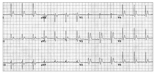
ECG Examples


Clinical Pearls
References
- Drew BJ, Ackerman MJ, Funk M, et al. Prevention of torsade de pointes in hospital settings: a scientific statement from the American Heart Association and the American College of Cardiology Foundation (landmark scientific statement). Circulation. 2010;121(8):1047–1060. https://doi.org/10.1161/CIRCULATIONAHA.109.192704
- Rautaharju PM, Surawicz B, Gettes LS, et al. AHA/ACCF/HRS recommendations for the standardization and interpretation of the electrocardiogram: part IV: the ST segment, T and U waves, and the QT interval. J Am Coll Cardiol. 2009;53(11):982–991. https://doi.org/10.1016/j.jacc.2008.12.014
- Vandenberk B, Vandael E, Robyns T, et al. Which QT correction formulae to use for QT monitoring? J Am Heart Assoc. 2016;5(6):e003264. https://doi.org/10.1161/JAHA.116.003264
- Zeppenfeld K, Tfelt-Hansen J, de Riva M, et al. 2022 ESC Guidelines for the management of patients with ventricular arrhythmias and the prevention of sudden cardiac death. Eur Heart J. 2022;43(40):3997–4126. https://doi.org/10.1093/eurheartj/ehac262
- Wigginton JG, Agarwal S, Bartos JA, et al. Part 9: Adult advanced life support: 2025 American Heart Association guidelines for cardiopulmonary resuscitation and emergency cardiovascular care. Circulation. 2025;152(16 suppl 2):S538–S577. https://doi.org/10.1161/CIR.0000000000001376
- Lasa JJ, Dhillon GS, Duff JP, et al. Part 8: Pediatric advanced life support: 2025 American Heart Association and American Academy of Pediatrics guidelines for cardiopulmonary resuscitation and emergency cardiovascular care. Circulation. 2025;152(16 suppl 2):S479–S537. https://doi.org/10.1161/CIR.0000000000001368
- Schwartz PJ, Priori SG, Spazzolini C, et al. Genotype-phenotype correlation in the long-QT syndrome: gene-specific triggers for life-threatening arrhythmias (landmark cohort study). Circulation. 2001;103(1):89–95. https://doi.org/10.1161/01.CIR.103.1.89
- Gollob MH, Redpath CJ, Roberts JD. The short QT syndrome: proposed diagnostic criteria. J Am Coll Cardiol. 2011;57(7):802–812. https://doi.org/10.1016/j.jacc.2010.09.048
- American Regent, Inc. Droperidol injection: prescribing information. DailyMed; revised 2023. https://dailymed.nlm.nih.gov/dailymed/drugInfo.cfm?setid=147e033d-d997-4ef6-8bb5-a9ba372590b2
- Mylan Institutional LLC. Methadone hydrochloride injection: prescribing information. DailyMed; revised 2026. https://dailymed.nlm.nih.gov/dailymed/drugInfo.cfm?setid=092d78eb-6423-495c-bf0d-e6532bea7138
- National Association of State EMS Officials (NASEMSO). National Model EMS Clinical Guidelines, Version 3.0. Cardiac Arrest, p. 119. Published March 2022. https://nasemso.org/content.aspx?page_id=22&club_id=157064&module_id=701974
Medical Disclaimer
- For Educational Purposes Only: This content is intended for educational reference and should not be used for clinical decision-making.
- Not a Substitute for Professional Judgment: Always consult your local protocols, institutional guidelines, and supervising physicians.
- Verify Before Acting: Users are responsible for verifying information through authoritative sources before any clinical application.