

Bedside Snapshot
- Normal (Adult)
- 27–33 pg (1.68–2.05 fmol); some labs use 26 pg as the lower limit in women. Newborns run higher; young children lower.
- Critical Values
- No universal critical value: interpret by trend and clinical context. Act on the hemoglobin and the patient, not the MCH.
- High Suggests
- B12 or folate deficiency
- Alcohol, liver disease, drugs
- Myelodysplasia, reticulocytosis
- Artifact: cold agglutinins, lipemia
- Low Suggests
- Iron deficiency (chronic blood loss)
- Thalassemia trait
- Anemia of inflammation
- Sideroblastic anemia (rare)
- Act Now If
- Low MCH with bleeding or a falling hemoglobin: treat the bleed and anemia. High MCH with numbness, ataxia or confusion: suspect B12 deficiency; do not delay treatment.
- Watch Out For
- A normal MCH does not exclude deficiency: combined iron and B12/folate deficiency can average out. Check RDW and the smear.
Reference & Critical Ranges
Reference ranges
| Population | Conventional | SI | Notes |
|---|---|---|---|
| Adult | 27–33 pg | 1.68–2.05 fmol | Typical. Some labs set the lower limit at 26 pg (1.61 fmol) in women. |
| Pregnancy | 27–33 pg | 1.68–2.05 fmol | No separate range: hemodilution lowers hemoglobin, not MCH. |
| Older adult | 27–33 pg | 1.68–2.05 fmol | Same as adult. Age does not explain a new abnormal value. |
| Neonate, term (day 1) | About 36 pg | About 2.24 fmol | Higher in preterm infants: about 40 pg at ≤25 weeks. Untransfused neonates. |
| Infant 0–<1 y | 22.9–32.7 pg | 1.42–2.03 fmol | Falls through the first year. |
| Child 1–<3 y | 22.7–29.0 pg | 1.41–1.80 fmol | Lowest values of life; the adult cut-off of 27 pg does not apply. |
| Child 3–<6 y | 25.2–29.3 pg | 1.56–1.82 fmol | — |
| Child 6–<13 y | 25.4–30.8 pg | 1.58–1.91 fmol | — |
| Adolescent 13–<21 y | 25.9–32.4 pg | 1.61–2.01 fmol | No sex difference; rises toward adult values. |
Severity & critical values
| Level | Conventional | SI | What it means |
|---|---|---|---|
| Markedly low | <25 pg | <1.55 fmol | Iron deficiency or thalassemia trait (including α⁰ carrier states). In pregnancy, check iron and follow the antenatal hemoglobinopathy screening pathway. |
| Low (hypochromic) | 25–26.9 pg | 1.55–1.67 fmol | Early or mild iron deficiency, α⁺ thalassemia trait, anemia of inflammation. |
| Normal | 27–33 pg | 1.68–2.05 fmol | Does not exclude combined iron and B12/folate deficiency or early deficiency. |
| High | >33 pg | >2.05 fmol | Almost always larger cells (macrocytosis): B12 or folate deficiency, alcohol, liver disease, drugs, reticulocytosis; or artifact. |
| Critical | None defined | — | Act on the hemoglobin, bleeding and symptoms, not on MCH. |
Conversion: Multiply pg by 0.0621 to get fmol per cell. Labs in the US and UK report MCH in pg.
Example: Take Hb 14 g/dL and an RBC count of 4.5 ×10⁶/µL. Divide 14 × 10 by 4.5 to get 31.1 pg, which is normal.
Interpretation: An MCV of 90 fL with an MCHC of 33 g/dL gives 29.7 pg. MCHC sits in a narrow band, so MCH follows MCV.
Causes of Abnormal Results
Low (hypochromic, MCH <27 pg)
A low MCH means the marrow built red cells with too little hemoglobin. Three things cause it: not enough iron, not enough globin chains, or iron the cell cannot use. Almost always the MCV is low too (microcytosis).
| Mechanism | Common causes | Bedside clue |
|---|---|---|
| Absolute iron deficiency | Chronic GI blood loss (including colorectal and gastric cancer), menstrual loss, pregnancy, poor intake, celiac disease, gastrectomy or bariatric surgery | Melena or positive history of bleeding, heavy periods, pica; low ferritin; man or postmenopausal woman with no obvious cause |
| Functional iron deficiency | Chronic kidney disease, heart failure, inflammatory bowel disease, chronic inflammation | Ferritin normal or high but transferrin saturation low; known chronic disease |
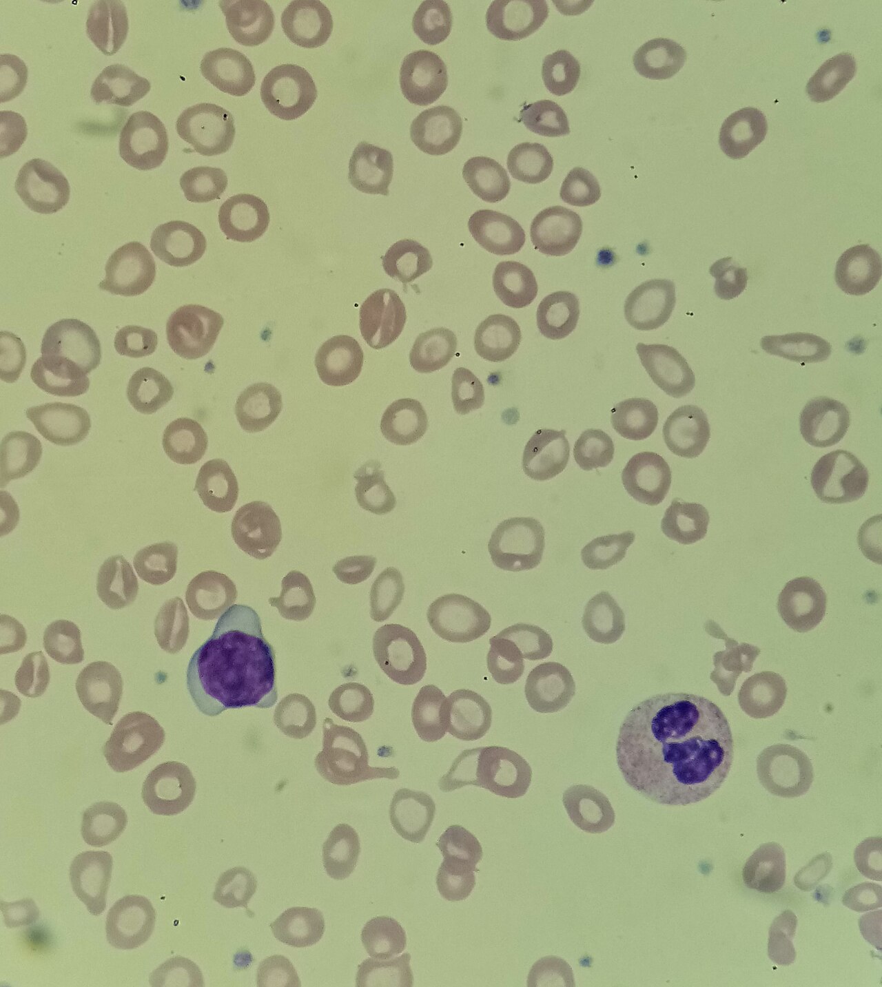
| Reduced globin synthesis | α- or β-thalassemia trait, hemoglobin Lepore, hemoglobin E or C trait | MCV and MCH low out of proportion to a mild or absent anemia; normal ferritin; family origin or family history |
| Anemia of inflammation (chronic disease) | Infection, autoimmune disease, malignancy | Usually normochromic; mildly low MCH with normal or high ferritin |
| Defective heme synthesis | Sideroblastic anemia (inherited or acquired); lead poisoning | Iron studies not low; ring sideroblasts on marrow; basophilic stippling on smear with lead |
High (MCH >33 pg)
Red cells have a ceiling on how much hemoglobin they can hold, which is why MCHC has a limit. So a high MCH almost always means bigger cells (macrocytosis), not "hyperchromic" cells. Mild rises are common and often benign. Find the cause before you put a label on it.
| Mechanism | Common causes | Bedside clue |
|---|---|---|
| Impaired DNA synthesis (megaloblastic) | Vitamin B12 deficiency (autoimmune gastritis, gastrectomy, ileal disease, vegan diet), folate deficiency (poor diet, alcohol, pregnancy, malabsorption) | Numbness, ataxia, cognitive change, glossitis; hypersegmented neutrophils; high LDH and indirect bilirubin |
| Drugs | Hydroxyurea, methotrexate, azathioprine, zidovudine and other antiretrovirals, valproic acid, phenytoin, trimethoprim, metformin | Medication list; MCV can exceed 130 fL with hydroxyurea or antiretrovirals |
| Alcohol and liver disease | Alcohol use disorder, cirrhosis | Macrocytosis (MCV 100–110 fL) often before anemia; raised GGT |
| Marrow disorders | Myelodysplastic syndrome | Older adult; other cytopenias (low WBC or platelets) |
| Young red cells | Reticulocytosis after hemolysis or bleeding | Reticulocyte count >100,000/µL; jaundice, dark urine, recent bleed |
| Endocrine | Hypothyroidism | Check TSH |
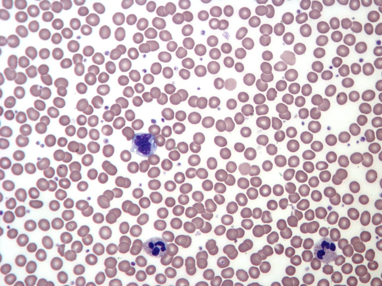
| Artifact | Cold agglutinins, lipemia, marked leukocytosis, in-vitro hemolysis | MCHC implausibly high; analyzer flag; normal smear |
Interpretation & Bedside Approach
Step-by-step approach
- Is it real? Check the MCHC and the analyzer flags. An MCHC above about 37 g/dL with a high MCH points to cold agglutinins, lipemia or a hemolyzed sample. Ask the lab to warm or correct the sample and rerun it.
- How sick is the patient? MCH is never the emergency. Look first at the hemoglobin, the vital signs, active bleeding and neurologic signs.
- Read MCH with MCV, MCHC, RDW and the RBC count. MCH and MCV normally move together. When they split apart, think spherocytosis, artifact or a mixed population of cells.
- Low MCH: check iron first. A ferritin below 15 µg/L confirms empty iron stores. Below 45 µg/L, iron deficiency is likely. Inflammation pushes ferritin up, so a ferritin above 150 µg/L makes absolute iron deficiency unlikely.
- Low MCH with normal iron studies: request hemoglobin analysis (high-performance liquid chromatography or electrophoresis) for thalassemia trait or a hemoglobin variant. Especially when the family origin is suggestive.
- High MCH: check B12, folate, reticulocyte count, liver tests and TSH. Review drugs and alcohol, and look at the smear. Other cytopenias on top of that make it a hematology referral (myelodysplastic syndrome).
- Trend beats a single value. On oral iron, a hemoglobin rise of 1 g/dL (10 g/L) or more within 2 weeks supports iron deficiency. MCH lags behind. Cells made before treatment keep circulating for their full lifespan of about 120 days.
Pattern recognition
| Condition | MCV and MCH | MCHC | Distinguishing clue |
|---|---|---|---|
| Iron deficiency | Low | Normal or low | Low ferritin; responds to iron |
| Thalassemia trait | Low, out of proportion to the anemia | Normal or low | Normal ferritin; abnormal hemoglobin analysis (β trait: raised HbA₂) |
| Anemia of inflammation | Normal or mildly low | Normal | Ferritin normal or high (acute-phase reactant) |
| B12 or folate deficiency | High | Normal | Hypersegmented neutrophils; high LDH and indirect bilirubin |
| Alcohol, liver, drugs, hypothyroidism | High (usually mild) | Normal | History, GGT, TSH, medication list |
| Hereditary spherocytosis | MCV normal or low; MCH normal | High | Spherocytes on smear; signs of hemolysis; family history |
| Acute blood loss (first hours) | Normal | Normal | Indices unchanged; hemoglobin falls later |
| Artifact (cold agglutinin, lipemia) | MCH falsely high | Falsely high, often >37 g/dL | Analyzer flag; corrects after warming or plasma replacement |
Special Populations
| Population | What changes | Clinical implication |
|---|---|---|
| Neonates & infants | MCH is highest at birth (about 36 pg at term, higher if preterm) and falls through the first year. | Use age-specific limits; a high neonatal MCH is normal. |
| Children & adolescents | Lowest at 1–3 years (lower limit 22.7 pg); climbs toward adult values through adolescence. | Do not apply the adult 27 pg cut-off. A toddler with a low-for-age MCH: check diet and ferritin. |
| Pregnancy | No separate range; plasma expansion lowers hemoglobin but not MCH. Iron needs rise. | Low MCH: check ferritin. If MCH is below 27 pg and either biological parent is of high-risk family origin or the origin is unknown, request hemoglobinopathy testing (hemoglobin analysis). |
| Older adults | Same range. Macrocytosis becomes more common. | New high MCH with other cytopenias: myelodysplastic syndrome is a leading cause; refer. |
| CKD / dialysis | Anemia is usually normocytic; functional iron deficiency is common. | A falling MCH suggests iron-restricted red cell production (absolute or functional iron deficiency). |
| Liver disease / alcohol | Macrocytosis with mild MCH rise, often before anemia. | Improves with abstinence; consider folate deficiency. |
| Other | Gastrectomy or bariatric surgery: iron and B12 deficiency (MCH may be low, high or normal). Recent transfusion: donor cells mask the patient's indices. | Interpret a pre-transfusion CBC whenever possible. |
Interfering Factors & Pitfalls
MCH is calculated from two measured values: hemoglobin and the RBC count. Anything that falsely raises the hemoglobin or falsely lowers the RBC count pushes MCH up. MCV is different. MCH does not depend on cell volume, so osmotic swelling leaves it alone.
Collection & handling
| Factor | Effect | Why | Avoid / fix |
|---|---|---|---|
| In-vitro hemolysis (traumatic draw) | ↑ | Lysed cells drop out of the RBC count, but their hemoglobin is still measured | Redraw with a clean venipuncture |
| Clotted sample | Unreliable | Cells trapped in the clot | Mix the EDTA tube gently right after the draw; redraw |
| Delayed processing | Minimal | MCV can drift with storage; MCH depends little on sample age | Prefer MCH over MCV on an aged sample |
Medications & supplements
| Factor | Effect | Why | Avoid / fix |
|---|---|---|---|
| Hydroxyurea, methotrexate, antiretrovirals, antiepileptics | ↑ (true) | Impaired DNA synthesis; macrocytosis | Expected on hydroxyurea; document baseline |
| Recent red cell transfusion | Toward normal | Donor cells dilute the patient's own cells | Use the pre-transfusion CBC |
| Iron or B12 therapy | Gradual normalization | New cells are normal; old cells persist | Judge early response by hemoglobin and reticulocytes, not MCH |
Physiologic, biologic & assay
| Factor | Effect | Why | Avoid / fix |
|---|---|---|---|
| Cold agglutinins | ↑ (false) | Red cell clumps counted as single cells: RBC count falls | Warm sample to 37 °C (98.6 °F) and rerun; MCHC flag |
| Lipemia (parenteral lipid emulsions, propofol, hypertriglyceridemia) | ↑ (false) | Turbidity raises measured hemoglobin | Plasma replacement or hemoglobin by another method |
| Marked leukocytosis | ↑ (false) | Turbidity raises hemoglobin; very high WBC can enter the RBC count | Lab correction; review smear |
| Cryoglobulins, paraproteins | ↑ (false) | Turbidity or particles counted as cells | Warm sample; lab review |
Common misinterpretations
- "High MCH means hyperchromic cells": High MCH means larger cells. True hyperchromia is limited to a few conditions, hereditary spherocytosis among them, and it shows up in the MCHC.
- "Normal MCH rules out deficiency": Combined iron and B12/folate deficiency can average out to a normal MCH. B12 deficiency can also cause neurologic damage while the indices look normal.
- "Low MCH means iron deficiency": Thalassemia trait gives the same indices. Check ferritin before you give iron long term.
- "Children's low MCH is abnormal": Pediatric limits sit lower. Use age-specific intervals.
Physiology & Test Basics
What it measures
MCH (mean corpuscular hemoglobin) is the average mass of hemoglobin in one red cell, in picograms (1 pg = 10⁻¹² g). Three siblings sit beside it. MCV is average cell size. MCHC is hemoglobin concentration inside the cell. RDW is how much cell size varies.
Physiology & regulation
- Building hemoglobin: Every red cell precursor needs iron, heme synthesis and α- and β-globin chains. Short on any one of them and you get small, pale cells with low MCH.
- Building DNA: B12 and folate drive cell division. When division stalls, the precursors keep growing. They leave larger cells with more hemoglobin, and that shows up as a high MCH.
- A fixed ceiling: Cells hold hemoglobin only up to a fixed concentration. So MCH rises only when cells get bigger.
- A lagging marker: MCH averages every circulating cell, including cells made up to about 120 days ago. It moves slowly after the cause starts or the treatment begins.
- Age: Fetal and newborn cells run large. MCH falls in infancy, then climbs back through childhood.
How it is measured
- Specimen: Whole blood goes in a lavender-top (K₂EDTA) tube. Fill it to the line and invert it gently. MCH comes with every automated CBC, and no fasting is needed.
- Method: The analyzer calculates it from the measured hemoglobin and RBC count. Nobody measures it directly.
- Point of care: Handheld hemoglobin meters do not report MCH. It takes a full cell counter.
Why it matters
MCH helps you find the cause of an anemia. A low MCH in a bleeding patient means the loss has gone on long enough to drain the iron stores. A high MCH with neurologic signs points to B12 deficiency. That one is treatable before the damage is permanent.
Clinical Pearls
References
- MedlinePlus, U.S. National Library of Medicine. (2026). RBC indices. MedlinePlus Medical Encyclopedia. https://medlineplus.gov/ency/article/003648.htm
- Snook J, Bhala N, Beales ILP, et al. (2021). British Society of Gastroenterology guidelines for the management of iron deficiency anaemia in adults. Gut, 70(11), 2030–2051. https://doi.org/10.1136/gutjnl-2021-325210
- NHS England. (2024). Interpretation and reporting of antenatal screening results. In Sickle cell and thalassaemia screening: handbook for antenatal laboratories. https://www.gov.uk/government/publications/sct-screening-handbook-for-antenatal-laboratories/interpretation-and-reporting-of-antenatal-screening-results
- National Institute for Health and Care Excellence. (2024). Vitamin B12 deficiency in over 16s: diagnosis and management (NICE guideline NG239). https://www.nice.org.uk/guidance/ng239
- Tahmasebi H, Higgins V, Bohn MK, Hall A, Adeli K. (2020). CALIPER hematology reference standards (I): improving laboratory test interpretation in children (Beckman Coulter DxH 900). American Journal of Clinical Pathology, 154(3), 330–341. https://doi.org/10.1093/ajcp/aqaa059
- Christensen RD, Jopling J, Henry E, Wiedmeier SE. (2008). The erythrocyte indices of neonates, defined using data from over 12,000 patients in a multihospital health care system (landmark reference-interval study). Journal of Perinatology, 28(1), 24–28. https://doi.org/10.1038/sj.jp.7211852
- Nagao T, Hirokawa M. (2017). Diagnosis and treatment of macrocytic anemias in adults. Journal of General and Family Medicine, 18(5), 200–204. https://doi.org/10.1002/jgf2.31
- Zandecki M, Genevieve F, Gerard J, Godon A. (2007). Spurious counts and spurious results on haematology analysers: a review. Part II: white blood cells, red blood cells, haemoglobin, red cell indices and reticulocytes (landmark review). International Journal of Laboratory Hematology, 29(1), 21–41. https://doi.org/10.1111/j.1365-2257.2006.00871.x
Medical Disclaimer
- For Educational Purposes Only: This content is intended for educational reference and should not be used for clinical decision-making.
- Not a Substitute for Professional Judgment: Always consult your local protocols, institutional guidelines, and supervising physicians.
- Verify Before Acting: Users are responsible for verifying information through authoritative sources before any clinical application.