

Bedside Snapshot
- Normal (Adult)
- 32–36 g/dL (320–360 g/L)
Newborns and children: similar, mean about 34 g/dL; use the lab's age-specific range - Critical Values
- No universal critical value: interpret by trend and clinical context. Analyzers flag about >36–37.5 or <28 g/dL for lab review (typical; lab-specific).
- High Suggests
- Artifact first: cold agglutinins, lipemia, hemolyzed sample, hyponatremia
True: intravascular hemolysis, warm AIHA, hereditary spherocytosis, sickle cell or HbSC disease - Low Suggests
- Iron deficiency (most common), thalassemia (often only mildly low), sideroblastic anemia
Artifact: glucose ≥600 mg/dL, sample stored 1–4 days, hypernatremia - Act Now If
- High MCHC with pink plasma, falling Hb, dark urine or jaundice: treat as active hemolysis; stop any running transfusion, send hemolysis labs, DAT and smear; notify medical control or the receiving team
- Watch Out For
- MCHC >36 g/dL with a normal smear is usually artifact (cold agglutinins, lipemia, hemolysis); have the lab warm or correct the sample before a spherocytosis workup
Reference & Critical Ranges
Reference ranges
| Population | Conventional | SI | Notes |
|---|---|---|---|
| Adult | 32–36 g/dL | 320–360 g/L | Same for men and women |
| Pregnancy | Same as adult | Same as adult | A low MCHC with a low MCV points to iron deficiency, which is common in pregnancy |
| Older adult | Same as adult | Same as adult | New hypochromia needs an iron and blood-loss workup |
| Neonate 0–28 d | Mean about 34 g/dL | Mean about 340 g/L | Does not vary with gestational age; somewhat higher values can occur in healthy neonates, so use the lab's neonatal interval |
| Infant and child | Similar to adult | Similar to adult | Some labs use a slightly lower limit in infancy; use the lab's age-specific interval |
Severity & critical values
| Level | Conventional | SI | What it means |
|---|---|---|---|
| Low (hypochromia) | <32 g/dL | <320 g/L | Iron deficiency most likely; thalassemia trait is often normal or only mildly low |
| Marked low (lab flag) | <28 g/dL | <280 g/L | Advanced iron deficiency or interference; many analyzers flag for review |
| High | >36 g/dL | >360 g/L | Exclude artifact first, then spherocytes, intravascular hemolysis or sickle cell/HbSC disease |
| High (lab flag) | >36–37.5 g/dL | >360–375 g/L | Analyzer-specific review threshold; the lab checks the sample and may correct and rerun |
| Critical | None universal | None universal | No panic value; interpret by trend and clinical context (typical; lab-specific) |
Conversion: g/dL × 10 = g/L
Normal: 32–36 g/dL. Example: Hb 14 g/dL and Hct 42% give 33.3 g/dL.
Causes of Abnormal Results
High (hyperchromia >36 g/dL)
Before you believe a high value, rule out a bad result. Most high values come from the sample or the analyzer, not the patient.
| Mechanism | Common causes | Bedside clue |
|---|---|---|
| Spurious: red cell clumping lowers RBC count and Hct | Cold agglutinins (Mycoplasma pneumonia, cytomegalovirus, cold agglutinin disease) | MCV also high, RBC count low; clumps on smear; corrects when the tube is warmed to 37 °C (98.6 °F) |
| Spurious: turbidity or color falsely raises Hb | Lipemia (markedly elevated triglycerides; threshold is analyzer-specific, often above 1,000–2,000 mg/dL), lipid infusions, marked hyperbilirubinemia, IgM paraprotein, cryoglobulins | Milky or deep-yellow plasma; Hb out of step with Hct |
| Spurious: cells lyse in the tube, Hb still counted | In vitro hemolysis (difficult draw, syringe aspiration, rough handling) | Pink plasma with a normal reticulocyte count; K⁺, LDH and AST rise in parallel |
| Spurious: cells shrink in the analyzer | Hyponatremia, underfilled EDTA tube | Low Na⁺ (MCHC can approach 40 g/dL); short-filled tube |
| True: free plasma Hb adds to measured Hb | Intravascular hemolysis: hemolytic transfusion reaction, mechanical heart valve, toxins or severe infection | Pink or red plasma, dark urine, falling Hb, high LDH, low haptoglobin |
| True: spherocytes keep Hb but lose membrane | Warm autoimmune hemolytic anemia (AIHA), hereditary spherocytosis (HS), ABO hemolytic disease of the newborn | Spherocytes on smear; direct antiglobulin test (DAT) positive in AIHA and ABO disease, negative in HS |
| True: dehydrated (dense) red cells | Sickle cell disease, HbSC disease, hereditary xerocytosis | Known hemoglobinopathy; MCHC can be well above 36 g/dL |
| True: heat-damaged cells | Severe burns | Very small cells and fragments after major burns |
Low (hypochromia <32 g/dL)
| Mechanism | Common causes | Bedside clue |
|---|---|---|
| Too little iron to build Hb | Iron deficiency (chronic GI or menstrual blood loss, pregnancy, poor intake, malabsorption) | Low MCV and MCH; pale cells with wide central pallor; low ferritin |
| Too little globin chain | Thalassemia trait and thalassemia disease | MCV very low for the degree of anemia; RBC count normal or high; MCHC often normal or only mildly low |
| Defective heme synthesis | Sideroblastic anemia (congenital or acquired) | Microcytic, hypochromic cells in many cases; ring sideroblasts on marrow |
| Spurious: cells swell in the analyzer, raising Hct | Glucose ≥600 mg/dL (often IV-line contamination), sample stored 1–4 days at room temperature, hypernatremia | High MCV with low MCHC; check glucose, Na⁺ and collection time |
| Spurious: small white cells counted as red cells | Extreme lymphocytosis or lymphoblasts (chronic or acute lymphoid leukemia) | Very high WBC; double population on the red cell histogram |
Interpretation & Bedside Approach
Step-by-step approach
- Is it real? Check the whole CBC. Hct should be about 3 × Hb (±3). A mismatch, an analyzer flag, or lipemic, icteric or pink plasma means the lab should correct and rerun. Nobody treats the number until then.
- How sick is the patient? Tachycardia, hypotension, chest pain, dark urine, new jaundice or fever with a high MCHC points to active hemolysis. A stable patient with chronic hypochromia gets an outpatient workup.
- Acute vs chronic and the trend: Pull up the prior CBCs. A sudden high MCHC after normal values favors artifact or new hemolysis. A recent transfusion mixes donor and patient cells.
- Read it with the other indices: MCV (size), MCH (Hb per cell), RDW (size variation) and the reticulocyte count. Use the pattern table below.
- Classify: A low MCHC goes to iron studies (ferritin, transferrin saturation). If iron is normal, it goes to thalassemia testing. A confirmed high MCHC goes to smear review, reticulocytes, LDH, indirect bilirubin, haptoglobin and DAT.
- Escalate or dispose: A suspected hemolytic transfusion reaction or massive hemolysis is an emergency. Iron deficiency anemia in men and postmenopausal women needs a GI evaluation. AGA 2020 recommends bidirectional endoscopy.
Patterns with the other indices
| Pattern | MCV | MCHC | Suggests |
|---|---|---|---|
| Microcytic, hypochromic | ↓ | ↓ | Iron deficiency; thalassemia if the RBC count is high |
| Microcytic, MCHC normal | ↓ | N | Thalassemia trait; check ferritin and RBC count |
| Normocytic, normochromic | N | N | Acute blood loss, CKD, anemia of inflammation |
| Macrocytic | ↑ | N | B12 or folate deficiency, liver disease, alcohol |
| High MCV, high MCHC, low RBC count | ↑ | ↑ | Cold agglutinins: warm the sample and rerun |
| High MCV, low MCHC | ↑ | ↓ | Glucose contamination, old sample or hypernatremia (artifact) |
| High MCHC, high RDW, spherocytes | N or ↓ | ↑ | Hereditary spherocytosis or AIHA: send a DAT |
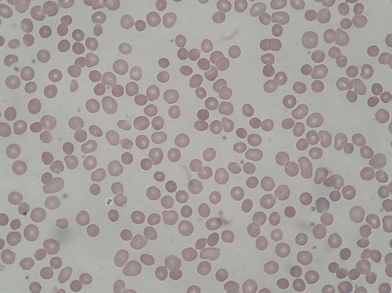
Spherocytes on the smear: immune or hereditary?
- DAT positive: warm AIHA, a delayed hemolytic transfusion reaction, or in a newborn, ABO hemolytic disease.
- DAT negative: hereditary spherocytosis is likely. Per the British Society for Haematology (BSH) guideline, spherocytes, a raised MCHC, reticulocytosis and a family history need no further tests. If the picture is equivocal, the screening tests are eosin-5′-maleimide (EMA) binding or cryohemolysis.

Special Populations
| Population | What changes | Clinical implication |
|---|---|---|
| Neonates & infants | Mean about 34 g/dL regardless of gestational age; somewhat higher values can occur in healthy neonates; heel-stick samples can read higher | In a jaundiced newborn, MCHC ≥36 g/dL raises suspicion of HS but is not diagnostic; check the DAT and smear, and if equivocal use EMA binding or cryohemolysis |
| Children & adolescents | Similar to adult | HS often presents with anemia, jaundice, splenomegaly or gallstones; a negative family history does not exclude it (recessive and new mutations) |
| Pregnancy | No clinically meaningful difference | Low MCHC with low MCV: check ferritin; iron deficiency is common |
| Older adults | No clinically meaningful difference | New iron deficiency anemia: look for GI blood loss |
| CKD / dialysis | Anemia is usually normochromic | Ferritin is less reliable; add transferrin saturation before calling iron status |
| Sickle cell and HbSC disease | Dense, dehydrated cells raise MCHC, sometimes well above the usual range | Expected in these patients once interference is excluded; compare with the patient's baseline |
| Recently transfused | Indices reflect a mix of donor and patient cells | Use the pre-transfusion CBC for an HS or iron workup |
Interfering Factors & Pitfalls
Collection & handling
| Factor | Effect | Why | Avoid / fix |
|---|---|---|---|
| In vitro hemolysis | ↑ | Lysed cells lower RBC count and Hct while their Hb is still measured | Redraw with good technique; avoid forceful syringe transfer |
| Underfilled EDTA tube | ↑ | Excess EDTA shrinks red cells | Fill to the line |
| Draw above a dextrose infusion | ↓ | Glucose-loaded cells swell in the diluent: falsely high MCV and Hct | Draw from the other arm, or follow line-draw discard policy |
| Lipid infusion running | ↑ | Lipid turbidity raises measured Hb | Draw away from the infusion; lab can replace plasma |
| Delay at room temperature (1–4 days) | ↓ | Red cells swell with storage | Analyze promptly or recollect |
| Capillary sample (neonates) | ↑ | Hemoconcentration and lower MCV | Confirm an abnormal value on a venous sample |
Physiologic / biologic
| Factor | Effect | Why | Avoid / fix |
|---|---|---|---|
| Cold agglutinins | ↑ (with ↑ MCV, ↓ RBC) | Clumps count as one cell or are excluded | Warm to 37 °C for 10–30 min and rerun; for high titers, keep a new tube at 37 °C until analysis |
| Lipemia, icterus, IgM paraprotein, cryoglobulins | ↑ | Turbidity or color falsely raises Hb | Plasma replacement, dilution or optical Hb |
| Hyponatremia | ↑ | Cells shrink in the higher-osmolality diluent | Interpret with Na⁺; cannot be corrected in the lab |
| Hypernatremia or glucose ≥600 mg/dL | ↓ | Cells swell in the diluent | Spun (micro) Hct and recalculate |
| Hyperleukocytosis | ↑ or ↓ | Large granular cells raise Hb (↑); small lymphoid cells are counted as red cells (↓) | Dilution or optical counts |
Assay & device-specific
| Factor | Effect | Why | Avoid / fix |
|---|---|---|---|
| Impedance analyzers | False + or False − | MCHC is always calculated (Hb ÷ Hct), so any Hb or Hct error lands in it | Labs use an out-of-range MCHC as a validation check |
| Lysis-resistant HbS or HbC cells | ↑ | Unlysed cells add turbidity to the Hb reading | Longer lysis or dilution and rerun |
Common misinterpretations
- “MCHC above 37 is impossible”: Wrong. Real values can run past the usual range in HS, sickle cell and HbSC disease. Extreme values still need a check for interference.
- A normal MCHC rules out iron deficiency: It doesn't. Check ferritin.
- High MCHC on a pink sample is always a bad draw: Not always. Intravascular hemolysis in the body raises MCHC too.
- Thalassemia always lowers MCHC: No. Thalassemia trait usually has a normal or near-normal MCHC.
Physiology & Test Basics
What it measures
MCHC is the average hemoglobin concentration inside a red cell. It tells you how densely the cell is packed. Its siblings measure cell size (MCV), hemoglobin mass per cell (MCH) and size variation (RDW). Low MCHC means pale, under-filled cells (hypochromia). High MCHC means dense cells, like spherocytes or dehydrated cells, or a measurement error.
| Index | Measures | Units | Think of it as |
|---|---|---|---|
| MCV | Cell volume | fL | How big is the cell? |
| MCH | Hb mass per cell | pg | How much Hb is in the cell? |
| MCHC | Hb concentration in cells | g/dL | How densely packed is the Hb? |
| RDW | Variation in cell size | % | How uneven are the cells? |
Physiology & regulation
- Hb supply: Run short of iron, globin chains or heme and MCHC falls.
- Cell water: Cation content sets how much water a red cell holds (Na⁺/K⁺ pump, PIEZO1 and Gardos channels, K⁺–Cl⁻ cotransport). Lose K⁺ and water, and the cell dehydrates and MCHC rises (sickle cell, xerocytosis).
- Membrane loss: Spherocytes lose surface area but keep their Hb. The concentration rises.
- Narrow range: MCHC barely varies between healthy people. That is why labs use it to catch analyzer interference.
How it is measured
Draw it in a lavender-top (K₂EDTA) tube, fill it to the line and invert it gently. No fasting is needed. Every automated CBC reports MCHC. Impedance analyzers measure Hb by spectrophotometry after lysis, count and size the red cells, then calculate MCHC = Hb ÷ Hct. Optical analyzers can also derive cellular Hb concentration directly. The reference method divides spectrophotometric Hb by a spun Hct.
Why it matters
MCHC points you toward iron deficiency when it is low. It points toward spherocytes and hemolysis when it is high. It is also often the first clue that the CBC is unreliable. In the ED, ICU or on transport, a sudden high MCHC with pink plasma can be the earliest lab sign of a hemolytic transfusion reaction or massive hemolysis.
Clinical Pearls
References
- Girard, S., Berda-Haddad, Y., Brouzes, C., et al. (2025). Breaking free from MCHC interferences? French-speaking Cellular Haematology Group (GFHC) review of causes, rising trends and practical solutions. International Journal of Laboratory Hematology, 47(5), 798–807. https://doi.org/10.1111/ijlh.14536
- Gulati, G., Uppal, G., & Gong, J. (2022). Unreliable automated complete blood count results: Causes, recognition, and resolution. Annals of Laboratory Medicine, 42(5), 515–530. https://doi.org/10.3343/alm.2022.42.5.515
- Bolton-Maggs, P. H. B., Langer, J. C., Iolascon, A., Tittensor, P., & King, M.-J. (2012). Guidelines for the diagnosis and management of hereditary spherocytosis: 2011 update (current BSH guideline). British Journal of Haematology, 156(1), 37–49. https://doi.org/10.1111/j.1365-2141.2011.08921.x
- Achenjang, N.-S., Jadczak, E., Ryan, R. M., & Nock, M. L. (2025). Hereditary spherocytosis: Review of presentation at birth. Children, 12(9), 1207. https://doi.org/10.3390/children12091207
- Ko, C. W., Siddique, S. M., Patel, A., et al. (2020). AGA clinical practice guidelines on the gastrointestinal evaluation of iron deficiency anemia. Gastroenterology, 159(3), 1085–1094. https://doi.org/10.1053/j.gastro.2020.06.046
- MedlinePlus, U.S. National Library of Medicine. (2026). RBC indices (normal values). https://medlineplus.gov/ency/article/003648.htm
Medical Disclaimer
- For Educational Purposes Only: This content is intended for educational reference and should not be used for clinical decision-making.
- Not a Substitute for Professional Judgment: Always consult your local protocols, institutional guidelines, and supervising physicians.
- Verify Before Acting: Users are responsible for verifying information through authoritative sources before any clinical application.