

Bedside Snapshot
- Normal (Adult)
- 35–45 mmHg (4.7–6.0 kPa)
Pregnancy: about 27–32 mmHg (3.6–4.3 kPa) - Critical Values
- <20 or >70 mmHg (<2.7 or >9.3 kPa); typical, lab-specific
- High Suggests
- Opioid or sedative hypoventilation
- COPD or tiring asthma
- Neuromuscular weakness, airway obstruction
- Obesity hypoventilation; uncontrolled O₂ in retainers
- Seizures or exercise (high CO₂ output)
- Low Suggests
- Compensation: DKA, shock, toxins
- Hypoxemia: PE, pneumonia, early asthma
- Sepsis, salicylate, pregnancy
- Methylxanthines (theophylline)
- Overventilation by bag or ventilator
- Act Now If
- PaCO₂ >45 mmHg with pH <7.35, a rising PaCO₂ with drowsiness, or a normal PaCO₂ in severe acidosis or asthma: support ventilation (BVM, NIV, airway), naloxone if opioid, EtCO₂; notify receiving/medical control
- Watch Out For
- Oxygen hides hypoventilation: SpO₂ stays normal while PaCO₂ climbs. Watch rate, mental status and EtCO₂.
Reference & Critical Ranges
Reference ranges
| Population | Conventional | SI | Notes |
|---|---|---|---|
| Adult (arterial) | 35–45 mmHg | 4.7–6.0 kPa | Same target for men and women; some labs quote slightly different bounds by sex |
| Pregnancy | About 27–32 mmHg | About 3.6–4.3 kPa | Progesterone-driven hyperventilation; HCO₃⁻ falls to about 18–22 mEq/L in compensation. A PaCO₂ approaching 40 mmHg suggests CO₂ retention |
| Older adult | Same as adult | Same as adult | — |
| Children | Same as adult | Same as adult | Use the lab's neonatal range in the first weeks of life |
| Umbilical cord, arterial | 35–70 mmHg (one provincial lab standard) | 4.7–9.3 kPa | Birth only; reflects placental gas exchange, not the newborn's ventilation |
| Peripheral venous (VBG) | 35–50 mmHg (one provincial lab standard) | 4.7–6.7 kPa | Usually higher than arterial; not interchangeable with PaCO₂ |
Severity & critical values
| Level | Conventional | SI | What it means / typical findings |
|---|---|---|---|
| Critical (low) | <20 mmHg | <2.7 kPa | Extreme hyperventilation: usually compensation for severe metabolic acidosis or overventilation by bag or ventilator. Light-headedness, paresthesias, carpopedal spasm; cerebral vasoconstriction |
| Hypocapnia | 20–34 mmHg | 2.7–4.6 kPa | Hyperventilation: primary respiratory alkalosis (pH high) or compensation for metabolic acidosis (pH low). Read with pH and HCO₃⁻ |
| Normal | 35–45 mmHg | 4.7–6.0 kPa | Adequate alveolar ventilation for current CO₂ production. Not reassuring in severe metabolic acidosis or a tiring asthmatic |
| Hypercapnia | >45 to 70 mmHg | >6.0 to 9.3 kPa | Hypoventilation. Acute if pH is low with a near-normal HCO₃⁻; chronic if pH is near normal with a high HCO₃⁻. COPD: pH ≤7.35 with PaCO₂ >45 mmHg meets the NIV threshold |
| Critical (high) | >70 mmHg | >9.3 kPa | Severe hypercapnia: headache, confusion, drowsiness, then stupor (CO₂ narcosis). Acute: needs ventilatory support now |
Conversion: mmHg × 0.133 = kPa; kPa × 7.5 = mmHg
A measured PaCO₂ above the range means the patient has added a respiratory acidosis, because they cannot keep up. Below the range, they have added a respiratory alkalosis.
Causes of Abnormal Results
High (hypercapnia, PaCO₂ >45 mmHg)
PaCO₂ climbs when alveolar ventilation falls behind CO₂ production. There are three ways that happens. The patient will not breathe, so the drive is the problem. The patient cannot breathe, so the pump or the airway is the problem. Or the patient is wasting breaths on dead space.
| Mechanism | Common causes | Bedside clue |
|---|---|---|
| Low respiratory drive | Opioids, sedatives, alcohol, brainstem stroke or injury, myxedema coma | Slow, shallow breathing; small pupils with opioids; rising EtCO₂ with a low rate |
| Obstructive lung disease | COPD exacerbation, severe asthma as the patient tires | Wheeze or silent chest, accessory muscles, drowsiness |
| Upper-airway obstruction | Foreign body, angioedema, epiglottitis, airway burns | Stridor, drooling, voice change |
| Neuromuscular weakness | Guillain–Barré syndrome, myasthenic crisis, high cervical cord injury, ALS, residual neuromuscular blockade | Weak cough, paradoxical abdominal motion; PaCO₂ rises late |
| Chest wall and obesity | Obesity hypoventilation syndrome, kyphoscoliosis, flail chest | High HCO₃⁻ at baseline, daytime sleepiness, morning headache |
| Dead space or exhaustion | Severe pneumonia, ARDS, pulmonary edema once the patient tires | Hypoxemia with rising PaCO₂; widening PaCO₂–EtCO₂ gap |
| Iatrogenic | Uncontrolled oxygen in CO₂ retainers, low ventilator minute ventilation, oversedation, deliberate permissive hypercapnia | PaCO₂ rises after an oxygen or ventilator change |
| High CO₂ production, fixed ventilation | Fever, sepsis, malignant hyperthermia, sodium bicarbonate in a ventilated patient | Rising EtCO₂ with unchanged settings |
A mild rise is also normal compensation for metabolic alkalosis. Expect 0.6–0.75 mmHg of rise for each 1 mEq/L that HCO₃⁻ goes up. It rarely climbs above 55 mmHg.
Low (hypocapnia, PaCO₂ <35 mmHg)
PaCO₂ falls when ventilation runs ahead of CO₂ production. Decide first whether it is compensation, with a low pH and a low HCO₃⁻, or a primary respiratory alkalosis, with a high pH.
| Mechanism | Common causes | Bedside clue |
|---|---|---|
| Compensation for metabolic acidosis | DKA, lactic acidosis (shock, sepsis), toxic alcohols, salicylate, kidney failure | Deep, rapid (Kussmaul) breathing; low HCO₃⁻; check Winter's formula |
| Hypoxemia and lung disease | Pulmonary embolism, pneumonia, early asthma, pulmonary edema, pneumothorax, high altitude | Low SpO₂, tachypnea, pleuritic pain |
| Central stimulation | Sepsis, salicylate toxicity, liver failure, stroke, meningitis or encephalitis, fever | Fever, confusion, tinnitus with salicylate |
| Pregnancy | Progesterone effect (expected) | PaCO₂ about 27–32 mmHg with a mildly low HCO₃⁻ |
| Pain and anxiety | Acute pain, panic | Diagnosis of exclusion only |
| Iatrogenic | Overventilation by bag-valve mask or ventilator, especially after ROSC or in head injury | EtCO₂ <35 mmHg; fast or forceful bagging |
Interpretation & Bedside Approach
Step-by-step approach
- Is it real? Confirm it's an arterial sample. Venous PCO₂ usually runs higher, and it agrees too poorly to stand in for arterial. Air bubbles lower PaCO₂. A delay at room temperature raises it.
- How sick is the patient? Look at mental status, rate and depth, work of breathing, SpO₂ and EtCO₂. The pH sets the urgency, not the PaCO₂ alone.
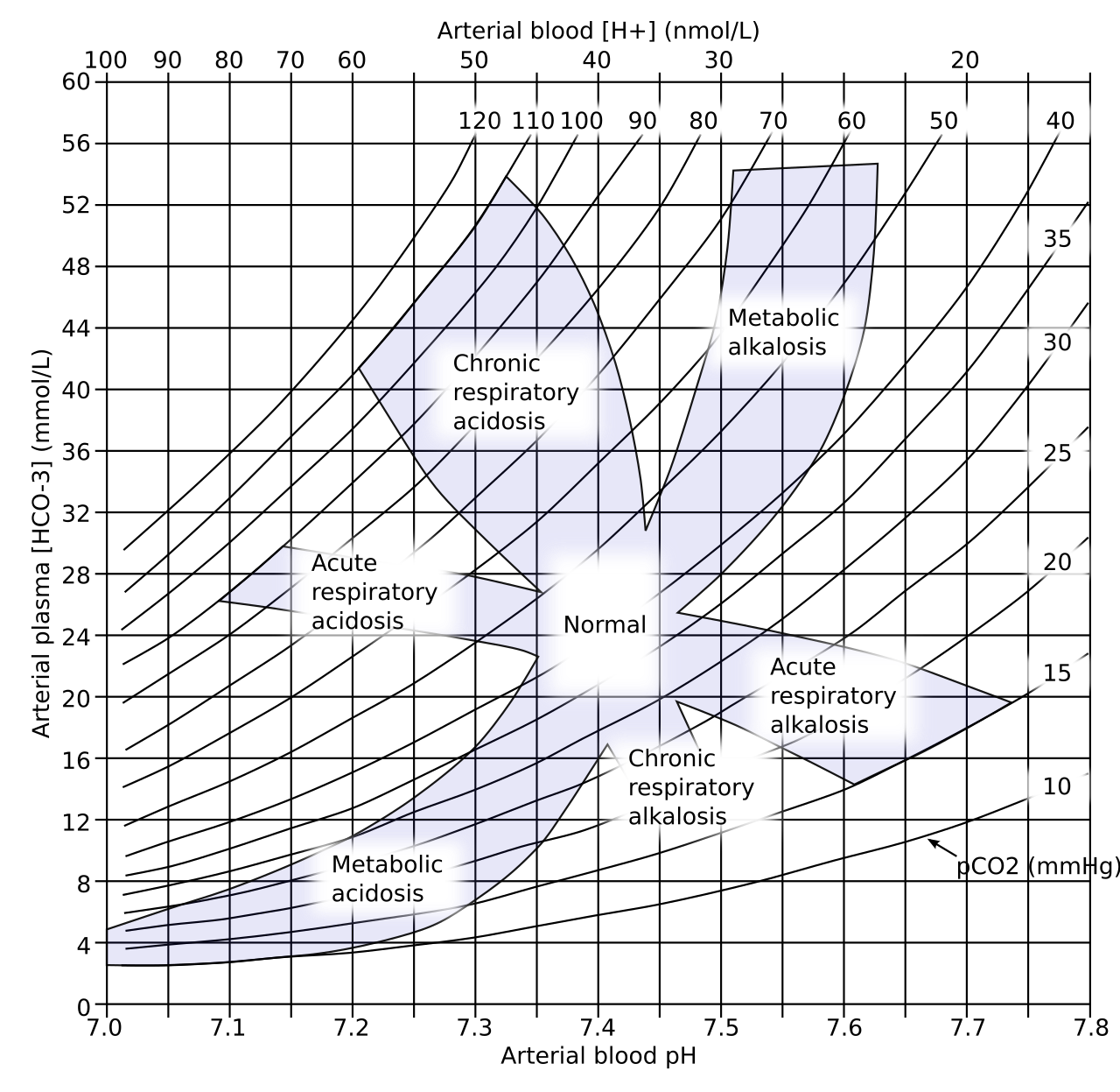
- Acute, chronic or acute-on-chronic? Check the HCO₃⁻ and pH against the expected compensation below, then against the patient's previous gases. A trend tells you more than a single value.
- Calculate. Use Winter's formula in metabolic acidosis. Use the A–a gradient to tell pure hypoventilation from lung disease.
- Classify. A primary respiratory disorder moves the pH and PaCO₂ in opposite directions. Compensation moves them the same way. For a COPD exacerbation with acute hypercapnic respiratory failure, ERS/ATS 2017 recommends NIV. The entry criteria (pH ≤7.35, PaCO₂ >45 mmHg, RR >20–24/min despite standard therapy) follow GOLD and BTS/ICS guidance.
- Escalate. Rising PaCO₂ with a falling pH, drowsiness or exhaustion calls for ventilatory support and a higher level of care. Notify the receiving facility or medical control.
Expected compensation
| Primary disorder | Expected change | Timing |
|---|---|---|
| Respiratory acidosis, acute | HCO₃⁻ rises 1–2 mEq/L per 10 mmHg rise in PaCO₂ | Minutes (buffering) |
| Respiratory acidosis, chronic | HCO₃⁻ rises 3–4 mEq/L per 10 mmHg rise in PaCO₂ | 3–5 days (kidney) |
| Respiratory alkalosis, acute | HCO₃⁻ falls 1–2 mEq/L per 10 mmHg fall in PaCO₂ | Minutes to hours |
| Respiratory alkalosis, chronic | HCO₃⁻ falls 4–5 mEq/L per 10 mmHg fall in PaCO₂ | 2–3 days |
| Metabolic acidosis | PaCO₂ = (1.5 × HCO₃⁻) + 8 ± 2 mmHg | Minutes to hours |
| Metabolic alkalosis | PaCO₂ rises 0.6–0.75 mmHg per 1 mEq/L rise in HCO₃⁻ (rarely above 55) | Hours |
Treat these as rough rules of thumb. A value well outside them points to a second, mixed disorder.
Common patterns
| Pattern | Suggests |
|---|---|
| High PaCO₂, low pH, HCO₃⁻ near normal | Acute respiratory acidosis: overdose, airway obstruction, acute neuromuscular failure |
| High PaCO₂, pH near normal, high HCO₃⁻ | Chronic (compensated) CO₂ retention: COPD, obesity hypoventilation |
| High PaCO₂ and high HCO₃⁻ but pH lower than the patient's baseline | Acute-on-chronic respiratory failure: COPD exacerbation |
| Low PaCO₂, low HCO₃⁻, low pH | Metabolic acidosis with compensation; check Winter's formula and the anion gap |
| Low PaCO₂, high pH, HCO₃⁻ normal or low | Primary respiratory alkalosis: hypoxemia, sepsis, PE, pregnancy, pain |
| PaCO₂ above Winter's prediction | Added respiratory acidosis: tiring, sedated or failing patient |
On room air at sea level, FiO₂ × (Patm − 47) comes to about 150 mmHg. If the gradient is normal, or small, while PaCO₂ is high and PaO₂ is low, you're looking at pure hypoventilation from a drug or a neuromuscular cause. A wide gradient points to lung disease.
Management Priorities
Emergency: Acute hypercapnic respiratory failure (PaCO₂ >45 mmHg with pH <7.35, or rising PaCO₂ with drowsiness)
- Support ventilation now: Open the airway. If breathing is inadequate, assist with a bag-valve mask and watch for visible chest rise. Don't wait on a blood gas.
- Reverse what is reversible: For opioid toxicity, titrate naloxone to breathing. For COPD or asthma, give bronchodilators and steroids. Relieve any airway obstruction.
- Controlled oxygen: When hypercapnia risk exists (COPD, obesity hypoventilation, neuromuscular or chest-wall disease), aim for SpO₂ 88–92%. Never withhold oxygen from a hypoxemic patient. Check a blood gas 30–60 min after any increase.
- Noninvasive ventilation (NIV): Bilevel NIV is the tool for a COPD exacerbation with acute hypercapnic respiratory failure, and ERS/ATS 2017 backs it. Entry criteria such as pH ≤7.35 and PaCO₂ >45 mmHg follow GOLD and BTS/ICS guidance. Paramedic/RN/CCT, per local protocol. Early gains in pH or respiratory rate predict success, and the lower the pH, the tighter the watch.
- Intubate when: the patient cannot protect the airway, is apneic or arresting, or gets worse despite NIV. In a chronic retainer, ventilate to the baseline pH and PaCO₂, not to 40 mmHg. Correct chronic hypercapnia over hours.
Emergency: Ventilating when PaCO₂ is the target (head injury, after ROSC, severe metabolic acidosis)
- Traumatic brain injury (prehospital TBI guideline, 3rd edition): Target about 10 breaths/min in adults on continuous capnography. Keep EtCO₂ at 35–45 mmHg and avoid dropping under 35. Hyperventilate only if active herniation shows, and then only to EtCO₂ 30–35 mmHg.
- After ROSC (2025 AHA Guidelines for CPR and ECC, Part 11, COR 1, LOE B-R): Hold PaCO₂ in the normal physiologic range, generally 35–45 mmHg, for adults who stay comatose. Mechanically ventilated adults can reasonably get a blood gas (COR 2b, LOE B-NR). After arrest, EtCO₂ can read falsely low, so arterial gases belong alongside it. ERC–ESICM 2025 lands on the same range. Under hypothermia, check PaCO₂ often and stick to a single temperature-correction method.
- Severe metabolic acidosis (DKA, salicylate, toxic alcohols): Low PaCO₂ is what's holding the pH up here. Avoid oversedation. When intubation can't be dodged, limit apnea and match the pre-intubation PaCO₂ or EtCO₂.
- Monitor & repeat: Draw a blood gas after each major ventilator change, and track EtCO₂ against the measured PaCO₂ gap. Lung-protective ventilation in ARDS or severe asthma can tolerate a higher PaCO₂, so long as the pH holds above the protocol floor.
Medications
Doses vary by protocol; follow local protocols and medical direction.
| Drug | Adult dose | Pediatric dose (max) | Onset / key notes |
|---|---|---|---|
| Naloxone: opioid hypoventilation | 0.2–2 mg IV/IO/IM or 2–4 mg IN, every 2–3 min, titrated to adequate ventilation, not full arousal (no fixed max; no response after 10 mg: question the diagnosis). In opioid-dependent patients, expect precipitated withdrawal, agitation and possible pulmonary edema: use the lowest effective dose, and some protocols start at 0.04–0.4 mg IV titrated to breathing. Iatrogenic: 0.1–0.2 mg IV every 2–3 min | 0.1 mg/kg IV/IO, every 2–3 min (max 2 mg/dose) | IV: within 2 min. Ventilate first. Often shorter-acting than the opioid: watch for falling RR and rising EtCO₂ |
| Oxygen: hypercapnia risk | Titrate to SpO₂ 88–92%: Venturi 24% (about 2 L/min) or 28% (about 4 L/min), per the device's labeled flow; or nasal cannula 1–2 L/min (titrate to effect: no fixed max) | Titrate to the target SpO₂ in the pediatric protocol; individualize in chronic lung or neuromuscular disease (no fixed max) | Lower the flow if SpO₂ >92%; blood gas 30–60 min after any increase |
Definitive care & disposition
- ICU or step-down: NIV with a pH that is not improving, any intubated hypercapnic patient, neuromuscular weakness with a rising PaCO₂ (a late sign).
- Chronic retainers: Write down the baseline PaCO₂, HCO₃⁻ and target SpO₂ so the next team starts with them. A patient with prior hypercapnic failure may carry an oxygen alert card.
- Do not give sodium bicarbonate to correct a respiratory acidosis. It doesn't touch the underlying problem, and it can raise PaCO₂ unless ventilation goes up. Ventilation is the treatment.
Doses are typical references. Follow local protocols, medical direction and institutional policy.
Field & Transport Considerations
Point-of-care testing
Cartridge blood-gas analyzers measure PCO₂ at 37 °C (98.6 °F). Respect the cartridge temperature and storage limits, and trend on one device. In the field, waveform EtCO₂ stands in for PaCO₂. It normally reads a few mmHg below PaCO₂, but the gap widens with dead space (low cardiac output, PE, COPD, ARDS). A normal EtCO₂ does not rule out hypercapnia. SpO₂ tells you about oxygenation, not ventilation.
Before and during interfacility transport
| Setting | Consideration |
|---|---|
| Before departure | Timed blood gas with FiO₂ and ventilator or NIV settings; paired EtCO₂ to know the patient's gap; baseline PaCO₂ if a chronic retainer |
| Ventilator or NIV switch | Match the sending minute ventilation; repeat a blood gas after the switch if available |
| Sedation and opioids en route | Falling rate with rising EtCO₂ is hypoventilation; reassess before more doses |
| Altitude and flight | Spontaneously breathing patients hyperventilate as cabin altitude rises, so PaCO₂ drifts down; keep the SpO₂ target and watch EtCO₂ |
| Redirect | Hypercapnic patient tiring without ventilatory support: nearest facility that can secure the airway |
Special Populations
| Population | What changes | Clinical implication |
|---|---|---|
| Neonates & infants | Umbilical cord arterial PCO₂ is normally higher (up to about 70 mmHg); use neonatal ranges in the first weeks | Read cord gases against cord ranges, not adult ones |
| Children & adolescents | Adult range applies. Prehospital TBI guidance sets EtCO₂ 35–45 mmHg for adults. For a child with a pulse and inadequate breathing, the rate is 20–30 breaths/min (1 breath every 2–3 s); during CPR with an advanced airway, a reasonable target is 20–30 breaths/min, adjusted for age (2025 AHA Guidelines for CPR and ECC, Part 8, COR 2b, LOE C-LD; the rate with a pulse and inadequate breathing is in Part 6, COR 2a, LOE C-EO) | After cardiac arrest, target a PaCO₂ appropriate to the child's underlying condition, and limit both hypercapnia and hypocapnia (2025 AHA Guidelines for CPR and ECC, Part 8, COR 2b, LOE C-LD). The supporting text uses normocapnia (35–45 mmHg), or the baseline value in a chronic retainer; see pediatric vital signs for normal rates |
| Pregnancy | PaCO₂ about 27–32 mmHg with HCO₃⁻ about 18–22 mEq/L; compensation formulas were not validated in pregnancy | PaCO₂ approaching 40 mmHg in a breathless pregnant patient (especially asthma) is CO₂ retention |
| Older adults | No clinically meaningful difference in PaCO₂; the A–a gradient widens with age | Interpret oxygenation with age in mind; the PaCO₂ range is unchanged |
| Chronic CO₂ retainers | COPD, obesity hypoventilation, neuromuscular disease: baseline PaCO₂ high with a high HCO₃⁻ | Treat the pH and the change from baseline; SpO₂ target 88–92%; avoid rapid correction |
| High altitude | Hypoxic drive lowers PaCO₂ in residents at altitude; pregnant residents run lower still | Use local ranges for residents at altitude |
Interfering Factors & Pitfalls
Collection & handling
| Factor | Effect | Why | Avoid / fix |
|---|---|---|---|
| Air bubbles in the syringe | ↓ | CO₂ diffuses into air (room-air PCO₂ is near zero); PaO₂ drifts toward about 150 mmHg | Expel air at once, cap, mix gently |
| Delay at room temperature | ↑ | Cells keep producing CO₂; rise measurable after about 30 min | Analyze within 30 min; do not batch |
| Excess liquid heparin or underfilled syringe | ↓ | Dilution with fluid that contains almost no CO₂ | Use dry (lyophilized) heparin syringes; fill to the stated volume |
| Flush in an arterial line sample | ↓ | Saline dilution | Clear the line of IV solution before drawing |
| Venous blood drawn by mistake | ↑ | Venous PCO₂ is higher | Confirm arterial flow; compare with SpO₂ and PaO₂ |
Medications & supplements
| Factor | Effect | Why | Avoid / fix |
|---|---|---|---|
| Sodium bicarbonate (ventilated patient) | ↑ | Bicarbonate is converted to CO₂ that must be exhaled | Raise minute ventilation; recheck the gas |
| Supplemental oxygen in a CO₂ retainer | ↑ | Excess oxygen raises PaCO₂ in at-risk patients | Titrate to SpO₂ 88–92%; recheck in 30–60 min |
Physiologic / biologic
| Factor | Effect | Why | Avoid / fix |
|---|---|---|---|
| Hypothermia (therapeutic or accidental) | Value depends on correction | Analyzer measures at 37 °C | Use one method (temperature-corrected or not) consistently |
Assay & device-specific
| Factor | Effect | Why | Avoid / fix |
|---|---|---|---|
| EtCO₂ used for PaCO₂ | EtCO₂ reads low | Dead space and low cardiac output widen the gap | Calibrate the trend with a blood gas |
| Peripheral venous gas | Not interchangeable | Venous–arterial PCO₂ agreement is wide | Use a VBG for pH and trend; get an arterial gas when the number matters |
Common misinterpretations
- "SpO₂ is normal, so breathing is fine": Oxygen can hold the SpO₂ up while PaCO₂ keeps rising. The oximeter looks fine. Ventilation may not be.
- "PaCO₂ 40 is normal": Not in severe metabolic acidosis or acute asthma. There, it signals fatigue.
- "CO₂ on the BMP is PaCO₂": The CO₂ on a chemistry panel is bicarbonate. PaCO₂ is the dissolved gas on the blood gas.
- "Bring the retainer back to 40": Overcorrecting causes posthypercapnic alkalosis.
Physiology & Test Basics
What it measures
PaCO₂ is the partial pressure of carbon dioxide dissolved in arterial blood. It is the respiratory half of acid–base balance, and it measures alveolar ventilation directly. The other parts of the blood gas each have their own page: pH, bicarbonate and base excess (the metabolic half), and PaO₂ (oxygenation). The CO₂ on a chemistry panel is bicarbonate, not PaCO₂.
Physiology & regulation
- Ventilation sets it: PaCO₂ is proportional to CO₂ production divided by alveolar ventilation. Cut alveolar ventilation in half, and PaCO₂ roughly doubles.
- Dead space: Rapid, shallow breaths move air that never reaches perfused alveoli. Minute ventilation can look high while PaCO₂ rises anyway.
- Acid–base: CO₂ + H₂O ⇌ H₂CO₃ ⇌ H⁺ + HCO₃⁻. Push PaCO₂ up, and the pH falls within minutes.
- Control: Central chemoreceptors sense PaCO₂ and set the pace of breathing, breath by breath.
- Brain blood flow: PaCO₂ is a powerful regulator. Low PaCO₂ constricts cerebral vessels, which is why hyperventilation lowers ICP and can cause ischemia. High PaCO₂ dilates them.
- Kidney: For a sustained change, the kidney compensates over days by retaining or excreting HCO₃⁻.
How it is measured
- Specimen: Arterial whole blood, from the radial artery or an arterial line, in a pre-heparinized blood-gas syringe. Check collateral circulation before a radial stick. Expel air, cap, mix gently.
- Method: A CO₂ electrode on a lab or point-of-care blood-gas analyzer does the measuring. The result is reported at 37 °C, and the analyzer then calculates HCO₃⁻ from pH and PCO₂.
- Timing and turnaround: No fasting needed. Analyze within 30 min. At the point of care, results come back in minutes.
Why it matters
PaCO₂ tells you whether the patient is ventilating enough. Overdose, COPD and asthma exacerbations, neuromuscular failure and exhaustion all show up here before arrest. The moment you take over a patient's breathing, PaCO₂ becomes a treatment target. That holds in head injury, after cardiac arrest and in severe acidosis.
Clinical Pearls
References
- Lewis, J. L., III. (2025). Acid-base disorders. In Merck Manual Professional Version. Merck & Co., Inc. https://www.merckmanuals.com/professional/endocrine-and-metabolic-disorders/acid-base-regulation-and-disorders/acid-base-disorders
- Lewis, J. L., III. (2025). Respiratory acidosis. In Merck Manual Professional Version. Merck & Co., Inc. https://www.merckmanuals.com/professional/endocrine-and-metabolic-disorders/acid-base-regulation-and-disorders/respiratory-acidosis
- Ortega, V. E., & Chiarella, S. E. (2025). Asthma. In Merck Manual Professional Version. Merck & Co., Inc. https://www.merckmanuals.com/professional/pulmonary-disorders/asthma-and-related-disorders/asthma
- Rochwerg, B., Brochard, L., Elliott, M. W., Hess, D., Hill, N. S., Nava, S., et al. (2017). Official ERS/ATS clinical practice guidelines: noninvasive ventilation for acute respiratory failure. European Respiratory Journal, 50(2), 1602426. https://doi.org/10.1183/13993003.02426-2016
- O'Driscoll, B. R., Howard, L. S., Earis, J., Mak, V., & British Thoracic Society Emergency Oxygen Guideline Group. (2017). BTS guideline for oxygen use in adults in healthcare and emergency settings. Thorax, 72(Suppl 1), ii1–ii90. https://doi.org/10.1136/thoraxjnl-2016-209729
- Hawryluk, G. W. J., Lulla, A., Bell, R., Jagoda, A., Mangat, H. S., Bobrow, B. J., & Ghajar, J. (2023). Guidelines for prehospital management of traumatic brain injury 3rd edition: executive summary. Neurosurgery, 93(6), e159–e169. https://doi.org/10.1227/neu.0000000000002672
- Nolan, J. P., Sandroni, C., Cariou, A., Cronberg, T., D'Arrigo, S., Haywood, K., et al. (2025). European Resuscitation Council and European Society of Intensive Care Medicine guidelines 2025: post-resuscitation care. Resuscitation, 215(Suppl 1), 110809. https://doi.org/10.1016/j.resuscitation.2025.110809
- Molano Franco, D., Viruez-Soto, A., & Luna, Y. (2026). Evaluating acid–base status and perfusion: arterial and venous blood gases during pregnancy. Revista Colombiana de Obstetricia y Ginecología, 77(1). https://doi.org/10.18597/rcog.4357
- American Heart Association. (2025). Part 11: Post-cardiac arrest care. In 2025 American Heart Association Guidelines for Cardiopulmonary Resuscitation and Emergency Cardiovascular Care. https://cpr.heart.org/en/resuscitation-science/cpr-and-ecc-guidelines/post-cardiac-arrest-care
- American Heart Association and American Academy of Pediatrics. (2025). Part 8: Pediatric advanced life support. In 2025 American Heart Association and American Academy of Pediatrics Guidelines for Cardiopulmonary Resuscitation and Emergency Cardiovascular Care. https://cpr.heart.org/en/resuscitation-science/cpr-and-ecc-guidelines/pediatric-advanced-life-support
- Alberta Precision Laboratories. (2021, July 12). Changes to blood gas reference intervals and critical values [Laboratory bulletin; effective July 21, 2021]. https://www.albertahealthservices.ca/assets/wf/lab/if-lab-hp-lab-bulletin-changes-to-blood-gas-reference-intervals-and-critical-values.pdf
Medical Disclaimer
- For Educational Purposes Only: This content is intended for educational reference and should not be used for clinical decision-making.
- Not a Substitute for Professional Judgment: Always consult your local protocols, institutional guidelines, and supervising physicians.
- Verify Before Acting: Users are responsible for verifying information through authoritative sources before any clinical application.