

Bedside Snapshot
- Indications
- Unresponsive, not breathing normally, no pulse: start CPR
- Pulse absent or uncertain after no more than 10 s: start CPR
- Infant and child arrest: same sequence, pediatric depth and ratio
- Child or infant pulse under 60/min with poor perfusion: start compressions
- Contraindications / Abort
- Signs of life: normal breathing or a palpable pulse
- Unsafe scene for rescuer or patient
- Valid DNR, obvious death or medical control direction
- Critical Steps
- Adult: 5–6 cm depth, full recoil, 30:2
- Child/infant, two rescuers: 15:2 (prepuberty)
- Pauses under 10 s; rotate compressor every 2 min
- Confirm & Monitor
- Waveform EtCO₂ during CPR; low value means check compressions
- Rhythm and pulse check every 2 min
- Chest rise with each breath
Key Numbers
High-quality CPR keeps blood moving to the brain and heart until defibrillation or return of circulation. Every pause costs perfusion, so keep interruptions under 10 seconds.
| Parameter | Value |
|---|---|
| Compression rate (all ages) | 100–120 compressions/min |
| Adult depth | At least 5 cm (2 in), no more than 6 cm (2.4 in); full chest recoil |
| Child and infant depth | At least one-third of the AP chest diameter Child about 5 cm (2 in); infant about 4 cm (1.5 in) |
| Ratio, no advanced airway | Adult: 30:2, one or two rescuers Child and infant, one rescuer: 30:2 Child and infant, two rescuers (prepuberty): 15:2 |
| Ventilation, advanced airway | Adult: 1 breath every 6 s (about 10/min) Child and infant: 1 breath every 2–3 s (20–30/min) |
| Each breath | About 1 second, with visible chest rise |
| Pause limit | Under 10 s for any pulse check, shock or airway step |
| Compressor rotation | Every 2 min, or sooner if compressions weaken |
Overview & Principles
Compressions keep blood moving. Rescue breaths or an advanced airway handle the oxygen. Start the moment cardiac arrest is recognized. Keep going until defibrillation, return of circulation or a valid stop decision.
How It Works
- Compressions: Push hard and fast over the lower half of the sternum. Then let the chest recoil fully.
- Ventilation: Give each breath over about 1 second. Watch for visible chest rise. Avoid excessive volume.
- Feedback: You judge rate, depth and recoil by feel. A feedback device or waveform EtCO₂ helps where you have one.
- Team rotation: Change the compressor every 2 minutes. Keep the switch under 10 seconds.
Where It Fits
The 2025 AHA algorithms run CPR alongside rhythm analysis, defibrillation and drugs. Compressions stop only for the rhythm check, a shock or a brief airway step. Shockable rhythm? Shock, then 2 minutes of CPR. Non-shockable? CPR and drugs, no shock.
Indications & Contraindications
Indications
- Cardiac arrest: Unresponsive, not breathing normally, no pulse. Agonal gasps do not count as normal breathing.
- Pulse check: Pulse absent or uncertain after no more than 10 seconds? Start CPR.
- Infant and child: Same sequence. Pediatric depth, ratio and ventilation rates apply.
- Infant or child with a pulse: Heart rate under 60/min with poor perfusion, even after oxygenation and ventilation? Start compressions.
- Pulse present, not breathing normally: Rescue breathing, not compressions, while the pulse is adequate. 1 breath every 2–3 s for a child or infant, every 6 s for an adult.
Absolute Contraindications
- None when cardiac arrest is confirmed: Start CPR without waiting for a full assessment.
Relative Contraindications & Cautions
- Unsafe scene: Make the scene safe first, without a long delay. Use local protocol for scene safety.
- Known DNR or obvious death: Before you stop, follow the valid DNR or the local death-determination rule.
- Spinal injury: Keep the head and neck in line through compressions and airway work. Never delay CPR for a collar.
Abort / Stop Criteria
- Signs of life: normal breathing or a palpable pulse with a rhythm.
- Rescuer safety: Stop only when a hazard cannot be controlled. Hand CPR over the moment another rescuer arrives.
- Valid stop: a documented DNR, obvious death or medical control direction.
Scope & Team Roles
| Provider Level | Typical Role | Notes |
|---|---|---|
| EMT | Performs | CPR, AED with pediatric attenuator where carried, bag-valve mask |
| EMR | Performs | CPR and AED within the national EMS scope; compression-only CPR for an untrained bystander |
| AEMT | Performs | CPR and ventilation; advanced airway and drugs per protocol |
| Paramedic | Performs | CPR, defibrillation and advanced airway per protocol |
| RN (ED/ICU) | Performs | ACLS-trained RNs lead CPR and assist with airway and drugs per facility policy |
| Critical Care Transport (CFRN / CTRN / FP-C / CCP-C) | Performs | CPR during transport; advanced airway and drugs per agency protocol |
| Physician / APP | Performs | Directs the resuscitation, decides on stop and advanced therapy |
Team Roles During the Procedure
- Compressor: maintains rate and depth, and calls for rotation at 2 minutes.
- Airway and ventilation: gives breaths over about 1 second and keeps the airway open.
- Monitor and recorder: watches EtCO₂, rhythm and time; keeps the timeline.
- Team leader: calls each rhythm check and shock, and directs the drugs.
Pre-Procedure Assessment
Recognition
- Responsiveness and breathing: unresponsive, with no normal breathing.
- Pulse check (trained rescuers): No more than 10 seconds. An absent or uncertain pulse means start CPR.
- Scene check: safety, bystander CPR already started, and the time of collapse.
- Lay rescuers and untrained bystanders: Don't check a pulse. Start CPR once the person is unresponsive and not breathing normally. Untrained? Compression-only CPR.
Anatomy & Landmarks
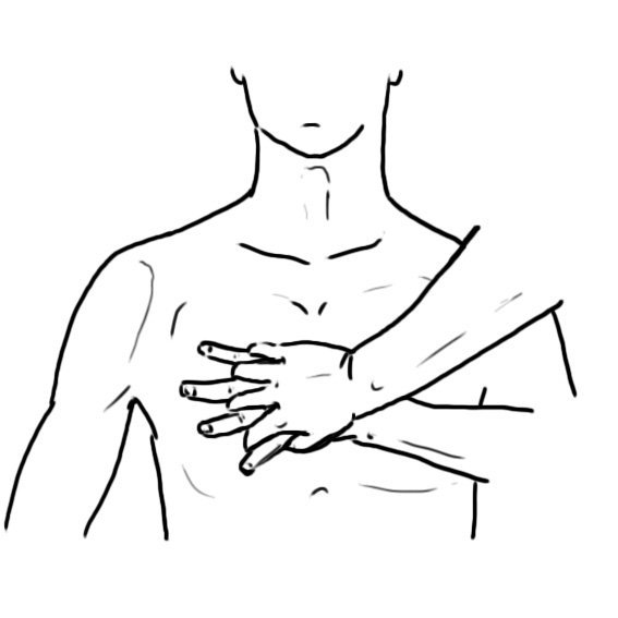
Hand placement is the landmark for CPR. Compress directly over the lower half of the sternum. Hand position changes with the patient's size.
| Patient | Hand Position | Depth | Avoid If |
|---|---|---|---|
| Adult | Heel of one hand on the lower half of the sternum; second hand on top | 5–6 cm (2–2.4 in) | Pressing the xiphoid or upper abdomen |
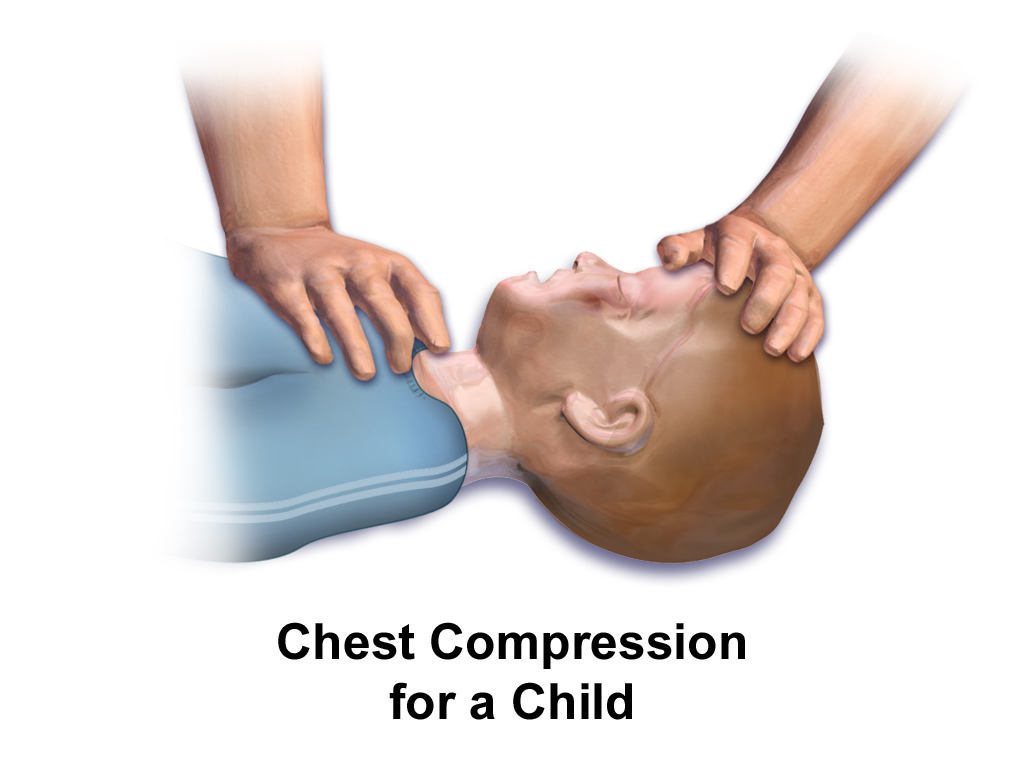
| Child | Heel of one or both hands on the lower half of the sternum | About one-third of AP diameter (about 5 cm) | Hands too high on the chest or on the abdomen |
| Infant | Two thumbs encircling the chest, or the heel of one hand, on the lower half of the sternum | About 4 cm (1.5 in) | Two-finger technique, which is no longer recommended |
Adult Hand Position
- Find the point: the lower half of the sternum, in the center of the chest.
- Place the hands: heel of one hand on the point, second hand on top, fingers lifted off the chest.
- Lock the elbows: keep the arms straight and the shoulders over the hands.
Pediatric
Same landmark for a child, with one or two hands sized to the chest. Depth comes from the AP diameter. Don't use the adult number.
Infant Hand Position
- Choose the technique: two thumbs encircling the chest, or the heel of one hand.
- Place the thumbs or hand: on the lower half of the sternum.
- Compress the chest: about 4 cm deep, with full recoil between compressions.
Pediatric
Forget the two-finger technique for infant compressions. It is no longer recommended. Use two thumbs encircling the chest, or the heel of one hand.
Equipment & Preparation
Equipment
- Bag-valve mask: adult and pediatric sizes, with reservoir and oxygen.
- AED: adult pads and a spare set; pediatric pads or attenuator where available.
- Monitoring: EtCO₂ capnography with an airway adapter, and a timer or metronome.
- Feedback device: where available, to guide rate and depth.
- Suction and advanced airway: Yankauer suction and the advanced airway kit per protocol.
Sizing
- Adult: adult pads and 5–6 cm depth.
- Child: pediatric pads or attenuator per device label; depth about one-third of AP diameter (about 5 cm).
- Infant: pediatric attenuator per device label; depth about 4 cm.
Patient Positioning & Preparation
- Surface: move the patient supine onto a firm, flat surface; a soft bed absorbs compression.
- Exposure: expose the chest and remove any clothing that blocks the pads.
- Start: begin CPR at once and send for the AED; do not wait for the equipment.
Pre-Procedure Checklist
- Scene safe for the team and patient
- Patient on a firm, flat surface and chest exposed
- AED on, adult pads and pediatric attenuator available
- Bag-valve mask with reservoir and oxygen connected
- Suction ready and airway kit within reach
- EtCO₂ adapter ready and timer or metronome running
- Team roles named: compressor, airway, monitor, leader
Step-by-Step Procedure
Phase 1: Recognize and Call
- Confirm unresponsiveness: tap and shout, then check for normal breathing. Agonal gasps do not count.
- Call for help: activate the emergency response and send for the AED and equipment.
- Check the pulse: no more than 10 seconds. Start CPR if absent or uncertain.
Phase 2: Compress and Ventilate
- Start compressions: hand position per the Anatomy section; push at 100–120/min to the target depth.
- Allow full recoil: lift the hands off the chest between compressions without losing position.
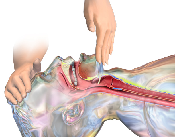
- Give the breaths: 30:2 for an adult with no advanced airway; 15:2 for a child or infant with two rescuers. Open the airway for each breath: head tilt-chin lift, or jaw thrust if a spinal injury is suspected (see Figure 3).
- Keep the pauses short: keep every interruption, including the rhythm check and shock, as short as possible and under 10 seconds.
- Rotate the compressor: switch every 2 minutes, with the switch kept under 10 seconds.
Phase 3: Analyze, Shock and Rotate
- Apply the AED or monitor as soon as it arrives: keep compressing while the pads go on, then analyze at once.
- Shock only a shockable rhythm, then repeat: follow the device prompts. Repeat the analysis every 2 minutes, with each pause under 10 seconds.
- Resume compressions at once: after any shock, restart compressions without a pulse check.
- Plan the next cycle: rotate the compressor and continue CPR until the next rhythm check or a valid stop.
Alternative Technique: Infant with One Hand
When two thumbs won't work, use the heel of one hand on the lower half of the sternum. The rest of the sequence stays the same.
Confirmation & Monitoring
Primary Confirmation
Watch the waveform EtCO₂ the whole time CPR runs. A value that stays low means the compressions need work. A sudden, sustained rise suggests return of circulation.
Secondary Confirmation
- Palpable pulse at the rhythm check, no more than 10 seconds, with an organized rhythm.
- Visible chest rise with each breath, and no gastric distension.
- Feedback device readings for rate and depth where available.
| Finding | Success | Failure / Next Step |
|---|---|---|
| EtCO₂ | Rising or stable value during CPR | Persistently low (under 10 mmHg) or falling: check compressions, then reversible causes. A low value alone is not a reason to stop CPR. |
| Pulse at rhythm check | Palpable with an organized rhythm | Absent or uncertain: continue CPR and the algorithm |
| Compression depth | 5–6 cm adult; about 4–5 cm child and infant | Too shallow or too deep: correct position and depth |
| Chest rise | Visible with each breath | No rise: reposition the airway and check the seal |
Ongoing Monitoring
- Continuous ECG and EtCO₂ during CPR; SpO₂ once circulation returns.
- Rhythm and pulse check at every 2-minute cycle, with the pause timed.
- Compressor fatigue, rate and depth reassessed at each rotation.
Troubleshooting
| Problem | Likely Cause | Fix |
|---|---|---|
| Compressions too shallow | Fatigue, soft surface, arms bent | Rotate the compressor; firm surface; straight arms; full push |
| Hands on the abdomen or xiphoid | Hand position too low | Move the hands to the lower half of the sternum |
| Incomplete recoil | Leaning on the chest | Lift hands fully between compressions, without losing position |
| Low EtCO₂ during CPR | Poor compressions or a low-flow cause | Check depth and rate; check for pneumothorax, hypovolemia and PE per protocol |
| Chest does not rise | Airway obstruction or poor mask seal | Reposition the airway; two-person mask seal; consider advanced airway |
| Gastric distension | Excessive volume or rate | Give breaths over about 1 second; avoid forceful bagging |
| Long pauses | Multiple checks at once | Pair the pulse check with the rhythm check; minimize any other pause |
If Compressions Still Fail
- Check the basics: depth, rate, recoil and hand position.
- Check the airway: open, sealed and ventilating, with chest rise.
- Check the reversible causes: per the arrest algorithm and local protocol.
Complications
| Complication | Recognition | Prevention | Management |
|---|---|---|---|
| Immediate: rib or sternal fracture | Crepitus or a new chest deformity during CPR | Correct hand position; depth within range | Continue CPR; reassess after return of circulation |
| Immediate: gastric distension | Rising abdomen with poor chest rise | Breaths over about 1 second; avoid forceful bagging | Reduce breath volume and rate; reposition the airway. Do not press on the abdomen. Place an OG tube only after the airway is secured, per protocol. |
| Immediate: rescuer fatigue | Falling depth or rate on the feedback device | Rotate every 2 minutes | Rotate at once; keep pauses short |
| Delayed: liver or spleen laceration | Abdominal pain or bleeding after return of circulation | Keep the hands on the lower sternum | Imaging and surgical review per receiving team |
| Delayed: regurgitation and aspiration | Vomit or wet airway during or after CPR | Avoid excessive ventilation; suction early | Suction; turn to side during transfers; reassess airway |
Special Populations
Pediatrics
- Depth: child about 5 cm (at least one-third of AP diameter); infant about 4 cm.
- Ratio: 15:2 with two rescuers (prepuberty); 30:2 with one rescuer or after puberty.
- Advanced airway: compressions continuous; 1 breath every 2–3 s (20–30/min).
- Infant technique: two thumbs encircling the chest, or heel of one hand; two-finger technique is no longer recommended.
- AED: pediatric attenuator or pads when available, applied immediately per the 2025 pediatric algorithm.
- Bradycardia: with a pulse under 60/min and poor perfusion despite oxygenation and ventilation, start compressions.
- Choking: infant: 5 back blows alternating with 5 chest thrusts; child: 5 back blows alternating with 5 abdominal thrusts.
- Newborn: a separate resuscitation sequence; see the neonatal resuscitation page and the 2025 Part 5 guideline.
See pediatric cardiac arrest, equipment sizing, Broselow and pediatric airway.
Drowning and Opioid Arrest
- Respiratory cause: ventilation has priority; give rescue breaths or bag-valve-mask ventilation with compressions per the algorithm.
- Opioids: give naloxone for suspected opioid toxicity, per protocol.
Older Adults
- Fragile chest: rib fractures are more likely; keep depth within range, but never push deeper to compensate.
- Surface: a firm surface is still required; a bed or chair is a poor compression base.
Pregnancy
- Displacement: relieve aortocaval compression with manual left uterine displacement (left lateral tilt is the alternative); the uterine-height threshold is in the AHA 2015 pregnancy statement.
- Algorithm: the adult CPR and defibrillation sequence applies; see maternal cardiac arrest.
Post-Procedure Care & Transport
Immediate Aftercare
- Reassess pulse, breathing, SpO₂ and blood pressure at return of circulation.
- Check the chest wall for fracture and the abdomen for distension or pain.
Ongoing Maintenance
- Airway: keep the airway patent and the advanced airway confirmed with waveform EtCO₂.
- Rhythm: watch for recurrent VF and treat per protocol.
- Post-arrest care: targets per the receiving facility; see post-cardiac arrest care.
Transport Considerations
- Each move interrupts compressions; plan transfers to keep pauses under 10 seconds, per local protocol.
- Use continuous ECG and EtCO₂ during transport and reassess after every move.
- Mechanical CPR devices can keep compressions going during moves where protocol allows; see mechanical CPR.
Document & Hand Off
- Time of collapse, time of CPR start and time of AED arrival
- Compressor rotations, rhythm checks and shocks with times
- Compression depth and rate if measured, and EtCO₂ values
- Airway used, breaths and ventilation method
- Return of circulation time and post-arrest vital signs
Clinical Pearls & Pitfalls
Pearls
- Depth is a range: 5–6 cm for adults. Shallow compressions under-perfuse. Deep ones add injury.
- Pauses decide survival: Count no-flow time, not minutes.
- Infant technique changed: Two-finger compressions are out for infants.
- EtCO₂ guides quality: A low value during CPR is your cue to check the compressions first.
Common Pitfalls
- Pulse check after every shock: Resume compressions at once. The pulse check waits for the rhythm check.
- Hands too low: Move them to the lower half of the sternum. Not the xiphoid.
- Adult ratio for a child: Before puberty, use 15:2 with two rescuers.
- Waiting for equipment: Don't wait on equipment. Start compressions, then bring the AED to the patient.
Education & Skill Maintenance
Skill Maintenance
- Practice depth, rate and recoil on a feedback manikin each training cycle.
- Rehearse the 2-minute cycle with a timer, including the rhythm check and rotation.
- Practice infant technique on an infant manikin with two thumbs and with one hand.
- Debrief each arrest for pause time, time to first compression and time to the first shock.
- Check that pediatric pads or the attenuator are on the AED before each shift.
References
- Kleinman ME, et al. Part 7: Adult Basic Life Support: 2025 American Heart Association Guidelines for Cardiopulmonary Resuscitation and Emergency Cardiovascular Care. Circulation. 2025. https://doi.org/10.1161/CIR.0000000000001369
- Joyner BL, et al. Part 6: Pediatric Basic Life Support: 2025 American Heart Association and American Academy of Pediatrics Guidelines for Cardiopulmonary Resuscitation and Emergency Cardiovascular Care. Circulation. 2025. https://doi.org/10.1161/CIR.0000000000001370
- International Liaison Committee on Resuscitation. Basic Life Support: 2025 International Liaison Committee on Resuscitation Consensus on Science With Treatment Recommendations. Circulation. 2025. https://doi.org/10.1161/CIR.0000000000001364
- American Heart Association. Part 1: Executive Summary: 2025 American Heart Association Guidelines for Cardiopulmonary Resuscitation and Emergency Cardiovascular Care. Circulation. 2025. https://doi.org/10.1161/CIR.0000000000001372
- American Heart Association. Part 10: Adult and Pediatric Special Circumstances of Resuscitation: 2025 American Heart Association Guidelines for Cardiopulmonary Resuscitation and Emergency Cardiovascular Care. Circulation. 2025. https://doi.org/10.1161/cir.0000000000001380
- American Heart Association. Cardiac Arrest in Pregnancy: A Scientific Statement From the American Heart Association. Circulation. 2015. https://doi.org/10.1161/cir.0000000000000300
- American Heart Association and American Academy of Pediatrics. Part 5: Neonatal Resuscitation: 2025 American Heart Association and American Academy of Pediatrics Guidelines for Cardiopulmonary Resuscitation and Emergency Cardiovascular Care. Circulation. 2025. https://doi.org/10.1161/cir.0000000000001367
Medical Disclaimer
- For Educational Purposes Only: This content is intended for educational reference and should not be used for clinical decision-making.
- Not a Substitute for Professional Judgment: Always consult your local protocols, institutional guidelines, and supervising physicians.
- Verify Before Acting: Users are responsible for verifying information through authoritative sources before any clinical application.