

Bedside Snapshot
- Indications
- Pulseless VF or pulseless VT: shock as the first treatment
- Persisting VF or VT (after 3 or more shocks): shock again per algorithm
- Pediatric pulseless VF or VT: same algorithm, pediatric energy
- Contraindications / Abort
- Asystole and PEA: not shockable; continue CPR and drugs
- Organized rhythm with a pulse: do not defibrillate
- Pads over an implanted generator: move them off, without delaying the shock
- Critical Steps
- Biphasic: manufacturer dose (120–200 J); if unknown, maximum available
- Child: 2 J/kg first, then 4 J/kg or more (max 10 J/kg or adult dose)
- Shock, then CPR for 2 min before the next rhythm check
- Confirm & Monitor
- Rhythm check after every 2 min of CPR
- Epinephrine 1 mg IV/IO every 3–5 min from shock 2 (asystole/PEA: ASAP)
- Amiodarone 300 mg, then 150 mg, or lidocaine 1–1.5 mg/kg, from shock 3
Key Numbers
Defibrillation ends VF or pulseless VT. Each shock is followed by 2 minutes of CPR, so delay between shock and compressions costs more than a slightly lower first dose.
| Parameter | Value |
|---|---|
| Biphasic shock, adult | Manufacturer dose, initial 120–200 J; if the device is unknown, use the maximum available |
| Monophasic shock, adult | 360 J |
| Second and later shocks, adult | Equivalent to the first; higher doses may be considered |
| Shocks, pediatric | First shock 2 J/kg; second shock 4 J/kg; later shocks 4 J/kg or more; maximum 10 J/kg or the adult dose |
| CPR after each shock | Resume compressions at once; rhythm check after 2 minutes of CPR |
| Epinephrine (VF/pVT, from shock 2) | Adult: 1 mg IV/IO every 3–5 min Peds: 0.01 mg/kg IV/IO (max 1 mg) every 3–5 min |
| Amiodarone (persisting VF/pVT) | Adult: 300 mg, then 150 mg (after shock 3) Peds: 5 mg/kg (max 300 mg first dose); later doses max 150 mg. Differs from FDA label: IV amiodarone safety and effectiveness are not established in children |
| Lidocaine (alternative) | Adult: 1–1.5 mg/kg, then 0.5–0.75 mg/kg Peds: 1 mg/kg IV/IO. Not established in children (FDA label) |
| Persisting VF/pVT | VF or pulseless VT persisting after 3 or more consecutive shocks. Vector change and double sequential defibrillation: usefulness not established (2025 AHA Guidelines for CPR and ECC, Part 9, COR 2b, LOE B-R) |
Overview & Principles
Defibrillation sends an unsynchronized shock through the chest. The goal is to depolarize enough myocardium to stop VF or pulseless VT. Only shockable rhythms get it. A shock will not restart a heart in asystole or PEA, and it does not take the place of CPR.
How It Works
- Shockable rhythms: VF and pulseless VT. Asystole and PEA cannot be shocked. Keep CPR going and give drugs.
- Unsynchronized shock: there is no timing against the R wave. Synchronized cardioversion is only for a tachycardia with a pulse (see cardioversion).
- Waveform: a biphasic device gives the manufacturer dose. A monophasic device gives 360 J in adults.
- Immediate CPR: every shock is followed by 2 minutes of compressions. The rhythm check comes after that.
Where It Fits
The 2025 AHA Adult Cardiac Arrest Algorithm splits at the first rhythm check. Shockable, VF or pulseless VT? Shock 1 goes out, then 2 minutes of CPR with IV/IO access. Still shockable? Shock 2 is followed by 2 minutes of CPR and epinephrine 1 mg every 3–5 minutes. Shock 3 is followed by 2 minutes of CPR with amiodarone or lidocaine. Asystole or PEA is the other branch. It gets CPR and epinephrine as soon as possible, with no shock.
Indications & Contraindications
Indications
- Pulseless VF or pulseless VT: the shock comes first. Deliver it as soon as you identify the rhythm.
- Persisting VF or pulseless VT (after 3 or more shocks): keep shocking per the algorithm, alongside drug therapy.
- Pediatric pulseless VF or VT: the same algorithm, run on pediatric energies (see Special Populations).
Absolute Contraindications
- None for a shockable rhythm with no pulse. Shock right away.
- Asystole and PEA: not shockable. A shock treats neither one.
- Organized rhythm with a pulse: do not defibrillate. Work the rhythm through its own algorithm.
Relative Contraindications & Cautions
- Standing water or wet surfaces: dry the chest fast and move the patient out of standing water before the shock.
- Implanted pacemaker or ICD: put the pads off the generator. Do it fast, and do not hold up the shock to do it.
- Transdermal medication patches: take off any patch under the pad site.
- Oxygen and contact: oxygen goes away from the chest. Then confirm nobody is touching the patient when the shock goes out.
- Severe hypothermia (core below 30 °C): shock once. If that fails, hold further shocks until the core reaches at least 30 °C (86 °F) (2025 AHA Guidelines for CPR and ECC, Part 10, COR 2b, LOE C-LD for adults; C-EO for children). Holding epinephrine until 30 °C may be reasonable (COR 2b, LOE C-EO).
Consider an Alternative
- Unstable tachycardia with a pulse: go to synchronized cardioversion. Not defibrillation.
- Symptomatic bradycardia with a pulse: transcutaneous pacing, not a shock.
Scope & Team Roles
| Provider Level | Typical Role | Notes |
|---|---|---|
| EMT | Performs | AED use; manual defibrillation not in scope. Confirm against state scope. |
| AEMT | Assists | Pads, CPR and monitoring; AED and drugs per protocol |
| Paramedic | Performs (protocol/credentialing required) | Manual defibrillation and arrest drugs per agency protocol |
| RN (ED/ICU) | Performs (protocol/credentialing required) | ACLS-certified RNs in most facilities; defibrillator use per facility policy |
| Critical Care Transport (CFRN / CTRN / FP-C / CCP-C) | Performs (protocol/credentialing required) | Same credentialing caveat as Paramedic |
| Physician / APP | Performs | Directs the arrest, orders drugs and decides on advanced therapies |
Team Roles During the Procedure
- Operator: reads the rhythm, sets the energy and presses SHOCK.
- Compressor: maintains CPR and resumes it at once after each shock.
- Airway and oxygen: ventilates and keeps oxygen clear of the chest.
- Medication clinician: gives epinephrine and antiarrhythmics on time and records each dose.
- Recorder: logs the time of each shock, energy, rhythm check and drug.
Pre-Procedure Assessment
Rhythm and Pulse
- Confirm the rhythm: look for VF or pulseless VT on the monitor or in the AED analysis. Asystole and PEA go down the non-shockable branch.
- Check for a pulse: an organized rhythm with a pulse gives you no reason to defibrillate.
- Start CPR during analysis: keep compressions going. Pause only as long as the analysis and the shock take.
Site and Device Check
- Implanted device: look for a pacemaker or ICD generator under the skin. Keep the pads off it.
- Patches and jewelry: clear transdermal patches and metal jewelry off the pad sites.
- Chest and surface: the chest must be dry, and the patient must be off any wet surface.
Anatomy & Landmarks
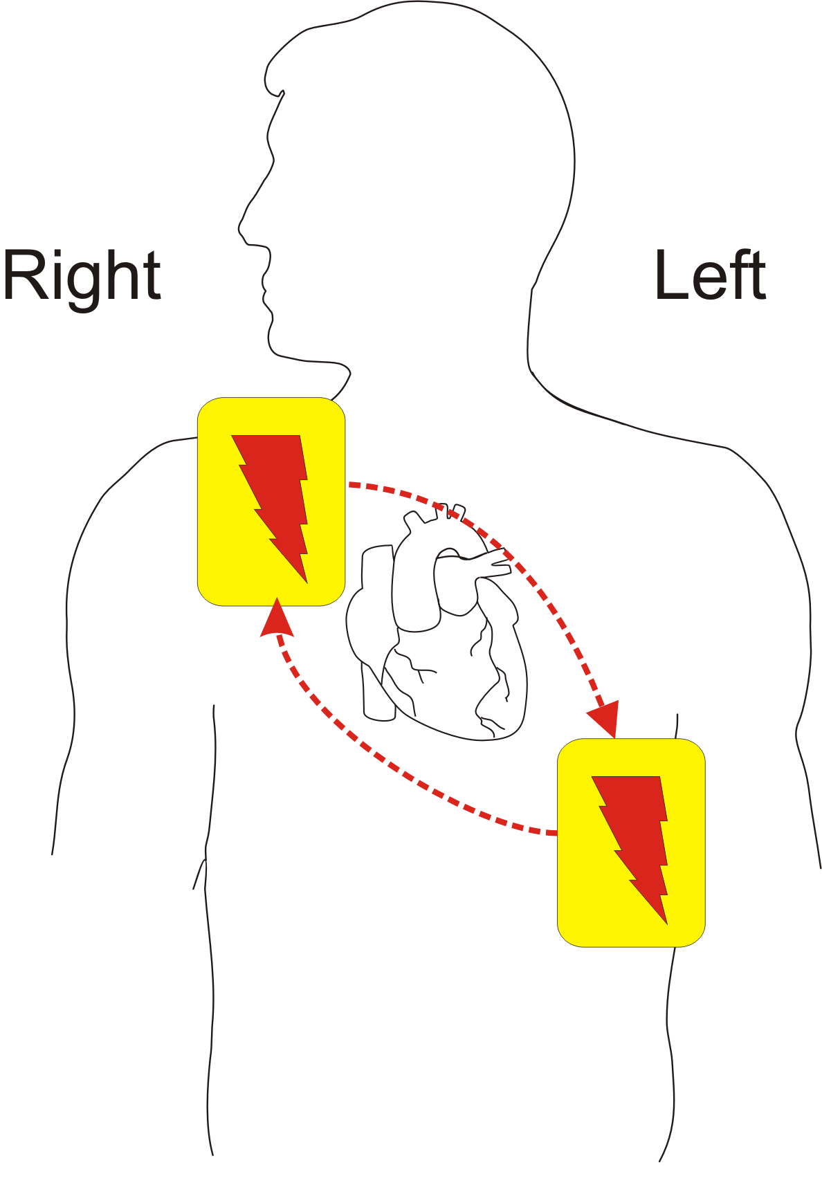
Two standard placements exist. Anterolateral comes first, as the usual choice. Anteroposterior works too, and both are reasonable in adults (2025 AHA Guidelines for CPR and ECC, Part 7, COR 2a, LOE C-LD). Move to the alternative if the first one fails you, or if a device sits under one pad.
| Placement | Pad Positions | Notes |
|---|---|---|
| Anterolateral | Sternal pad on the right upper chest, below the clavicle; apex pad on the left lateral chest | Standard first choice |
| Anteroposterior | One pad on the front of the chest over the heart; one on the back, below the left scapula | Reasonable alternative (2025 AHA Guidelines for CPR and ECC, Part 7, COR 2a, LOE C-LD) |
Anterolateral Pad Placement
- Place the sternal pad: right upper chest, below the clavicle.
- Place the apex pad: left lateral chest, in the midaxillary line. With large breast tissue, the pad must still touch skin. With a bra, adjust its position rather than remove it (2025 AHA Guidelines for CPR and ECC, Part 7, COR 2b, LOE C-EO).
- Check spacing: the pads must not touch each other or a gel bridge.
- Connect and confirm: attach the leads and confirm the device shows good contact.
Anteroposterior Pad Placement
- Place the anterior pad: front of the chest over the heart.
- Place the posterior pad: back, below the left scapula.
- Check the device position: keep neither pad directly over an implanted generator.
Pediatric
Pick the largest pediatric or self-adhering pads that still leave a gap of at least 1–2 cm (2025 AHA Guidelines for CPR and ECC, Part 6, COR 1, LOE C-EO). For children, either anteroposterior or anterolateral may be reasonable (COR 2b, LOE C-LD).
Equipment & Preparation
Equipment
- Defibrillator or AED: with a dose selector for manual use, and a pediatric dose setting or attenuator where available.
- Pads: adult pads and a spare set; pediatric pads or attenuator.
- Monitoring: continuous ECG and SpO₂; EtCO₂ when an advanced airway is in place.
- Drugs: epinephrine, amiodarone and lidocaine drawn up and labeled per protocol.
- Airway and CPR supplies: bag-valve mask, suction and oxygen.
Sizing
- Adult: adult pads with an electrode diameter more than 8 cm (2025 AHA Guidelines for CPR and ECC, Part 7, COR 2a, LOE C-LD). The energy is in Key Numbers.
- Pediatric: use pediatric pads or an attenuator, per the device label. Energy follows weight: 2 J/kg, then 4 J/kg. See weight-based dosing.
Patient Positioning & Preparation
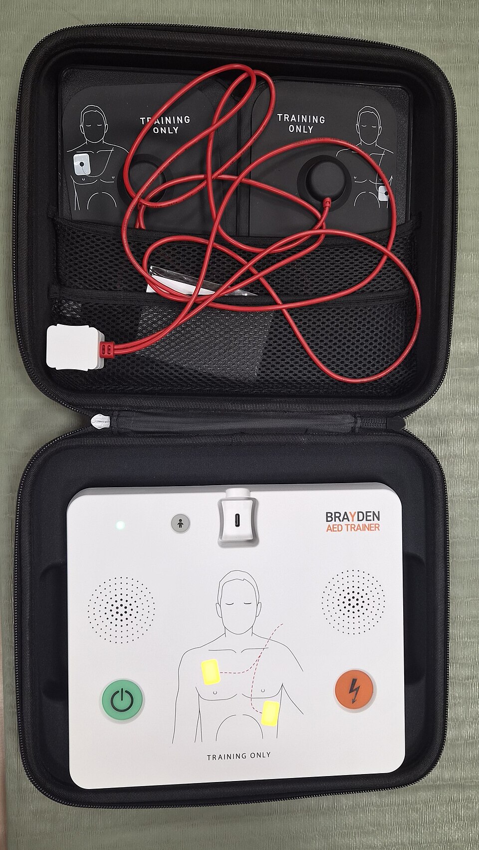
- Position: supine on a firm surface, chest exposed, with room left for CPR.
- Access: get IV or IO access in place, or start it during CPR, so drugs can go in.
Pre-Procedure Checklist
- Device on. Pads in the package, and the right size for this patient.
- Energy set for this patient: adult, or pediatric by kg.
- Pads away from any device. No patch under a pad.
- Chest dry. Patient off wet surfaces.
- Oxygen moved clear of the chest.
- Epinephrine and an antiarrhythmic drawn up and labeled.
- Team roles named. One person calls "clear."
Medications & Settings
Adult doses come from the 2025 AHA ACLS algorithm. Pediatric doses come from the 2025 AHA/AAP PALS algorithm. Where AHA/AAP 2025 states a pediatric dose, use it. If it does not, look to the NASEMSO National Model EMS Clinical Guidelines, Version 3.0 (2022), and then the US label. If none of them gives a dose, this page shows none. A pediatric maximum never goes above the adult maximum dose (NASEMSO Universal Care Guideline, p. 13). Doses are off-label unless marked labeled.
Energy Settings
| Rhythm | Adult | Pediatric | Notes |
|---|---|---|---|
| VF or pulseless VT, biphasic | Manufacturer dose (initial 120–200 J); if unknown, maximum available | 2 J/kg first, then 4 J/kg for the second shock and 4 J/kg or more after; max 10 J/kg or adult dose | Second and later shocks equivalent; higher may be considered |
| VF or pulseless VT, monophasic | 360 J | 2 J/kg first, then 4 J/kg or more | Use only where the device is monophasic |
Arrest Drugs
| Drug | Adult Dose | Pediatric Dose | Notes |
|---|---|---|---|
| Epinephrine | 1 mg IV/IO every 3–5 min | 0.01 mg/kg IV/IO every 3–5 min; max 1 mg | High-alert VF/pVT: from shock 2, then every 3–5 min. Asystole/PEA: as soon as possible. Never 1 mg to a patient with a pulse |
| Amiodarone | 300 mg IV/IO, then 150 mg per algorithm (after shock 3) | 5 mg/kg IV/IO max 300 mg first dose; later doses max 150 mg (2025 AHA PALS algorithm). Differs from FDA label: IV amiodarone safety and effectiveness are not established in children. | High-alert For persisting VF or pulseless VT |
| Lidocaine | 1–1.5 mg/kg IV/IO, then 0.5–0.75 mg/kg per algorithm | 1 mg/kg IV/IO. Not established in children (FDA label). | High-alert Alternative to amiodarone per the algorithm |
Step-by-Step Procedure
Phase 1: Recognize and Prepare
- Confirm pulselessness: check for a pulse and breathing, then start CPR and get the monitor or AED on.
- Analyze the rhythm: VF or pulseless VT is shockable; asystole and PEA are not.
- Set the energy: adult biphasic manufacturer dose, or 2 J/kg for a child's first shock, then 4 J/kg or more for later shocks.
- Place the pads: anterolateral first; keep them off any device and away from each other.
Phase 2: Shock and Resume CPR
- Clear the patient: announce "clear," confirm that no one touches the patient or bed, and check oxygen is away.
- Shock: press SHOCK. Resume CPR immediately after the shock, starting compressions without a pulse check.
- Give 2 minutes of CPR: then re-analyze the rhythm.
Phase 3: Drugs and Persisting VF
- Give epinephrine after the second shock: 1 mg IV/IO during the CPR period, then every 3–5 minutes (after initial defibrillation attempts have failed; 2025 AHA Guidelines for CPR and ECC, Part 9, COR 2a, LOE B-NR). In asystole or PEA, give it as soon as possible.
- Give an antiarrhythmic after the third shock if VF persists: amiodarone 300 mg, then 150 mg, or lidocaine 1–1.5 mg/kg, then 0.5–0.75 mg/kg (2025 AHA Guidelines for CPR and ECC, Part 9, COR 2b, LOE B-R).
- Consider an advanced airway and waveform capnography: ventilate without over-ventilating during CPR.
- Treat reversible causes: check for hypoxia, acidosis, electrolyte problems, hypothermia, tension pneumothorax, tamponade, toxins and thrombosis.
Alternative Technique: Persisting VF
When VF persists after 3 or more consecutive shocks, two options come up: a pad vector change (anteroposterior), or double sequential external defibrillation (DSD). The 2025 AHA Guidelines for CPR and ECC, Part 9, say their usefulness has not been established (COR 2b, LOE B-R). Neither one is a routine step (see Special Populations).
Confirmation & Monitoring
Primary Confirmation
The rhythm check comes after 2 minutes of CPR. A shockable rhythm gets another shock. A non-shockable rhythm changes the plan. So does an organized rhythm that comes back with a pulse.
Secondary Confirmation
- Waveform capnography runs continuously during CPR. If EtCO₂ is low or falling, reassess CPR quality.
- Once CPR stops, check arterial pressure or a pulse, per protocol.
- Watch the rhythm on the monitor between shocks.
| Finding | Success | Failure / Next Step |
|---|---|---|
| Rhythm | Organized rhythm with a pulse | VF or pulseless VT persists: shock again |
| Shock delivered | Device confirms the shock | No shock: check pads, leads and energy; restart CPR at once |
| Compressions | Resumed within seconds after the shock | Long pauses: correct, then resume |
| EtCO₂ | Not low or falling during CPR | Low or falling: reassess CPR quality |
Ongoing Monitoring
- Run continuous ECG and SpO₂, and EtCO₂ when you have it, during and after resuscitation.
- 12-lead ECG after return of circulation and at handoff.
- Reassess pulse, rhythm and pad skin at every move.
Troubleshooting
| Problem | Likely Cause | Fix |
|---|---|---|
| AED says no shock advised | Asystole, PEA or motion during analysis | Stop motion; resume CPR; re-analyze after 2 minutes |
| Shock not delivered | Poor pad contact or a gel bridge | Re-seat pads, dry the chest, separate the pads |
| Device shows poor contact | Wet skin, hair, or a patch under a pad | Dry and shave the site; remove the patch |
| Pads over a device | Pad sited on a generator | Reposition the pad per the device's instructions |
| VF persists after 3 shocks | Persisting VF/pVT | Antiarrhythmic and reversible causes. Vector change and DSD: usefulness not established (2025 AHA Guidelines for CPR and ECC, Part 9, COR 2b) |
| Organized rhythm returns, then VF | Recurrent VF | Back to the algorithm; amiodarone or lidocaine at the algorithm doses |
| Wrong energy on the device | Adult setting used for a child | Reset to pediatric before the next shock; document the error |
If It Still Fails
- Check the basics: Go back to fundamentals. Compression quality, pad position, electrolytes, oxygenation and reversible causes all count.
- Escalate: bring in an advanced airway and capnography. Consider the next therapy per protocol and medical direction.
- Document: log each shock with its energy, each drug dose and the time. Handoff depends on that record.
Complications
| Complication | Recognition | Prevention | Management |
|---|---|---|---|
| Immediate: skin burns | Redness or blistering under a pad | Good contact without gel bridges; remove patches | Burn care and documentation |
| Immediate: shock to a rescuer | Anyone touching the patient or bed during the shock | Clear the patient before every shock | Stop; check the rescuer; continue per protocol |
| Immediate: device or pad problem | Pad or device fault; no shock delivered | Check pads and cables before the shock | Change pads or device; continue CPR |
| Delayed: implanted device | ICD may have fired during the event | Keep pads off the generator | Notify the device team after any shock |
| Delayed: rib or sternal injury | Pain or crepitus after CPR | Correct depth and hand position | Analgesia and reassessment |
Special Populations
Pediatrics
- Energy: the first shock is 2 J/kg. (2–4 J/kg is reasonable; 2025 AHA Guidelines for CPR and ECC, Part 8, COR 2a, LOE C-LD.) The second shock is 4 J/kg, and later shocks are 4 J/kg or more. The ceiling is 10 J/kg or the adult dose (COR 2b, LOE C-LD).
- Pads: use pediatric pads or an attenuator when you have them, per the device label.
- Drugs: epinephrine 0.01 mg/kg (max 1 mg); amiodarone 5 mg/kg (max 300 mg first dose; later doses max 150 mg; differs from FDA label, where IV amiodarone is not established in children); lidocaine 1 mg/kg (not established in children on the FDA label). See pediatric cardiac arrest.
- Sizing: see equipment sizing and Broselow.
- Attenuator: under 8 years on an AED? Use a pediatric attenuator (2025 AHA Guidelines for CPR and ECC, Part 6, COR 1, LOE C-LD). A trained professional may use a manual defibrillator instead (COR 2a, LOE C-LD). If neither is on hand, an AED without an attenuator may be considered (COR 2b, LOE C-EO).
Pregnancy
- Shock: run the adult algorithm. The 2025 pregnancy recommendations set no different energy.
- CPR: when the fundal height is at or above the umbilicus, add manual left lateral uterine displacement to chest compressions (2025 AHA Guidelines for CPR and ECC, Part 10, COR 1, LOE C-LD). Put airway management first (COR 1, LOE C-LD).
- Resuscitative delivery: it is indicated when standard resuscitation does not bring back ROSC and the fundal height is at or above the umbilicus (2025 AHA Guidelines for CPR and ECC, Part 10, COR 1, LOE B-NR). Start preparing from the moment of arrest, aiming to finish delivery by 5 minutes (COR 1, LOE B-NR). Take the fetal monitors off. Do not perform fetal monitoring (2025 AHA Guidelines for CPR and ECC, Part 10, COR 3: No Benefit, LOE C-EO).
Implanted Devices
- Keep the pads off any pacemaker or ICD generator.
- Tell the device team after any shock. An ICD may have fired.
Persisting VF/pVT: Vector Change and DSD
Vector change and double sequential defibrillation (DSD) are not routine steps. Part 9 of the 2025 AHA Guidelines for CPR and ECC says their usefulness for VF or pulseless VT persisting after 3 or more consecutive shocks has not been established (COR 2b, LOE B-R). The DOSE VF randomized trial (2022) ran in six Canadian paramedic services and stopped early after 405 patients. Survival to hospital discharge was 30.4% with DSD, 21.7% with vector change and 13.3% with standard defibrillation. Part 9 adds two caveats. The survival difference held on intention-to-treat analysis, but not by treatment actually received. The secondary analysis found a DSD benefit only in incessant VF (17% of patients), not in VF that came back after a successful shock.
Post-Procedure Care & Transport
Immediate Aftercare
- Get a 12-lead ECG after return of circulation. Compare it with the arrest rhythm.
- Look at the skin under the pads. Check the pulse, SpO₂, blood pressure and mental status.
Ongoing Maintenance
- Rhythm: watch for VF coming back, and treat per protocol.
- Reversible causes: fix electrolytes, oxygenation and perfusion.
- Post-arrest care: targeted temperature and hemodynamic goals, per the receiving facility's protocol.
Transport Considerations
- Transport on continuous monitoring, with the defibrillator and pads within reach.
- Check pulse, rhythm and breathing again after every move.
- Send the drug record with the patient, so the receiving team has it.
Document & Hand Off
- Time of arrest. Time of each shock, its energy, and the device used.
- Pad position, and any implanted device.
- The rhythm before and after each shock.
- Each drug dose and its time, epinephrine and any antiarrhythmic included.
- The time of return of circulation, and the post-arrest vital signs.
Clinical Pearls & Pitfalls
Pearls
- Shock, then CPR: compressions restart at once after each shock. The 2-minute interval is when you check the rhythm.
- Equivalent doses: a second or later adult shock is not automatically a bigger one.
- Child dosing: 2 J/kg for the first shock, then 4 J/kg or more. The ceiling is 10 J/kg or the adult dose.
- Non-shockable rhythms: asystole and PEA get CPR and drugs. They do not get shocks.
Common Pitfalls
- Pulse check after each shock: restart compressions first. Check the pulse only at the 2-minute rhythm check.
- Adult energy for a child: set the pediatric dose before the first shock.
- Shocking a pulse: only a shockable rhythm with no pulse gets a shock.
- Pads touching or over a device: pull the pads apart and move them off the generator. Do it without holding up the shock.
Education & Skill Maintenance
Skill Maintenance
- Drill pad placement and the clear-and-shock sequence on a training AED every quarter.
- Run the 2-minute CPR cycle with a rhythm check, timed on a clock.
- After every cardiac arrest, debrief the time to first shock and the time back to compressions.
- Confirm the device has pediatric pads, attenuators and a pediatric energy setting.
References
- Wigginton JG, Agarwal S, Bartos JA, Coute RA, Drennan IR, Haamid A, et al. Part 9: Adult Advanced Life Support: 2025 American Heart Association Guidelines for Cardiopulmonary Resuscitation and Emergency Cardiovascular Care. Circulation. 2025;152(16 suppl 2):S538–S577. https://doi.org/10.1161/cir.0000000000001376. https://cpr.heart.org/en/resuscitation-science/cpr-and-ecc-guidelines/adult-advanced-life-support
- American Heart Association. 2025 Adult Cardiac Arrest Algorithm (ACLS), long description (250725). https://cpr.heart.org/-/media/CPR-Files/CPR-Guidelines-Files/2025-Accessible/Algorithm-ACLS-CA-LngDscrp-250725-Ed.pdf
- American Heart Association and American Academy of Pediatrics. 2025 PALS Pediatric Cardiac Arrest Algorithm, long description (250729). https://cpr.heart.org/-/media/CPR-Files/CPR-Guidelines-Files/2025-Accessible/Algorithm-PALS-CA-LngDscrp-250729-Ed.pdf
- Cheskes S, Verbeek PR, Drennan IR, McLeod SL, Turner L, Pinto R, et al. Defibrillation strategies for refractory ventricular fibrillation. N Engl J Med. 2022;387:1947–1956. (DOSE VF trial). https://doi.org/10.1056/NEJMoa2207304
- Nichol G, Atkins DL, Koster RW, Kudenchuk PJ, Cabañas JG, et al. Scientific priorities related to the use of double sequential external defibrillation in patients with refractory cardiac arrest: report from a multistakeholder thinktank. J Am Heart Assoc. 2025. https://doi.org/10.1161/jaha.125.044130
- Cao D, Arens AM, Chow SL, Easter SR, Hoffman RS, Lagina AT III, Lavonas EJ, Patil KD, Sutherland LD, Tijssen JA, Wang GS, Zelop CM, Rodriguez AJ, Drennan IR, McBride ME. Part 10: adult and pediatric special circumstances of resuscitation: 2025 American Heart Association Guidelines for Cardiopulmonary Resuscitation and Emergency Cardiovascular Care. Circulation. 2025;152(16 suppl 2):S578–S672. https://doi.org/10.1161/CIR.0000000000001380. https://cpr.heart.org/en/resuscitation-science/cpr-and-ecc-guidelines/adult-and-pediatric-special-circumstances-of-resuscitation
- Lasa JJ, Dhillon GS, Duff JP, Hayes J, Kamath-Rayne BD, Levy A, et al. Part 8: Pediatric Advanced Life Support: 2025 American Heart Association and American Academy of Pediatrics Guidelines for Cardiopulmonary Resuscitation and Emergency Cardiovascular Care. Circulation. 2025;152(16 suppl 2):S479–S537. https://doi.org/10.1161/cir.0000000000001368. https://cpr.heart.org/en/resuscitation-science/cpr-and-ecc-guidelines/pediatric-advanced-life-support
- Kleinman ME, Buick JE, Huber N, Idris AH, Levy M, Morgan SG, et al. Part 7: Adult Basic Life Support: 2025 American Heart Association Guidelines for Cardiopulmonary Resuscitation and Emergency Cardiovascular Care. Circulation. 2025;152(16 suppl 2):S448–S478. https://doi.org/10.1161/cir.0000000000001369. https://cpr.heart.org/en/resuscitation-science/cpr-and-ecc-guidelines/adult-basic-life-support
- Joyner BL, Dewan M, Bavare A, de Caen A, DiMaria K, Donofrio-Odmann J, Fosse G, Haskell S, Mahgoub M, Meckler G, Requist J, Schexnayder SM, Olech Smith M, Werho D, Raymond TT. Part 6: pediatric basic life support: 2025 American Heart Association and American Academy of Pediatrics Guidelines for Cardiopulmonary Resuscitation and Emergency Cardiovascular Care. Circulation. 2025;152(16 suppl 2):S424–S447. https://doi.org/10.1161/CIR.0000000000001370. https://cpr.heart.org/en/resuscitation-science/cpr-and-ecc-guidelines/pediatric-basic-life-support
- National Association of State EMS Officials (NASEMSO). National Model EMS Clinical Guidelines, Version 3.0. March 2022. Universal Care Guideline, Patient Care Goals, item 5 (p. 13). https://nasemso.org/content.aspx?page_id=22&club_id=157064&module_id=701974
- Nexterone (amiodarone hydrochloride) injection, US prescribing information. Baxter Healthcare Corporation. Label effective 2024-04-16. DailyMed setid e9108958-b8d7-4fba-87c3-9a32990de551 (section 8.4, Pediatric Use). https://dailymed.nlm.nih.gov/dailymed/drugInfo.cfm?setid=e9108958-b8d7-4fba-87c3-9a32990de551
- Lidocaine hydrochloride and 5% dextrose injection, US prescribing information. Baxter Healthcare Corporation. Label effective 2026-02-20. DailyMed setid f21512ea-c7c3-4766-b976-b2c0b5616bf0 (Pediatric Use and Dosage). https://dailymed.nlm.nih.gov/dailymed/drugInfo.cfm?setid=f21512ea-c7c3-4766-b976-b2c0b5616bf0
Medical Disclaimer
- For Educational Purposes Only: This content is intended for educational reference and should not be used for clinical decision-making.
- Not a Substitute for Professional Judgment: Always consult your local protocols, institutional guidelines, and supervising physicians.
- Verify Before Acting: Users are responsible for verifying information through authoritative sources before any clinical application.