

Bedside Snapshot
- Recognize
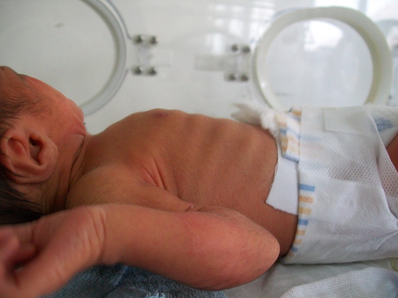
- Distress: rising rate and effort, retractions, nasal flaring, accessory muscles.
Failure: effort tires, SpO₂ falls, consciousness decreases. - Red Flags
- Inadequate breathing with a pulse
- Decreased consciousness
- Heart rate below 60/min with cardiopulmonary compromise
- SpO₂ below 94% on oxygen
- First Actions
- Open the airway. Oxygen when SpO₂ is below 94%. Treat the cause. Bag-mask ventilate at 20–30/min when breathing is inadequate.
- Key Doses
- Albuterol 0.15 mg/kg nebulized (min 2.5 mg)
- Dexamethasone 0.6 mg/kg (max 16 mg)
- Epinephrine 0.01 mg/kg IV/IO (max 1 mg)
- Naloxone 0.1 mg/kg (max 2 mg)
- Don't Miss
- Epiglottitis; foreign-body aspiration; anaphylaxis; opioid depression. Oximetry misreads with carboxyhemoglobin, methylene blue or poor perfusion
- Escalate & Transport
- Early PICU or pediatric-ready ED call. Advanced airway if bag-mask fails. Monitor SpO₂ and EtCO₂ in transport; structured handoff
Overview & Pediatric Differences
Distress and failure sit on one continuum, and they need different responses. Distress is compensation. The child raises the respiratory rate and works harder to meet oxygen needs. Failure is the point where breathing is no longer adequate, so oxygenation and ventilation fall short (2025 AHA Guidelines for CPR and ECC, Part 6). Cardiac arrest in children usually does not start with a primary heart problem. It follows progressive respiratory failure or shock (Part 6). So you fix the breathing before any drug.
Why Kids Are Different
- Breathing control is immature: premature babies slide into distress from mild causes. Irregular breathing in the first days of life can mean a blunted response to hypoxia and hypercapnia.
- Narrow, collapsible airways: airway resistance climbs as the radius falls. The walls of the pharynx, larynx and trachea collapse more easily than an adult's.
- Little collateral ventilation: in infants, the channels between alveoli are not fully developed. Bronchial obstruction causes atelectasis, and the result is ventilation–perfusion mismatch.
- Muscles tire quickly: horizontal ribs and a horizontal diaphragm insertion make each breath less efficient. Fewer fatigue-resistant fibers mean they deteriorate faster as the rate climbs.
For the arrest pathway that follows failing breathing, see the pediatric cardiac arrest page.
Recognition & Severity
Sort distress from failure with three things: the work of breathing, oxygenation and level of consciousness. Each column is a stage, not an age.
| Feature | Distress (Compensated) | Failure (Decompensated) | Pre-Arrest |
|---|---|---|---|
| Breathing effort | Raised rate and effort; retractions, nasal flaring and accessory muscle use | Effort tires as the muscles fatigue; breathing falls behind the body's needs | Inadequate breathing with a pulse |
| Respiratory rate | Raised for age (ranges on the vital signs page) | Rate alone can mislead; judge the depth and effort of each breath | Ventilate at 20–30/min |
| Oxygenation | SpO₂ below 94%, or distress: start oxygen | SpO₂ falls despite oxygen | Hypoxia with poor perfusion |
| Consciousness | — | Decreased consciousness | Decreased consciousness with poor perfusion |
| Perfusion | No signs of poor perfusion | Signs of poor perfusion; see the shock page | Heart rate below 60/min with cardiopulmonary compromise: start CPR if it persists despite oxygen and ventilation |
Age-Specific Presentation
- Neonate and young infant: premature babies drift into distress from mild causes. Irregular breathing in the first days of life is common. It does not reassure you on its own.
- Under 1 year and 1–8 years: with an advanced airway, the ventilation rate is 30/min under 1 year and 20–30/min at 1–8 years.
- 8–18 years: with an advanced airway in a healthcare setting, 10–20/min at clinical discretion.
Differential Diagnosis
A small group of causes explains most respiratory failure in children. Each row pairs a cause with the feature that tells it apart.
| Condition | Distinguishing Features | Key Test or Finding |
|---|---|---|
| Asthma exacerbation | Wheeze, chest tightness, accessory muscle use | Initial SpO₂ below 91% raises the chance of a chest X-ray |
| Bronchiolitis | Rapid breathing, wheeze and crackles, nasal flaring, retractions | Guidelines advise against routine chest X-ray |
| Croup | Barking cough with stridor | Response to nebulized epinephrine and corticosteroid |
| Epiglottitis and bacterial tracheitis | Severe sore throat, drooling, muffled voice; high fever and toxic appearance | Epiglottitis has no barking cough; tracheitis responds poorly to nebulized epinephrine |
| Pneumonia | Fever, rapid breathing, crackles, retractions | Diagnosis is clinical; chest X-ray is often requested |
| Foreign-body aspiration | Sudden coughing or choking, then wheeze | Unilateral reduction of breath sounds; possible cyanosis |
| Anaphylaxis | Sudden distress, wheeze, stridor, rash, facial swelling, shock | Give IM epinephrine first (see the epinephrine IM page) |
| Opioid or sedative toxicity | Respiratory depression with decreased consciousness | Response to naloxone; ventilate while it works |
Rapid breathing also shows up with sepsis and shock, pulmonary edema and cardiac causes. For those, go to the shock page.
Management by Severity
Manage by stage. Escalate when the child does not respond or the work of breathing gets worse.
Distress (Compensated)
- Oxygen: give oxygen when SpO₂ is below 94% or the child shows respiratory distress, and titrate to 94–99% or the child's own normal level (Paramedic/RN/CCT, per local protocol).
- Treat the cause: nebulized albuterol for bronchospasm, with ipratropium for moderate to severe asthma; nebulized epinephrine and dexamethasone for moderate to severe croup; IM epinephrine first for anaphylaxis.
- Reassess: repeat the work of breathing, SpO₂ and perfusion after each intervention.
Foreign-body obstruction: sudden choking, then a cough, wheeze or one-sided breath sounds. A forceful cough is moving air, so encourage it. Weak or silent cough? Treat it as airway obstruction at once (current choking first aid, per local protocol).
For cause-specific drugs and thresholds, use the croup and bronchiolitis pages.
Failure (Decompensated)
- Position and suction: open the airway (sniffing position, with a shoulder roll under an infant's shoulders), suction secretions, and give oxygen.
- Ventilate: with a pulse but inadequate breathing, bag-mask ventilate at 20–30 breaths per minute with oxygen (2025 AHA Guidelines for CPR and ECC, Part 6, COR 2a, LOE C-EO).
- Noninvasive support as a bridge: for an alert child whose breathing is failing, consider high-flow nasal cannula or CPAP/NIV per local protocol. It buys time while the advanced airway is prepared.
- Advanced airway: intubate for failing effort, falling consciousness, or no response to noninvasive support. Once it is in place, give a breath every 2–3 seconds with continuous capnography. In severe asthma, slow the rate and lengthen the expiratory time to avoid air trapping.
- Naloxone for opioid depression: give it to support breathing. Restoring alertness is not the goal.
Pre-Arrest: Bradycardia With Cardiopulmonary Compromise
- Confirm: heart rate below 60/min with cardiopulmonary compromise (acutely altered mental status, signs of shock or hypotension) that persists despite effective oxygen and ventilation (2025 AHA Guidelines for CPR and ECC, Part 8, PALS Bradycardia With a Pulse algorithm).
- Start CPR: if the heart rate stays below 60/min with cardiopulmonary compromise despite effective oxygenation and ventilation, start chest compressions and follow the pediatric cardiac arrest page (2025 AHA Guidelines for CPR and ECC, Part 8, COR 1, LOE C-EO).
- Epinephrine: 0.01 mg/kg IV/IO (0.1 mL/kg of 0.1 mg/mL; max 1 mg), repeated every 3–5 minutes, when bradycardia with cardiopulmonary compromise persists after correction of other factors such as hypoxia (2025 AHA Guidelines for CPR and ECC, Part 8, COR 1, LOE C-EO). AHA 2025 prefers IV/IO over the endotracheal route. The endotracheal dose (0.1 mg/kg of 1 mg/mL, the Korean 2025 guideline (reference 2)) is only for the intubated child whose IV/IO access is difficult or unavailable.
- Atropine: 0.02 mg/kg IV/IO (minimum 0.1 mg, maximum 0.5 mg per dose; may repeat once) only for bradycardia with cardiopulmonary compromise due to increased vagal tone or atrioventricular block, not due to hypoxia (2025 AHA Guidelines for CPR and ECC, Part 8, COR 1, LOE C-LD).
- Pacing is a later option: emergency transcutaneous pacing may be considered (2025 AHA Guidelines for CPR and ECC, Part 8, COR 2b, LOE C-EO) for complete heart block or sinus node dysfunction that does not respond to CPR. Pacing does not replace oxygen, ventilation or CPR.
Medications & Dosing
Respiratory and Croup
| Drug | Dose & Route | Max | Notes |
|---|---|---|---|
| Oxygen | Distress or failure: give when SpO₂ is below 94% or the child is in distress; titrate to 94–99% or the child's own normal level (for example, cyanotic heart disease) After ROSC: 94–99% and normocapnia Not weight-based |
No fixed max: lowest FiO₂ that holds the target |
Excess oxygen can harm the child after ROSC as well as hypoxemia. Confirm with a blood gas when oximetry and the child disagree |
| Albuterol (nebulized) | 0.15 mg/kg nebulized (minimum 2.5 mg) every 20 min for 3 doses, then 0.15–0.3 mg/kg (up to 10 mg per dose) every 1–4 h as needed = 0.03 mL/kg of 0.5% (5 mg/mL), diluted to 3 mL; minimum 0.5 mL |
Child: no maximum stated for the first 3 doses; up to 10 mg per dose after that (EPR-3). Adult: 2.5–5 mg for the first 3 doses, then 2.5–10 mg |
Children 12 years and under (NAEPP EPR-3, 2007). Anaphylaxis: give only after IM epinephrine |
| Ipratropium (nebulized, with albuterol) | 0.25–0.5 mg every 20 min for 3 doses, mixed with albuterol |
Adult dose 0.5 mg per dose |
Moderate to severe asthma only. No added benefit once the child is admitted. Under 4 years with an MDI: valved holding chamber and face mask |
| Racemic epinephrine 2.25% or L-epinephrine 1 mg/mL (nebulized) | 0.5 mL of 2.25% racemic, or 5 mL of 1 mg/mL L-epinephrine, once; the same dose at any age or weight |
0.5 mL (11.25 mg) or 5 mL (5 mg) per dose; repeat per protocol |
Moderate to severe croup with a corticosteroid (CPS). Effect in 10–30 min, lasting about 1 h; observe 2–4 h before discharge. Never give the 1 mg/mL vial IV for croup |
| Dexamethasone | 0.6 mg/kg PO once; IM or IV if the child is vomiting or in significant distress |
16 mg single dose (common protocol cap) |
Any severity of croup; corticosteroids are the mainstay (CPS). Benefit is measured at 2 hours. Asthma courses: see the prednisone page |
| Naloxone | Pediatric overdose: 0.1 mg/kg IV/IO, max 2 mg (adult dose; AHA 2025 Part 10 Table 4) IM or IN per NASEMSO: 0.1 mg/kg, max 2 mg IM and 4 mg IN. Differs from FDA label: label starts 0.01 mg/kg, then 0.1 mg/kg. |
2 mg IV/IO/IM per dose (4 mg IN) |
For suspected opioid overdose with respiratory depression or arrest, give breaths or bag-mask ventilation first (2025 AHA Guidelines for CPR and ECC, Part 10, COR 1, LOE B-NR). Give naloxone for respiratory arrest with a definite pulse (COR 1, LOE B-NR). In cardiac arrest, naloxone is COR 2b and must not interrupt CPR (Part 10, COR 2b, LOE B-NR adults and C-EO children). Titrate to breathing. Respiratory depression from therapeutic opioid use: lower titrated doses of 1–5 mcg/kg. In opioid-dependent children, full-dose reversal can cause acute withdrawal. Endotracheal route only if IV/IO is not possible, followed by at least 5 mL saline and five breaths |
Bradycardia and Resuscitation
| Drug | Dose & Route | Max | Notes |
|---|---|---|---|
| Epinephrine 0.1 mg/mL (formerly 1:10,000), IV/IO High-alert |
0.01 mg/kg IV/IO (0.1 mL/kg of 0.1 mg/mL), every 3–5 min Endotracheal: 0.1 mg/kg (0.1 mL/kg of 1 mg/mL) only if intubated and IV/IO access is difficult or unavailable |
1 mg per dose |
Used with CPR for heart rate below 60/min with cardiopulmonary compromise despite oxygen and ventilation. Not a 1 mg/mL IV push. 2025 AHA Guidelines for CPR and ECC, Part 8, COR 1, LOE C-EO. Endotracheal dose is not stated in AHA 2025 (IV/IO preferred); from the Korean 2025 guideline (reference 2) |
| Atropine | 0.02 mg/kg IV/IO (minimum 0.1 mg) Endotracheal 0.04–0.06 mg/kg only if intubated and IV/IO access is not possible (Korean 2025 guideline, reference 2) |
0.5 mg per dose; may repeat once |
For bradycardia with cardiopulmonary compromise from increased vagal tone or atrioventricular block, not from hypoxia (2025 AHA Guidelines for CPR and ECC, Part 8, COR 1, LOE C-LD). Not a substitute for oxygen or ventilation. The 0.1 mg minimum and once-repeat rule are from the AHA 2025 PALS bradycardia algorithm |
Doses vary by protocol; follow local protocols and medical direction. When severe asthma does not respond to first-line treatment, magnesium sulfate is an EPR-3 adjunct (Evidence B). Its dose is on the magnesium sulfate page. For asthma steroid courses, use the prednisone and dexamethasone pages.
Monitoring, Reassessment & Disposition
| Parameter | Target | Notes |
|---|---|---|
| SpO₂ | 94–99% or the child's own normal level; give oxygen when SpO₂ is below 94% or the child is in distress. After ROSC: 94–99% (2025 AHA Guidelines for CPR and ECC, Part 8, COR 2b, LOE C-LD) | Oximetry can be inaccurate in shock or with carboxyhemoglobin or methylene blue |
| PaCO₂ | Normocapnia after ROSC | Check a blood gas after ROSC; avoid hypoventilation and hyperventilation |
| EtCO₂ | Continuous waveform capnography with an advanced airway | Use it to confirm ventilation, not as the only sign |
| Ventilation rate | 20–30/min with a pulse and inadequate breathing | Ten to 20 per minute at 8–18 years with an advanced airway in healthcare settings |
| Blood pressure | Systolic and mean arterial pressure above the 10th percentile for age after ROSC (2025 AHA Guidelines for CPR and ECC, Part 8, COR 1, LOE B-NR) | See the vital signs page for age ranges |
- Reassess after each intervention: work of breathing, SpO₂, mental status and perfusion.
- Croup after nebulized epinephrine: the child needs 2–4 hours of observation. Discharge only if symptoms have stayed away for that period (CPS).
- Asthma: once the child is admitted, stop ipratropium. It adds nothing (EPR-3). Keep albuterol going.
- Escalate: call for PICU or critical-care transport when failure does not respond to the first steps above.
Pitfalls & Safety Checks
| Pitfall | Consequence | Prevention |
|---|---|---|
| Trusting a normal SpO₂ | Missed hypoxia in shock, with carboxyhemoglobin or with methylene blue | Pair the number with the work of breathing and mental status; confirm with a blood gas |
| Expecting pacing to fix respiratory bradycardia | Delay in CPR and epinephrine while the child has cardiopulmonary compromise | Oxygenate and ventilate first; if HR stays below 60/min with cardiopulmonary compromise, start CPR and give epinephrine per the algorithm. Pacing is reserved for complete heart block or sinus node dysfunction unresponsive to CPR (2025 AHA Guidelines for CPR and ECC, Part 8, COR 2b) |
| Endotracheal epinephrine dosed like IV | Same 0.1 mL/kg volume but ten times the drug | Read the vial: 0.1 mg/mL for IV/IO, 1 mg/mL for endotracheal and IM |
| Expecting steroids to open the airway | Delayed nebulized epinephrine for stridor | Epinephrine acts in 10–30 minutes; dexamethasone benefit is measured at 2 hours (CPS) |
| Discharging croup early | Rebound stridor after the epinephrine wears off | Observe 2–4 hours; discharge only if symptoms have not recurred |
| Hyperoxemia | Harm from excess oxygen, most clearly after ROSC | Titrate to 94–99% or the child's normal level; after ROSC also keep PaCO₂ normal |
Special Populations
Neonates and Young Infants
- Premature babies drift into respiratory distress from mild causes.
- For neonatal resuscitation and the neonatal airway, follow the Neonatal Resuscitation Program (2025 AHA Part 5). You won't find neonatal doses or tube sizes on this page.
- Infants have less collateral ventilation. A small obstruction can cause atelectasis fast.
Adolescents (8–18 Years)
- With an advanced airway in a healthcare setting, ventilate at 10–20/min at clinical discretion.
- Switch to adult drug doses at the threshold your local protocol sets.
Opioid Exposure
- In opioid-dependent children, full-dose naloxone can trigger acute withdrawal. Titrate lower doses to effect.
- For respiratory depression from therapeutic opioids, go low: titrated naloxone at 1–5 mcg/kg, to effect.
- Keep supporting breathing while naloxone takes effect.
Prehospital & Transport Considerations
| Level | Typical Role in This Topic |
|---|---|
| EMT | Oxygen, positioning, suction, pulse oximetry and bag-mask ventilation; naloxone IN or IM where protocol allows |
| AEMT | Nebulized bronchodilators and nebulized epinephrine for croup where protocol allows |
| Paramedic | Advanced airway; CPR and IV/IO epinephrine and atropine for bradycardia with cardiopulmonary compromise; HFNC or CPAP where protocol allows; nebulized therapy, naloxone and steroids per protocol |
| RN (ED/ICU) | Continuous SpO₂ and EtCO₂ monitoring; titration of oxygen, HFNC or CPAP, and nebulized therapy; observation after croup treatment |
| Critical Care Transport | Ventilator-supported transport with an advanced airway, or HFNC/CPAP per protocol; infusion pumps and continuous capnography |
- Preparation: pre-drawn or colour-coded syringes, each labeled with drug, mg, mL and concentration. Check the epinephrine concentration on every syringe.
- Destination: a pediatric-ready ED. Go to a PICU when the child needs ventilation or vasoactive support.
- Restraint and caregivers: the child goes in a restraint on the stretcher, never on a caregiver's lap. Keep the caregiver close when protocol allows.
- Handoff: use a structured report such as IMIST-AMBO or MIST. Include the weight in kg and each drug's time, dose and route.
Transport and treatment depend on the cause, so use the croup and bronchiolitis pages.
Clinical Pearls
References
- Lasa JJ, Dhillon GS, Duff JP, et al. Part 8: Pediatric Advanced Life Support: 2025 American Heart Association and American Academy of Pediatrics Guidelines for Cardiopulmonary Resuscitation and Emergency Cardiovascular Care. Circulation. 2025;152(16 suppl 2):S479–S537. https://doi.org/10.1161/CIR.0000000000001368. Web version: https://cpr.heart.org/en/resuscitation-science/cpr-and-ecc-guidelines/pediatric-advanced-life-support
- Kim DK, Kim JT, Na JY, et al. 2025 Korean Guidelines for Cardiopulmonary Resuscitation: Part 8. Pediatric advanced life support. Clin Exp Emerg Med. 2026;13(suppl 1):S115–S141. doi:10.15441/ceem.26.103. https://pmc.ncbi.nlm.nih.gov/articles/PMC13277418/
- Lee JE, Kim M, Choi EK, et al. Updated pediatric cardiopulmonary resuscitation: a comprehensive review of the 2025 guidelines. Anesth Pain Med (Seoul). 2026;21(2):150–161. doi:10.17085/apm.26581. https://pmc.ncbi.nlm.nih.gov/articles/PMC13175904/
- Panetti B, Bucci I, Di Ludovico A, et al. Acute Respiratory Failure in Children: A Clinical Update on Diagnosis. Children (Basel). 2024;11(10):1232. doi:10.3390/children11101232. https://pmc.ncbi.nlm.nih.gov/articles/PMC11506303/
- Ortiz-Alvarez O, McKenzie K, Trottier ED; Canadian Paediatric Society. Acute management of croup in the emergency department. Canadian Paediatric Society practice point. Posted May 24, 2017; updated March 6, 2026. https://cps.ca/en/documents/position/acute-management-of-croup
- National Asthma Education and Prevention Program Expert Panel. Expert Panel Report 3: Guidelines for the Diagnosis and Management of Asthma. NIH Publication No. 07-4051. Bethesda, MD: National Heart, Lung, and Blood Institute; 2007 (foundational). https://www.nhlbi.nih.gov/sites/default/files/media/docs/EPR-3_Asthma_Full_Report_2007.pdf
- American Heart Association. 2025 American Heart Association Guidelines for Cardiopulmonary Resuscitation and Emergency Cardiovascular Care: Part 6, Pediatric Basic Life Support. 2025. https://cpr.heart.org/en/resuscitation-science/cpr-and-ecc-guidelines/pediatric-basic-life-support
- American Heart Association. 2025 American Heart Association Guidelines for Cardiopulmonary Resuscitation and Emergency Cardiovascular Care: Part 10, Adult and Pediatric Special Circumstances of Resuscitation. 2025. https://cpr.heart.org/en/resuscitation-science/cpr-and-ecc-guidelines/adult-and-pediatric-special-circumstances-of-resuscitation
Medical Disclaimer
- For Educational Purposes Only: This content is intended for educational reference and should not be used for clinical decision-making.
- Not a Substitute for Professional Judgment: Always consult your local protocols, institutional guidelines, and supervising physicians.
- Verify Before Acting: Users are responsible for verifying information through authoritative sources before any clinical application.