

Bedside Topline
- What It Does
- An EVD drains cerebrospinal fluid (CSF) from a lateral ventricle and reports intracranial pressure (ICP) through the same catheter. Parenchymal sensors report ICP only.
- Key Settings & Targets
- ICP: treat above 22 mmHg
- CPP: target 60–70 mmHg (CPP = MAP − ICP)
- Drain: zeroed and leveled at the protocol reference point; height by order
- Check Every Hour
- ICP and CPP trend, MAP, drain output and color, drain position and level, pupils and GCS, dressing and site.
- Top Alarm → First Move
- Flat or absent waveform: check the clamp, kinks, air and level, then the patient. A high ICP alarm: assess the patient first.
- Never
- Close the drain to drainage, or leave it open at an unordered height, without an order. Flush, reposition or re-zero the drain without an order.
- Escalate Now If
- ICP stays above 22 mmHg, CPP falls below 60 mmHg, pupils change, new posturing, falling GCS, or the drain stops draining while ICP rises.
Overview & How It Works
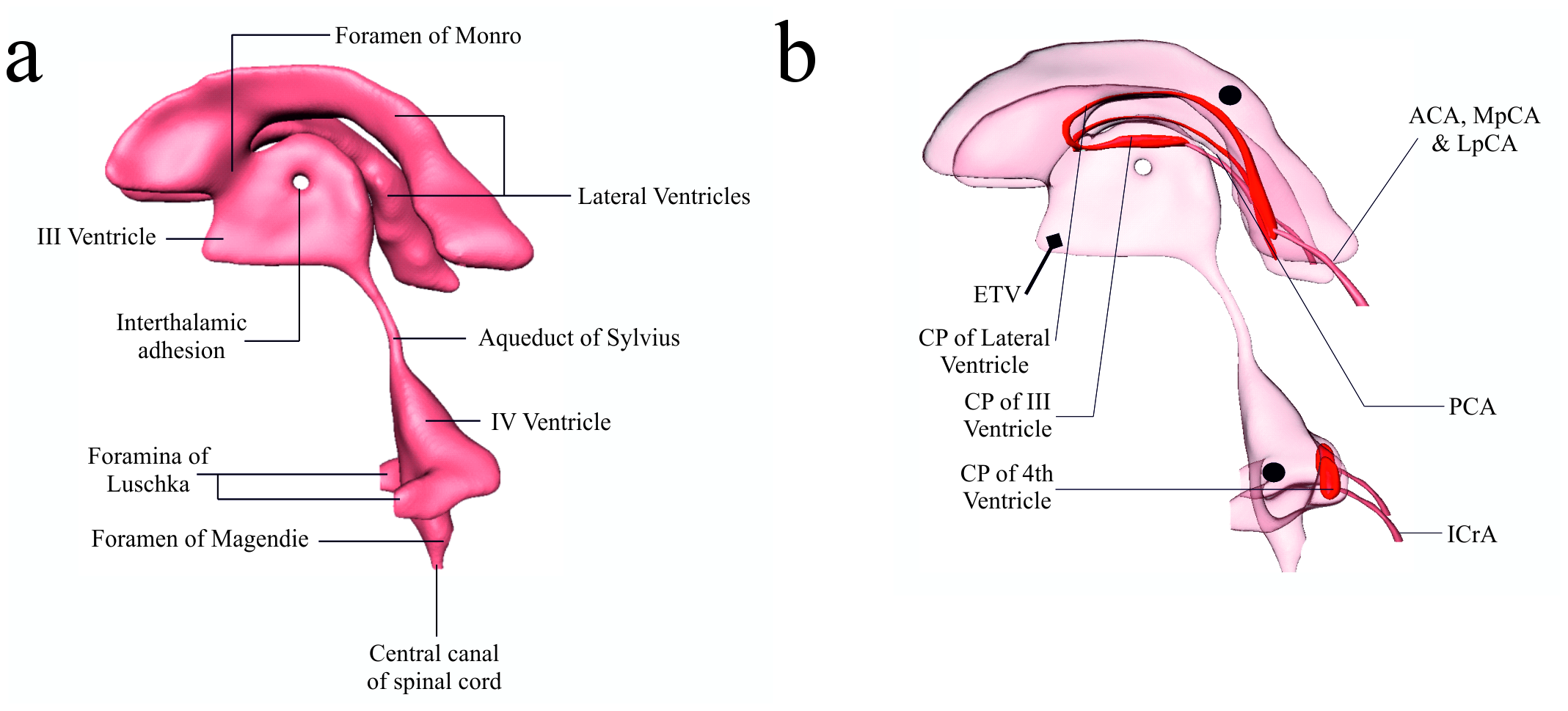
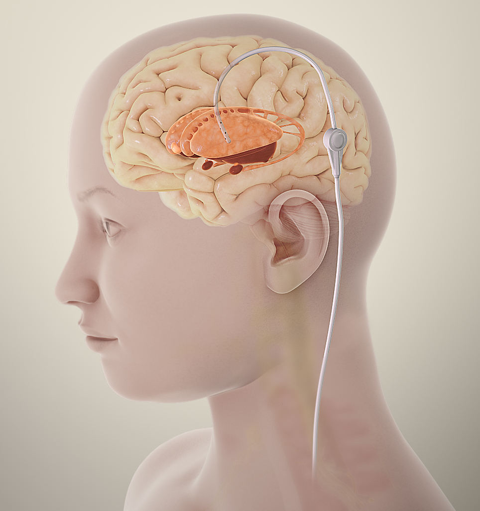
An external ventricular drain (EVD) is a catheter in a lateral ventricle. It drains cerebrospinal fluid (CSF) into a closed external collection system, and it reports intracranial pressure (ICP) through that same catheter. ICP monitoring records the pressure inside the skull. The team uses that number to keep cerebral perfusion pressure (CPP) in range.
Key Components
| Component | Where It Sits | What It Does |
|---|---|---|
| Ventricular catheter | Lateral ventricle, through a burr hole | Drains CSF and senses ICP |
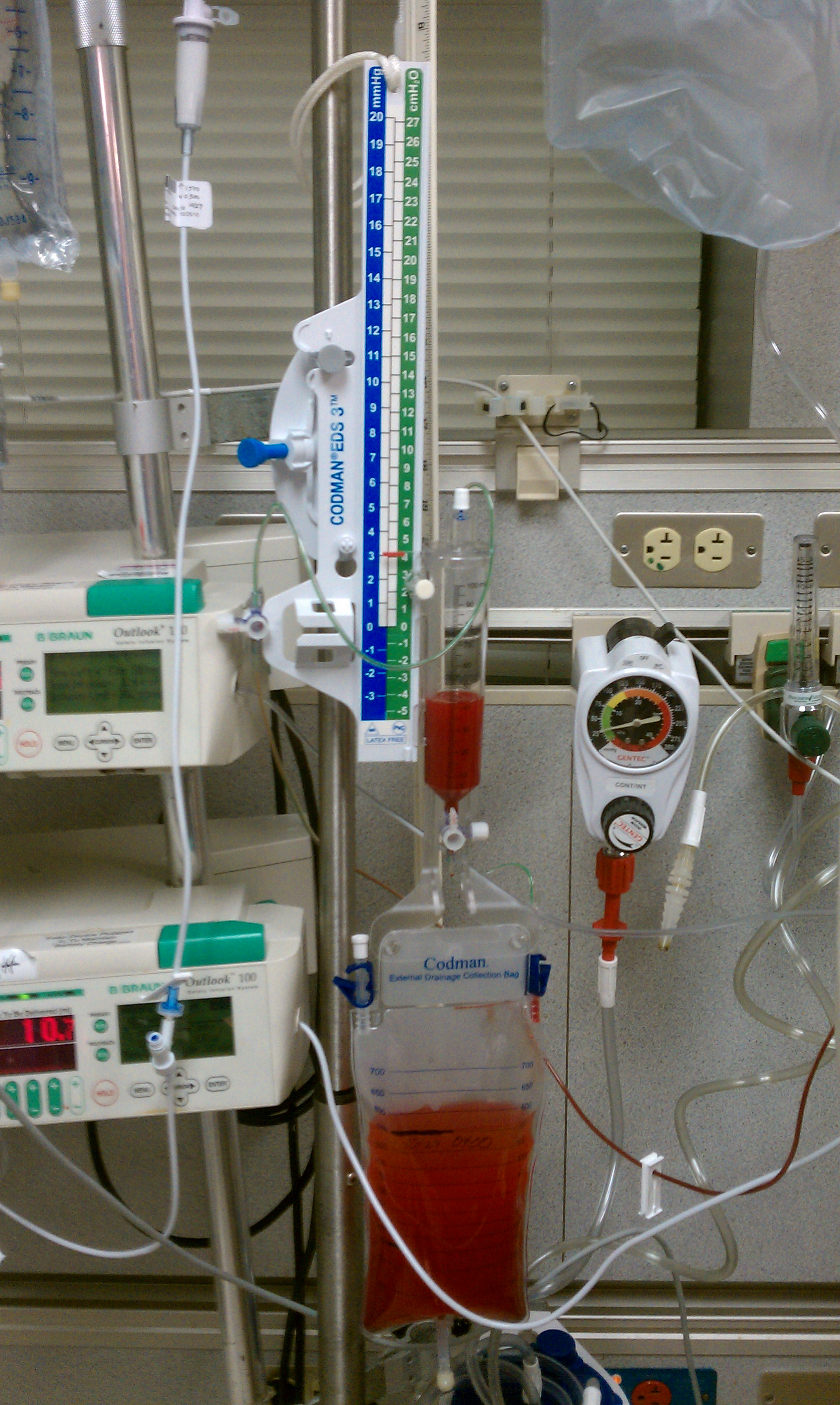
| Drip chamber and level scale | Bedside, at the reference level set by protocol | Sets the zero and shows pressure |
| Tubing and clamp | Between the catheter and the collection bag | Controls drainage; the clamp stops it |
| Collection bag | Below the drip chamber (per protocol) | Measures output and CSF color |
| Parenchymal sensor | Brain tissue, not the ventricle | Reports ICP only; it cannot drain CSF |
Flow Path
- Ventricle: CSF enters at the catheter tip.
- Catheter and tubing: CSF and the pressure signal both travel down to the drip chamber.
- Drip chamber: The level sets the zero. The column shows the pressure.
- Collection bag: Drained CSF gets measured, and sampled when ordered.
Physiology & Hemodynamic Effects
The skull is a fixed box. Blood, CSF and brain tissue share that space. Add volume to one and another has to give, or ICP climbs. The Monro-Kellie relationship and compensatory reserve set how much change the brain absorbs before ICP starts to climb.
What It Does
| Parameter | Effect | Why It Matters |
|---|---|---|
| ICP | ↑ when volume increases past compensatory reserve | Compresses brain tissue and vessels |
| CPP | ↓ as ICP rises with MAP unchanged | Less blood flow to the brain |
| Plateau waves | ↑ sudden ICP above 40 mmHg for at least 5 min | Associated with cerebral vasodilation and impaired autoregulation |
| CSF drainage | ↓ ICP by removing CSF volume | Continuous drainage may lower ICP burden more than intermittent use (BTF, Level III) |
| Hyperventilation | ↓ PaCO₂ lowers ICP only temporarily | Prolonged prophylactic use is harmful (see Setup) |
What It Depends On
- MAP: CPP = MAP − ICP. Lower the MAP and the pressure reaching the brain drops with it.
- Autoregulation: When static pressure autoregulation is intact, raising CPP can lower ICP (SIBICC).
- Drain patency and level: A wrong level or a blocked catheter changes the number. The patient has not changed.
- Temperature: SIBICC aims to avoid fever above 38 °C.
- Seizures: Seizures you cannot see can still raise ICP. SIBICC suggests considering EEG monitoring.
Types, Modes & Configurations
| Type | Route | Does | Key Limitation |
|---|---|---|---|
| EVD (ventriculostomy) | Lateral ventricle | ICP and CSF drainage | Infection risk; needs zeroing and a closed system |
| Parenchymal sensor | Brain tissue | ICP only | No CSF drainage; consider a ventriculostomy if drainage is needed |
Drain Modes
- Continuous drainage: The drain stays open at the set level. BTF (Level III) says this may lower the ICP burden better than intermittent use.
- Intermittent drainage: The drain opens to let CSF out, then closes again. Use only the mode the order sets.
Drain Positions
- Closed to drainage, open to the transducer: This is the position for reading ICP. The number shows on the monitor.
- Open to drainage: CSF is leaving the skull. While it drains, the ICP number is not valid. What you see is measured from the drip-chamber level.
- Closed to the transducer: The trace goes flat. There is no ICP to show.
Indications, Contraindications & Evidence
Indications
- Severe TBI with abnormal CT: ICP should be monitored in salvageable patients with GCS 3–8 after resuscitation and an abnormal CT (hematoma, contusion, swelling, herniation or compressed basal cisterns) (BTF 2007, 3rd edition, chapter VI).
- Severe TBI with normal CT: monitor if at least 2 of: age over 40 years, unilateral or bilateral motor posturing, or SBP below 90 mmHg (BTF 2007, 3rd edition, chapter VI).
- Initial GCS below 6: CSF drainage may be considered during the first 12 h (BTF, Level III).
- Hydrocephalus or other CSF diversion: The neurosurgical team owns that decision. The sources on this page set no criteria for it.
Contraindications
No numeric contraindication thresholds are set on this page. The neurosurgical team and local protocol decide. Clear these before placement, per protocol: coagulopathy or a low platelet count, anticoagulant or antiplatelet use, infection or skin breakdown at the insertion site, and a trajectory the imaging does not allow.
High-quality data on the best methods for EVD placement and management is lacking, says the Neurocritical Care Society consensus statement in the references. So these items rest on protocol and consensus, not on trial data.
Lumbar drains are outside the scope of this page. When only ICP is monitored, SIBICC does not recommend routine lumbar CSF drainage.
Limitations
- ICP alone does not show brain oxygenation. For patients with both an ICP and a brain oxygen monitor, SIBICC wrote a separate protocol.
- Change the zero point, the level or the clamp, and the ICP number changes with them.
Key Evidence
| Trial/Guideline (Year) | Population | Bottom Line |
|---|---|---|
| BTF Fourth Edition (2017) | Severe TBI | Treat ICP above 22 mmHg; CPP target 60–70 mmHg |
| SIBICC ICP algorithm (2019) | Adult severe TBI with an ICP monitor | Three-tier consensus algorithm; CSF drainage in Tier 1 |
| Hawryluk, Nielson and Huie, JAMA Neurology, 2020 | 523 neurocritical care patients, one center | Most recorded ICP was 8–9 mmHg; 19 mmHg was the threshold most linked to outcome. Single center; does not replace the guideline. |
Setup, Settings & Targets
| Parameter | Typical Target | If Below | If Above |
|---|---|---|---|
| ICP | At or below 22 mmHg | Continue monitoring | Treat above 22 mmHg per the tiered protocol |
| CPP | 60–70 mmHg | Below 60 mmHg: act now (minimum threshold, SIBICC) | Avoid pushing above 70 mmHg with fluids and pressors (BTF, Level III) |
| SBP | At or above 100 mmHg (age 50–69) or 110 mmHg (age 15–49 or over 70); BTF 4th ed., Level III, “may be considered” | Treat per protocol; BTF 4th ed. also says avoid SBP below 90 mmHg | Per protocol |
| PaCO₂ | Protocol-set; 32–35 mmHg only as mild Tier 2 hyperventilation | Not below 30 mmHg routinely; prolonged PaCO₂ 25 mmHg or lower is not recommended (BTF, SIBICC) | Per protocol |
| Core temperature | Avoid fever above 38 °C (SIBICC) | Per protocol | Treat fever per protocol |
Leveling & Zeroing
- Identify the reference: BTF names the midbrain as the zero point for an EVD. Many units use another bedside landmark. Confirm the one your protocol names.
- Level: set the drip chamber to the reference point.
- Zero: close the drain to air, zero, and open the drain to the ventricle.
- Record: document the baseline ICP and CPP. Re-level after position changes. Re-zero only per protocol, or when the trace is in doubt.
Initial Settings
- Drain height: set by order (protocol-dependent).
- Alarms: set ICP and CPP limits per order.
- Drainage: continuous or intermittent, per order. Read ICP only with the drain closed to drainage (see Drain Positions).
Waveforms & Console Interpretation
Normal
- Pulsatile trace: The ICP waveform tracks the arterial pulse.
Abnormal Patterns
| Pattern | What You See | Cause | Fix |
|---|---|---|---|
| Plateau wave | ICP above 40 mmHg for at least 5 min | Cerebral vasodilation | Treat per protocol; check CPP and drain |
| Flat trace | No pulsation | Transducer stopcock or drain clamp closed, kink, air, clot, wrong level | Check the stopcock and clamp position, tubing and level; re-zero only per order |
| Damped trace | Reduced amplitude | Air bubble, kink, low fluid | Check the tubing; clear air per protocol |
| No drainage with high ICP | Output stopped; ICP rising | Obstruction or the wrong height | Check the patient first, then the system |
Slow Waves
You may see slow B-waves (about 0.3 to 4 cycles per minute) in normal and abnormal states. Their size drops under general anesthesia and deep sedation. Read them with the sedation level in mind, and with neurosurgery.
Monitoring & Ongoing Care
| Frequency | What to Check | Action Threshold (typical; follow protocol) |
|---|---|---|
| Hourly | ICP and CPP trend, drain output and color, drain position and level | ICP above 22 mmHg or CPP below 60 mmHg: escalate |
| Every 1–2 h (typical) | Pupils and GCS | New asymmetry or falling GCS: call neurosurgery |
| Per protocol | Sodium and osmolality during hyperosmolar therapy | Interval and upper limits per order; SIBICC uses the same upper limits for mannitol and hypertonic saline |
| Daily | Dressing and site, collection system, CSF sample if ordered | Per protocol |
Positioning & Mobility
- Any change in head or body position means re-leveling. Re-zero only per protocol, or when the trace is in doubt.
- Head of bed: SIBICC's basic-care figure (Fig. 1) covers head-of-bed positioning for patients with an ICP monitor on ICU admission. Set the angle and head position per protocol.
- Secure tubing so it cannot pull on the catheter.
Anticoagulation & Hematology
| Strategy or Agent | Typical Target | Monitoring | Watch For |
|---|---|---|---|
| Pre-placement labs | Per neurosurgery protocol | Bleeding in the ventricle or along the tract | |
| Heparin (prophylaxis) | Low-dose UFH or LMWH with mechanical prophylaxis (BTF, Level III) | Per protocol | Expansion of intracranial hemorrhage (BTF) |
High-alert applies to IV heparin. Review anticoagulant and antiplatelet use before placement. Timing and reversal depend on protocol.
Bleeding & Platelets
- Thrombocytopenia and coagulopathy both raise the bleeding risk around the catheter.
- New blood in the drain: stop, call neurosurgery, and check the labs.
Key Medications & Drug Considerations
| Medication | Role Here | Typical Adult Dose / Target | Device-Specific Notes |
|---|---|---|---|
| Mannitol | Hyperosmolar therapy (Tier 1) | Dose and bolus rate per the mannitol page | Sodium and osmolality limits per protocol; SIBICC uses the same upper limits for mannitol and hypertonic saline |
| Hypertonic saline 3% High-alert | Hyperosmolar therapy (Tier 1) | Per protocol; same serum sodium and osmolality limits as mannitol (SIBICC) | Concentrated electrolyte; see the hypertonic saline page |
| Propofol High-alert | Sedation to lower ICP (Tier 1) | Per propofol page | BTF: high-dose propofol can cause significant morbidity |
| Norepinephrine High-alert | MAP support to keep CPP in range | Per norepinephrine page; titrate to CPP 60–70 mmHg | BTF: avoid aggressive attempts to keep CPP above 70 mmHg |
| Levetiracetam | Seizure control when activity is suspected | Per levetiracetam page | BTF: evidence versus phenytoin is insufficient |
Doses vary by protocol; follow local protocols and medical direction.
Alarms & Troubleshooting
| Alarm / Problem | What You See | Likely Causes | First Actions |
|---|---|---|---|
| High ICP | ICP above the limit | Cough, agitation, position change, plateau wave, obstruction | 1) Assess the patient and pupils 2) Check clamp, tubing, level 3) Treat per tier |
| Flat trace | No pulsation | Transducer stopcock or drain clamp closed, kink, air, clot, wrong level | 1) Check the stopcock and clamp position 2) Check kinks and air 3) Check level |
| No drainage | No output with rising ICP | Obstruction, kink, bag position | 1) Assess patient 2) Check the system 3) Call per protocol |
| Low CPP | CPP below 60 mmHg | MAP fall or ICP rise | 1) Restore MAP per order 2) Treat the ICP cause |
| Leak or disconnect | Wet dressing or loose connector | Loose connection, catheter movement | 1) Secure and clamp per order 2) Call neurosurgery |
Complications & Red Flags
| Complication | How You Recognize It | Immediate Action | Prevention |
|---|---|---|---|
| Hemorrhage at insertion | Blood in the drain; new neurologic change | Call neurosurgery | Pre-placement labs per protocol |
| Ventriculitis or infection | Fever, cloudy CSF | Send CSF per order; call neurosurgery | Antimicrobial-impregnated catheters may be considered (BTF, Level III) |
| Overdrainage | Sudden drop in ICP or output with symptoms | Follow the drain order; call | Use the drain mode and height from the order |
| Dislodgement | Catheter pulled or moved | Do not push back; cover; call | Secure tubing |
- Pupil change or new posturing
- Falling GCS or Cushing response
- Blood or cloudy CSF with fever
- Catheter dislodged or leaking
Emergencies: Arrest, Power Loss & Device Failure
Cardiac Arrest With an EVD
- Start CPR: Follow the current AHA adult advanced life support guidance (2025 AHA guideline, Part 9). Nothing about the drain holds up compressions.
- Drain: change the drainage clamp or height only if the order or protocol says so. EMS and transport crews keep to the existing order. They do not manage the EVD beyond it.
- Notify: call neurosurgery and the ICU team.
Herniation Signs
- Call: neurosurgery, now.
- Treat per tier: hyperosmolar therapy and CSF drainage, both per order (see the TBI page).
- Temporize: brief hyperventilation is a stopgap only, per neurosurgery and protocol. Lowering PaCO₂ below 30 mmHg as a routine matter is not recommended (SIBICC).
Dislodgement
- Do not push the catheter back.
- Cover the site under a sterile dressing.
- Call neurosurgery.
Power or monitor failure: check the connections and the level. With the drain open and level, it still drains by gravity. Protocol-dependent. No primary source was fetched.
Weaning & Discontinuation
Readiness Criteria
- ICP controlled per the neurosurgery plan, with no active herniation.
- CSF output and color acceptable for the order set.
Weaning Steps
- Clamp trial: per order, with ICP monitored during the trial.
- Re-check: pupils, GCS and ICP at the set intervals.
- Remove: only on the neurosurgical order.
Removal & Post-Removal Care
- Site care and dressing per protocol.
- Monitor for leak, bleeding and neurologic change.
Transport Considerations
Before You Move
- Get the clamp and level orders for transport before the move, and confirm them at the bedside.
- Secure the tubing and collection bag.
- Brief the receiving team on the baseline ICP and CPP.
If You Are Not the Device Specialist
- EMS and transport crews keep to the existing clamp and level order. They do not manage the EVD beyond it.
- Do not open, zero or re-level the drain without an order.
- Watch the pupils, GCS and the ICP trace.
- Call for help for any change in the trace or the patient.
During Transport
- A small US survey (30 responding hospitals; 37.5% response rate) found that 66.7% of hospitals clamp routinely during transport. Adherence to intrahospital transport recommendations was low (16.7%). Whether to clamp is an order decision.
- Overdrainage: When the patient or the drain moves, an open drain at an unordered height can drain CSF too fast. That risks ventricular collapse and subdural hematoma. Before any move, close the drain to drainage or level it exactly as ordered. After the move, re-level and reopen only as ordered. Do not leave an open drain at an unordered height during movement.
- In the EVD Aware survey, 14.4% of anesthesiologists reported monitoring ICP routinely during transport.
Air Medical & Altitude
This page does not cover altitude changes in gas volume. For those, go to the flight physiology page.
Handoff
- Baseline ICP and CPP, the drain height and clamp status.
- Output and color over the last hours; any alarms.
Special Populations
Pediatric & Neonatal
- ICP: In children, an ICP threshold of 20 mmHg is generally used. A lower threshold may suit infants and young children (BTF pediatric, 3rd edition).
- CPP: Keep CPP at a minimum of 40 mmHg. The suggested target is 40–50 mmHg, with age-specific thresholds (BTF pediatric, Level III).
- Drain practice: Follow the pediatric neurocritical care protocol. Neonatal EVD management and pregnancy sit outside this page's scope.
Other
With severe TBI and a normal CT, BTF counts age over 40 years as one feature. Pair it with at least one other, and it justifies ICP monitoring.
Bedside Pearls & Teaching Script
Systematic Walk-Through
- Indication and CT findings.
- Drain mode and level.
- Current ICP and CPP.
- Output and color.
- Pupils and GCS.
- Escalation plan.
Pearls
- Without a level or a drain position, a number is incomplete.
- Output and color belong to the ICP read.
- Hyperventilation is temporary. It is not prophylactic.
References
- Carney N, Totten AM, O'Reilly C, et al. Guidelines for the Management of Severe Traumatic Brain Injury, Fourth Edition. Neurosurgery. 2017;80(1):6-15. https://doi.org/10.1227/NEU.0000000000001432
- Hawryluk GWJ, Aguilera S, Buki A, et al. A management algorithm for patients with intracranial pressure monitoring: the Seattle International Severe Traumatic Brain Injury Consensus Conference (SIBICC). Intensive Care Med. 2019;45:1783-1794. https://doi.org/10.1007/s00134-019-05805-9
- Czosnyka M, Pickard JD, Steiner LA. Principles of intracranial pressure monitoring and treatment. Handb Clin Neurol. 2017;140:67-89. https://doi.org/10.1016/B978-0-444-63600-3.00005-2
- Hawryluk GWJ, Nielson JL, Huie JR, et al. Analysis of normal high-frequency intracranial pressure values and treatment threshold in neurocritical care patients. JAMA Neurol. 2020;77:1150-1158. https://doi.org/10.1001/jamaneurol.2020.1310
- Lalou DA, Czosnyka M, Donnelly J, et al. Influence of general anaesthesia on slow waves of intracranial pressure. Neurol Res. 2016;38(7):587-592. https://doi.org/10.1080/01616412.2016.1189200
- Liu X, Czosnyka M, Pickard JD, et al. Derangement of cerebral blood flow autoregulation during intracranial pressure plateau waves as detected by time and frequency-based methods. Acta Neurochir Suppl. 2016;122:233-238. https://doi.org/10.1007/978-3-319-22533-3_47
- Thamjamrassri T, Yuwapattanawong K, Chanthima P, et al. A narrative review of the published literature, hospital practices, and policies related to external ventricular drains in the United States: the EVDPoP study. J Neurosurg Anesthesiol. 2022;34:21-28. https://doi.org/10.1097/ANA.0000000000000694
- Lele AV, Mills B, Qiu Q, et al. International multicenter survey of perioperative management of external ventricular drains: results of the EVD Aware study. J Neurosurg Anesthesiol. 2020;32:132-139. https://doi.org/10.1097/ANA.0000000000000580
- Fried HI, Nathan BR, Rowe AS, et al. The insertion and management of external ventricular drains: an evidence-based consensus statement. A statement for healthcare professionals from the Neurocritical Care Society. Neurocrit Care. 2016;24(1):61-81. https://doi.org/10.1007/s12028-015-0224-8
- Kochanek PM, Tasker RC, Carney N, et al. Guidelines for the management of pediatric severe traumatic brain injury, third edition: update of the Brain Trauma Foundation guidelines. Pediatr Crit Care Med. 2019;20(3S):S1-S82. https://doi.org/10.1097/PCC.0000000000001735
- Brain Trauma Foundation; American Association of Neurological Surgeons; Congress of Neurological Surgeons. Guidelines for the management of severe traumatic brain injury. VI. Indications for intracranial pressure monitoring. J Neurotrauma. 2007;24(suppl 1):S37-S44. https://doi.org/10.1089/neu.2007.9990
- Wigginton J, Agarwal S, Bartos J, et al. Part 9: Adult advanced life support: 2025 American Heart Association guidelines for cardiopulmonary resuscitation and emergency cardiovascular care. Circulation. 2025;152(16 suppl 2). https://doi.org/10.1161/cir.0000000000001376
Medical Disclaimer
- For Educational Purposes Only: This content is intended for educational reference and should not be used for clinical decision-making.
- Not a Substitute for Professional Judgment: Always consult your local protocols, institutional guidelines, and supervising physicians.
- Verify Before Acting: Users are responsible for verifying information through authoritative sources before any clinical application.