

Bedside Snapshot
- Recognize
- Creatinine rise of 0.3 mg/dL within 48 hr or 1.5 times baseline within 7 days, or urine output under 0.5 mL/kg/hr for 6 hr per KDIGO 2012. Treat a hyperkalemic ECG change now.
- Red Flags
- K⁺ ≥6.5 mEq/L or any ECG change, severe acidemia, pulmonary edema, uremic signs (pericardial rub, confusion, bleeding), anuria
- First Actions
- 12-lead ECG and monitor; K⁺, glucose and blood gas; calcium for ECG change; insulin with dextrose; stop nephrotoxins; isotonic fluid if volume-responsive; treat the cause
- Key Doses
- Calcium gluconate 10% 3 g IV over 10 min for ECG change
- Regular insulin 10 units IV with dextrose 25 g (protocol-dependent; hypoglycemia risk is higher in AKI)
- Albuterol 10–20 mg nebulized, adjunct only
- Don't Miss
- Hyperkalemia from AKI, rhabdomyolysis or a drug (ACE inhibitor, ARB, NSAID, potassium), presenting as weakness or a slow pulse. Obstruction. Missed dialysis.
- Escalate & Transport
- Urgent RRT for life-threatening potassium, acidemia, pulmonary edema or uremic complications. Transport to an ICU or enhanced-care unit that can start dialysis.
Definition & Pathophysiology
Acute kidney injury (AKI) is a fast drop in kidney function. KDIGO 2012 defines it with creatinine and urine output. A rise of 0.3 mg/dL or more within 48 hours counts. So does a rise to 1.5 times baseline or more, known or presumed, within the prior 7 days. Urine output under 0.5 mL/kg/hr for 6 hours counts as well. KDIGO says the urine criteria are less well validated. Drugs, fluid balance and other factors have to be weighed too (KDIGO 2012, section 2.1). If kidney function stays abnormal for up to 90 days after the injury, that is acute kidney disease (AKD). After 90 days, the term is chronic kidney disease (CKD), per Zarbock 2025.
| Stage | Creatinine | Urine output |
|---|---|---|
| 1 | 1.5–1.9 times baseline, or rise of 0.3 mg/dL or more | Under 0.5 mL/kg/hr for 6–12 hr |
| 2 | 2.0–2.9 times baseline | Under 0.5 mL/kg/hr for 12 hr or more |
| 3 | 3.0 times baseline, or creatinine 4 mg/dL or more, or RRT started; under 18 years, eGFR below 35 mL/min/1.73 m² | Under 0.3 mL/kg/hr for 24 hr, or anuria for 12 hr |
Where is the injury? Before the kidney, inside it, or after it. Most cases come from one of those three places. Prerenal: kidney perfusion is reduced (hypovolemia, sepsis, shock, heart failure). Intrinsic: the tubules are damaged by ischemia or toxins, or the glomeruli, vessels or interstitium are diseased. Postrenal: the injury comes from urinary tract obstruction.
- Hypoperfusion: hemorrhage, dehydration, sepsis, cardiogenic or vasodilatory shock
- Nephrotoxins: NSAIDs, aminoglycosides, iodinated contrast, and some antibiotics
- Muscle breakdown: rhabdomyolysis and crush injury. Both release myoglobin and potassium.
- Obstruction: stones, tumor or retention. Kidney ultrasound finds it, per KDIGO 2012.
Recognition & Red Flags
Presentation
Early AKI is often silent. You find it on a lab result, in a dialysis or CKD patient who missed a session, or on an ECG that shows hyperkalemia. Creatinine rises late. A normal first value does not exclude AKI. Trend it, and track urine output.
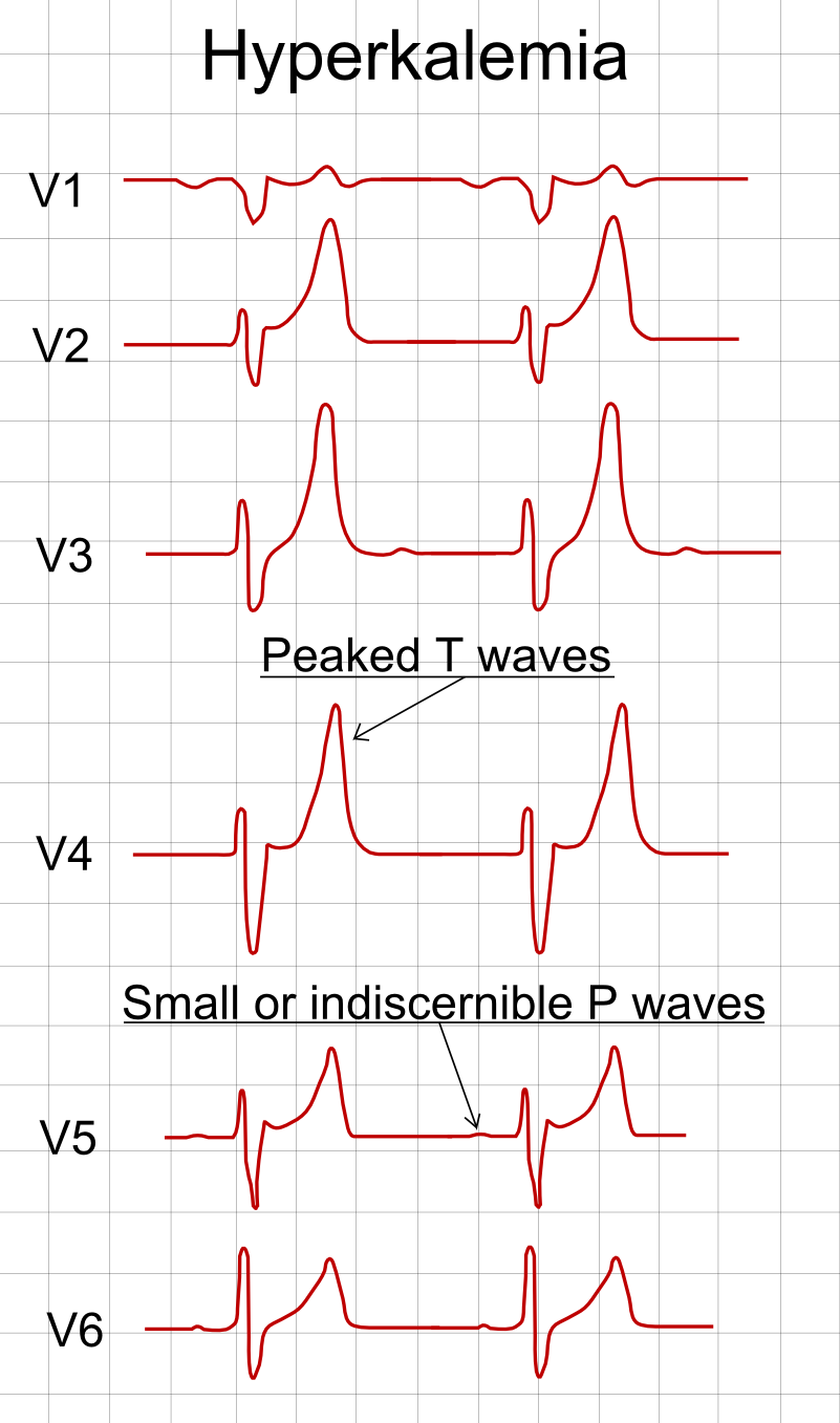
ECG pattern when potassium is high
Peaked T waves show up first. PR prolongation and small P waves follow. QRS widening is a red flag. Bradycardia, a sine-wave pattern or ventricular arrhythmia: those are late signs, and they are life-threatening. The whole pathway is on Hyperkalemia.
Red Flags
- Potassium: K⁺ ≥6.5 mEq/L, or any hyperkalemic ECG change at any K⁺ level
- Acidemia: severe metabolic acidosis, with deep or rapid breathing
- Pulmonary edema: dyspnea, crackles, hypoxemia, or failure to wean oxygen
- Uremic signs: pericardial rub, confusion or encephalopathy, bleeding, or a new neuropathy
- Anuria or oliguria: under 0.5 mL/kg/hr over several hours, or no urine at all
Assessment & Diagnostics
Confirm the AKI. Find the cause. Look for the life-threatening complications first. Do not hold treatment of an ECG change while you wait for a lab result.
| Check | Why | Act If |
|---|---|---|
| Urine output, measured hourly per kg | Staging and response to treatment per KDIGO 2012 | Under 0.5 mL/kg/hr for 6 hr: AKI. Under 0.3 mL/kg/hr for 24 hr, or anuria for 12 hr: stage 3 |
| Creatinine and baseline | Confirms AKI and sets the stage. Use the last known baseline | Rise of 0.3 mg/dL or more in 48 hr, or 1.5 times baseline in 7 days: AKI |
| Potassium and 12-lead ECG | Hyperkalemia is the most immediate lethal risk | K⁺ ≥6.5 mEq/L or any ECG change: treat now (see ALS Management) |
| Bicarbonate and blood gas pH | Acidemia can need dialysis when it is severe or refractory | Severe acidemia, or acidosis with volume overload or uremia: RRT per ED/ICU Management |
| Blood glucose | Must be known before insulin and during insulin therapy | Check before any insulin dose (see Insulin) |
| Volume status and perfusion | Reduced perfusion is the commonest prerenal cause per KDIGO 2012 | Hypovolemia or hypotension: isotonic or balanced crystalloid, then reassess |
| Kidney ultrasound | Excludes urinary tract obstruction per KDIGO 2012 | Obstruction, or no clear cause: urgent imaging and urology per local protocol |
Reach for urinalysis with sediment, serology and hematology when you suspect glomerular, vascular or interstitial disease, per KDIGO 2012.
Differential & Mimics
- Prerenal azotemia: BUN is up, and creatinine goes back to baseline once volume is restored. Fix the cause, then recheck creatinine.
- Acute on chronic kidney disease: Your baseline creatinine sets the stage. Pull an old result before you call the change AKI.
- Pseudohyperkalemia: The number is high because the sample was hemolyzed or delayed. A repeat on a free-flowing sample and a normal ECG separate it from true hyperkalemia (see Hyperkalemia).
- Acute glomerulonephritis or vasculitis: Active sediment, hematuria or rash point to a glomerular cause. Think glomerular, not volume.
- Cardiorenal or hepatorenal AKI: Heart failure or cardiogenic shock can injure the kidney (cardiorenal). Cirrhosis can too, but only after other causes are excluded (hepatorenal). Treat the underlying disease. Look for a nephrotoxin or obstruction first.
- Contrast-associated AKI: Creatinine rises after iodinated contrast. KDIGO 2012 still asks you to look for other causes of AKI (4.1.1, not graded).
BLS Management
- Think of AKI in high-risk patients: dialysis or CKD, a missed dialysis session, low urine output, crush or prolonged entrapment, dehydration, or a patient on an ACE inhibitor, ARB, NSAID or diuretic.
- Take the history: last dialysis session, urine output, medicines (including potassium supplements and salt substitutes), any crush or entrapment time, and recent contrast or infection. For crush, record the time of limb release. Potassium can rise fast after release, so that time matters.
- Monitor: pulse, blood pressure, SpO₂, respiratory effort and mental status. Record a 12-lead ECG if the protocol allows (12-lead ECG acquisition). In a dialysis patient, protect the fistula or graft arm: no blood pressure cuff, IV or blood draw on that arm.
- Support: oxygen for hypoxia and a position that eases breathing. Do not give NSAIDs for pain without medical direction.
- Call ALS early and report: potassium value and time, ECG findings, urine output, medicines, dialysis status and entrapment time. Request priority transport to a facility that can monitor the heart and start dialysis.
ALS Management
Doses vary by protocol; follow local protocols and medical direction. Any hyperkalemic ECG change comes first. Then support the patient while you work the cause. Avoid drugs that worsen the kidney.
- Monitoring and 12-lead (Paramedic/RN/CCT): continuous cardiac monitoring, and a 12-lead ECG when available, for any potassium concern or hypotension
- IV or IO access (Paramedic/RN/CCT): large-bore access, IO if IV fails (IO access)
- Calcium for ECG change (Paramedic/RN/CCT): calcium gluconate 10% 3 g (30 mL) IV over 10 min per the 2026 UKKA guideline (the label rate limit is 200 mg/min) for a hyperkalemic ECG change; repeat if the change persists. Monitor the ECG during each push and infusion. Watch the IV site for extravasation. Give with digoxin only if essential, and slowly with ECG monitoring. Never run it through the same line as bicarbonate or phosphate. For cardiac arrest or a peri-arrest wide QRS, follow the ACLS hyperkalemia pathway and local protocol (see Hyperkalemia). See Calcium Gluconate
- Shift potassium into cells (Paramedic/RN/CCT): regular insulin 10 units IV with dextrose 25 g. Insulin is an ISMP high-alert medication. Check glucose before the dose; if pre-dose glucose is under 126 mg/dL (7.0 mmol/L), add a glucose infusion per the Hyperkalemia page. In kidney failure, hypoglycemia risk peaks because insulin clearance falls. Use the 5-unit dose only where local protocol directs it. UKKA 2026 says more study is needed before 5 units can be recommended. Check glucose on schedule for at least 6 hours. See Insulin and Dextrose
- Albuterol adjunct (Paramedic/RN/CCT): albuterol 10–20 mg nebulized, as an adjunct only. Do not use it alone for severe hyperkalemia. See Albuterol
- Fluids for hypovolemia (Paramedic/RN/CCT): isotonic crystalloid rather than colloid in the absence of hemorrhagic shock (KDIGO 2012, 3.1.1, 2B); bleeding patients need blood products per the hemorrhage protocol. The Surviving Sepsis Campaign 2021 only weakly suggests balanced crystalloid over saline in sepsis (recommendation 33: weak recommendation, low-quality evidence). See Lactated Ringer's or Normal Saline. Reassess after each bolus; do not give fluid into pulmonary edema
- Vasopressor with fluids (Paramedic/RN/CCT): in vasomotor shock with, or at risk for, AKI, use a vasopressor together with fluids (KDIGO 2012, 3.1.2, 1C). See Norepinephrine
- Do not give (all levels): NSAIDs including ketorolac. Ketorolac carries a boxed warning for renal failure. Diuretics do not treat AKI. They are for volume overload only (KDIGO 2012, 3.4.2)
- Sodium bicarbonate (adjunct only): It is not a standalone potassium treatment. It does not replace insulin with dextrose or removal. For severe metabolic acidemia with AKI, it may be used per protocol (see ED & ICU Management for the septic shock criteria). See Sodium Bicarbonate
ED & ICU Management
- Protect the heart: IV calcium for any ECG change, even when dialysis is available. Calcium acts within minutes and lasts 30–60 minutes. It buys time. It does not remove potassium (see Hyperkalemia)
- Shift and remove: insulin with dextrose and glucose checks, then a removal step: dialysis for life-threatening potassium or acidemia
- Find and treat the cause: protocol-based management of hemodynamic and oxygenation parameters (KDIGO 2012, 3.1.3, 2C) in high-risk perioperative patients or septic shock. For sepsis with hypoperfusion, the suggestion is at least 30 mL/kg of crystalloid within the first 3 hr (weak recommendation, low-quality evidence: Surviving Sepsis Campaign 2021, recommendation 5). Individualize the volume. Reassess after each bolus
- Bicarbonate in septic AKI: for septic shock with severe metabolic acidemia (pH 7.2 or lower) and AKI (AKIN score 2 or 3), the Surviving Sepsis Campaign 2021 suggests sodium bicarbonate (recommendation 72: weak recommendation, low-quality evidence). For lactic acidemia from hypoperfusion, it suggests against bicarbonate to improve hemodynamics (recommendation 71)
- Stop nephrotoxins: avoid NSAIDs. Use an aminoglycoside only when no less nephrotoxic alternative exists (KDIGO 2012, 3.8.1). Gentamicin labeling carries a boxed warning for nephrotoxicity. Hold Metformin during AKI
- Iodinated contrast: give IV isotonic saline or bicarbonate before contrast for patients at increased risk (KDIGO 2012, 4.4.1, 1A). The ACR and NKF advise IV saline prophylaxis for AKI, or for eGFR under 30 mL/min/1.73 m² not on maintenance dialysis. At 30–44, the decision is case by case. Do not use prophylactic dialysis for contrast removal (KDIGO 2012, 4.5.1, 2C)
- Diuretic: Furosemide treats volume overload. It does not treat AKI itself (KDIGO 2012, 3.4.2)
- Dialysis indications (AEIOU: acidosis, electrolytes, intoxication, overload, uremia): start renal replacement therapy (RRT) emergently for life-threatening fluid, electrolyte or acid-base change (KDIGO 2012, 5.1.1). The life-threatening ones are hyperkalemia, acidemia, pulmonary edema and uremic complications (pericarditis, bleeding, encephalopathy). A dialyzable poison or drug overdose counts too, such as salicylate, ethylene glycol, methanol or metformin
- When to wait: In ICU AKI with no mandatory indication, a delayed start is as safe as an early one. STARRT-AKI (2020) randomized 3,019 critically ill patients. 90-day mortality was 43.9% with an accelerated start and 43.7% with a standard start. Of the standard arm, 61.8% still needed RRT. AKIKI2 (2021) found that waiting for a mandatory indication, or for a blood urea nitrogen of 140 mg/dL, gave no extra RRT-free days. In a multivariable analysis, the more-delayed arm had higher 60-day death (hazard ratio 1.65)
- Modality: CRRT rather than intermittent RRT for hemodynamic instability (KDIGO 2012, 5.6.2, 2B) or acute brain injury (5.6.3, 2B). Either is acceptable otherwise (Surviving Sepsis Campaign 2021, recommendation 67). See CRRT and Hemodialysis
- Anticoagulation during RRT: use anticoagulation unless bleeding risk is raised or coagulation is impaired (KDIGO 2012, 5.3.1.1, 1B). For CRRT, regional citrate is suggested over heparin, provided there is no contraindication to citrate, such as reduced liver function or lactic acidosis (KDIGO 2012, 5.3.2.2, 2B)
Transport Considerations
- Destination: a monitored ICU or enhanced-care unit that can start RRT without delay. For a dialysis patient with ECG change, choose the facility that can dialyze at once
- Time targets: repeat potassium and an ECG after each treatment. Insulin drops potassium within minutes. Glucose keeps falling for hours. Check glucose on schedule (see Hyperkalemia)
- Continue and monitor: continuous cardiac monitoring and urine output with the collection bag measured hourly. Keep the bag in view. A dialysis patient can turn oliguric in transit
- Handoff essentials: potassium values with times, ECG before and after treatment, calcium and insulin doses with times, glucose series, fluid totals, urine output, last dialysis session, contrast exposure, crush or entrapment time, and all medicines
- Equipment: cardiac monitor with 12-lead capability, calcium gluconate, regular insulin, dextrose, nebulizer for albuterol, and a glucometer
Special Populations
- Pediatrics: KDIGO 2012 applies to children. Small children may not reach a creatinine of 4 mg/dL, and creatinine can lag behind. In patients under 18, KDIGO stage 3 also counts an eGFR below 35 mL/min/1.73 m² (KDIGO 2012, Table 2). Lean on urine output per kg and the trend. Weight-based doses are on the weight-based dosing page. Use pediatric vital signs too. This page does not state a pediatric AKI-specific fluid or dialysis regimen
- Pregnancy: In pregnancy, look for an obstetric cause, including preeclampsia and HELLP syndrome (see Preeclampsia, Eclampsia and HELLP). RRT follows the same indications as in adults. No pregnancy-specific RRT regimen was found in the sources for this page
- Older Adults: Renal reserve is lower, and drugs accumulate more. Review metformin, NSAIDs and antibiotic dosing against current eGFR (see Metformin)
- Renal Impairment: AKI on CKD is the most common high-risk setting. Set the stage with the last known baseline creatinine. A dialysis patient who misses a session is a hyperkalemia risk; see Hyperkalemia
- Patients on ACE Inhibitors, ARBs, NSAIDs or Diuretics: Review these medicines, and hold them per local protocol and medical direction. Potassium-raising drugs add to the risk
Pitfalls & Safety Checks
- Error: waiting on a lab potassium before you treat an ECG change. Give IV calcium first. It acts within minutes
- Error: treating calcium or bicarbonate as the potassium-lowering step. Calcium only protects the heart. You need insulin with dextrose and a removal step
- Error: giving insulin with no glucose check, or with no dextrose. Check glucose before the insulin and on schedule for at least 6 hours
- Error: giving ketorolac or another NSAID to a volume-depleted patient. Volume depletion puts renal failure at risk, and ketorolac is contraindicated then (FDA labeling)
- Error: pouring fluid into a patient with pulmonary edema or oliguric AKI without reassessing. Give fluid in measured boluses. Reassess the lungs and oxygen after each one
- Error: continuing metformin, an aminoglycoside or contrast with no indication. Metformin builds up as kidney function falls (see Metformin)
- Error: using furosemide or mannitol to make urine, then calling the kidney recovered. Diuretics do not treat AKI (KDIGO 2012, 3.4.2)
Clinical Pearls
References
- KDIGO Acute Kidney Injury Work Group. KDIGO Clinical Practice Guideline for Acute Kidney Injury. Kidney International Supplements. 2012;2(1):1–138. https://kdigo.org/wp-content/uploads/2016/10/KDIGO-2012-AKI-Guideline-English.pdf
- Surviving Sepsis Campaign. Surviving Sepsis Campaign: International Guidelines for Management of Sepsis and Septic Shock 2021. Critical Care Medicine. 2021;49(11):e1063–e1143. https://doi.org/10.1097/CCM.0000000000005337
- STARRT-AKI Investigators. Timing of Initiation of Renal-Replacement Therapy in Acute Kidney Injury. New England Journal of Medicine. 2020;383:240–251. https://doi.org/10.1056/NEJMoa2000741
- AKIKI 2 Investigators. Comparison of two delayed strategies for renal replacement therapy initiation for severe acute kidney injury (AKIKI 2): a multicentre, open-label, randomised, controlled trial. Lancet. 2021;397(10281):1293–1300. https://doi.org/10.1016/S0140-6736(21)00350-0
- SMART Investigators. Balanced Crystalloids versus Saline in Critically Ill Adults. New England Journal of Medicine. 2018;378:829–839 (landmark trial). https://doi.org/10.1056/NEJMoa1711584
- American College of Radiology and National Kidney Foundation. Use of Intravenous Iodinated Contrast Media in Patients with Kidney Disease: Consensus Statements from the American College of Radiology and the National Kidney Foundation. Radiology. 2020;294:660–668. https://doi.org/10.1148/radiol.2019192094
- Zarbock A, et al. Preventing acute kidney injury and its longer-term impact in the critically ill. Intensive Care Medicine. 2025;51:1331–1347. https://doi.org/10.1007/s00134-025-08015-8
- Ketorolac Tromethamine injection, US prescribing information (boxed warning: renal risk), DailyMed; label effective 2022. https://dailymed.nlm.nih.gov/dailymed/drugInfo.cfm?setid=004588ba-e109-4a53-9c9d-a42dea9bb848
- Gentamicin Sulfate injection, US prescribing information (boxed warning: nephrotoxicity), DailyMed; label effective 2023. https://dailymed.nlm.nih.gov/dailymed/drugInfo.cfm?setid=09cf88af-59da-f147-e063-6294a90aac99
- Alfonzo A, et al. Clinical practice guidelines: Treatment of acute hyperkalaemia in adults (updated July 2026). UK Kidney Association; 2026. https://www.ukkidney.org/sites/default/files/documents/FINAL%20VERSION%20-%20UKKA%20CLINICAL%20PRACTICE%20GUIDELINE%20-%20MANAGEMENT%20OF%20HYPERKALAEMIA%20IN%20ADULTS%20-%20UPDATED%20JULY%202026_0.pdf
Medical Disclaimer
- For Educational Purposes Only: This content is intended for educational reference and should not be used for clinical decision-making.
- Not a Substitute for Professional Judgment: Always consult your local protocols, institutional guidelines, and supervising physicians.
- Verify Before Acting: Users are responsible for verifying information through authoritative sources before any clinical application.