

Bedside Snapshot
- Recognize
- Facial fracture, penetrating or blunt neck injury, or neck swelling after trauma, with a threat to the airway, bleeding or neurologic function. High-energy mechanism; penetrating neck wounds need a platysma and zone check.
- Red Flags
- Stridor, hoarseness, bubbling from the wound or tracheal deviation
- Expanding neck hematoma or airway obstruction
- Severe active bleeding, shock or absent radial pulse
- Tongue falling back after a bilateral mandible fracture
- First Actions
- Open and hold the airway (suction, jaw thrust, comfort position); high-flow oxygen; direct pressure or hemostatic gauze, impaled objects left in situ; spinal precautions for blunt mechanism with an indication; IV/IO; trauma-center transport
- Key Doses
- Tranexamic acid 1 g IV/IO over 10 min, then 1 g over 8 hr (significant hemorrhage, within 3 hr; per protocol)
- Fentanyl 1 mcg/kg IV/IO (max initial 100 mcg)
- Ketamine 1–2 mg/kg IV/IO for RSI induction; about 1 mg/kg in shock
- Don't Miss
- Delayed airway loss from swelling, hematoma or laryngotracheal injury; a neck wound not yet checked for platysma violation or imaged; a blind nasal tube in a midface or skull base fracture
- Escalate & Transport
- Escalate early for any airway threat, a hard sign or a failed intubation (surgical airway). Transport to a trauma center with ENT, maxillofacial and vascular surgery.
Definition & Pathophysiology
Three injury groups, one main threat: the airway. Facial fractures distort the upper airway and bleed into it. Neck injuries hit the larynx, trachea, pharynx and the great vessels beside them. Every section below gives you the mechanism, because the mechanism tells you what to do at the bedside.
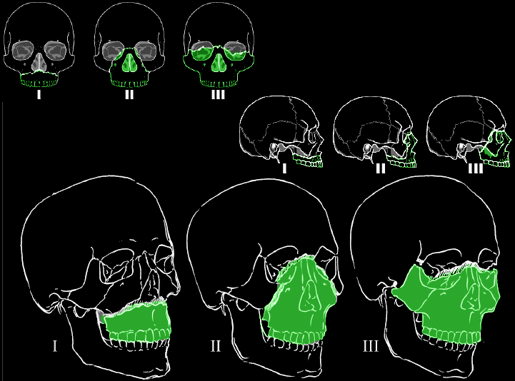
- Midface (Le Fort): Le Fort I runs horizontal through the lower maxilla, across the hard palate and alveolar processes. Le Fort II is pyramidal. It separates the maxilla and nasal bones from the skull base. Le Fort III is craniofacial disjunction: the whole facial skeleton comes away from the cranium. Le Fort III frequently brings cribriform plate damage, skull fracture and dural tear along for the ride.
- Mandible: think of it as a bony ring hinged at the temporomandibular joints. A bilateral or parasymphyseal fracture can let the tongue fall back and block the oropharynx.
- Panfacial: high-energy injury, with two or more regions hit across the frontal bone, midface and mandible. Airway and bleeding come first. The order of surgical repair gets planned later.
- Neck (penetrating or blunt): the internal jugular vein, cervical spine, larynx, external jugular vein, vertebral arteries, pharynx and vagus nerve are the structures hurt most often. In the neck, injury means hemorrhage, a growing hematoma, an aerodigestive leak or airway loss.
- Laryngotracheal injury: direct trauma to the larynx or trachea. Swelling and displacement of the upper airway can cause obstruction, and it gets worse after first contact.
Bleeding and airway obstruction kill early. The airway can look acceptable at first contact. Then edema, hematoma or blood builds up, and it still fails.
Recognition & Red Flags
Facial Fractures
- Swelling, subcutaneous emphysema, a mobile midface or mandible segment, altered sensation, epistaxis and clear fluid from the nose (CSF rhinorrhea): each one points to a fracture. Epistaxis and CSF rhinorrhea show up with Le Fort II and III.
- A bilateral or parasymphyseal mandible fracture can drop the tongue back into the pharynx. Check for tongue obstruction before you go looking for a lung problem.
- Blood from the mouth, nose or pharynx, plus a growing facial hematoma, can block the airway.
Penetrating and Blunt Neck Injury
The classic zones still tell you which structures are at risk. Don't trust them to pin the exact zone of injury, though. External wounds line up poorly with where the damage really is.
- Zone I: clavicles and sternal notch to the cricoid cartilage. Contains the innominate and subclavian vessels, the carotid origin, the vertebral artery, the brachial plexus, the trachea, the esophagus and the lung apex.
- Zone II: cricoid cartilage to the angle of the mandible. Contains the carotid and vertebral arteries, the internal jugular veins, the trachea, the esophagus and cranial nerves X, XI and XII.
- Zone III: angle of the mandible to the skull base. Contains the distal vertebral and carotid arteries, the pharynx and cranial nerves VII, IX, X, XI and XII.
Hard and Soft Signs
Hard signs send the patient to the operating room for exploration, and fast. The 2025 review of penetrating neck trauma says so:
- Expanding hematoma
- Severe active bleeding
- Shock not responding to fluids
- Absent or deficient radial pulse
- Vascular bruit or thrill
- Cerebral ischemia
- Airway obstruction
Soft signs alone don't mandate exploration. They send you to imaging and selective management:
- Hemoptysis, hematemesis or oropharyngeal blood
- Dyspnea or dysphonia
- Dysphagia
- Subcutaneous or mediastinal air, or a chest tube air leak
- Non-expanding hematoma
- Focal neurologic defect
Red Flags
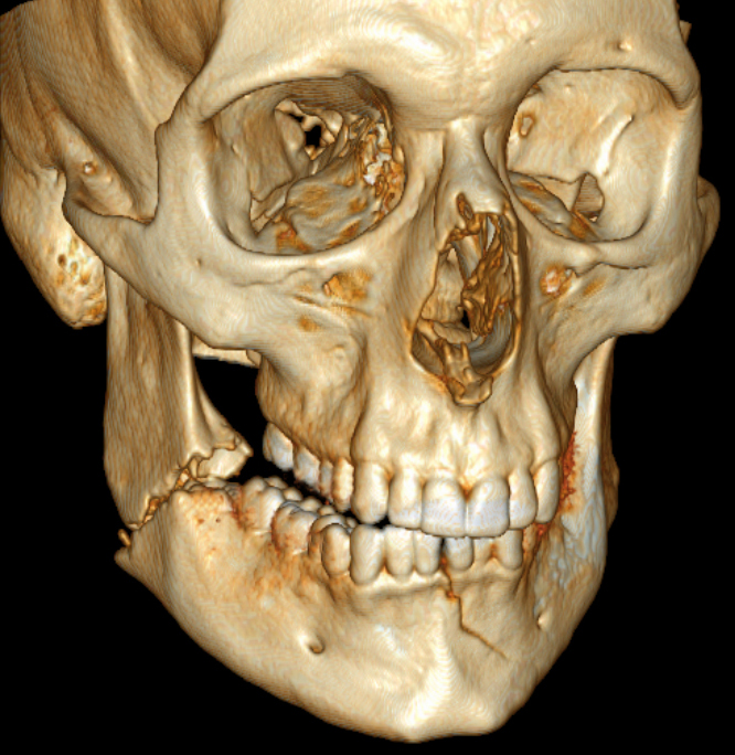
- Stridor, new hoarseness, bubbling from the wound, worsening tracheal deviation or subcutaneous emphysema: the airway is about to collapse. Intubate early.
- Expanding neck hematoma or airway obstruction (hard signs).
- Glasgow Coma Scale (GCS) 8 or less: a usual indication for intubation.
- Shock not responding to fluids, absent radial pulse or focal neurologic deficit.
Assessment & Diagnostics
| Check | Why | Act If |
|---|---|---|
| Airway: voice, stridor, drooling, tongue position, mouth opening | Shows impending collapse and tongue obstruction | Any sign of impending collapse: intubate early and plan the surgical airway before you need it |
| Neck inspection for platysma violation | Penetrating wounds: whether the wound breaks through the platysma | Patient cannot be examined (agitation, habitus, distracting injury): go straight to CT angiography |
| CT angiography (CTA) of the neck, with chest when needed | Vascular, aerodigestive and airway injury | Hard sign: operating room. Stable and positive: vascular and ENT plan. Near-miss result: further evaluation per local practice |
| Hard and soft signs | Sorts exploration from imaging and observation | Any hard sign: operating room. Soft signs only: imaging and selective management |
| CT of the face, mandible and skull base | Le Fort pattern, mandible, skull base and cribriform plate | Skull base, ethmoid or Le Fort II or III fracture: no blind nasal tube (see ALS Management) |
| Cervical spine imaging | Cervical injury occurs with facial and neck trauma | Per protocol; spinal precautions until imaging clears the spine after a blunt mechanism (see BLS Management) |
| Capnography and SpO₂ | Confirms tube position and ventilation (ETCO₂ is end-tidal carbon dioxide) | Falling ETCO₂ or SpO₂ after intubation: check the tube at once, then move to the next airway plan |
BLS Management
- Scene safety and mechanism: note the mechanism (high-energy impact, penetrating object, assault) and the time of injury.
- Airway first: suction blood and secretions. For a bleeding or drooling patient, drain the mouth by lateral positioning if spinal precautions allow; a conscious patient who protects the airway may sit forward instead. For a suspected mandible fracture, use a jaw thrust to lift the tongue and pull the mandible forward. Do not place a nasopharyngeal airway or nasogastric tube when a midface, skull base or cribriform plate fracture is suspected (CSF rhinorrhea, raccoon eyes, Battle sign); use an oropharyngeal airway if tolerated.
- Oxygen: give high-flow oxygen to all trauma patients, then titrate to SpO₂ per the Basic Airway Management protocol.
- Bleeding: apply direct pressure or hemostatic gauze to facial and neck wounds, with care, and avoid compressing both sides of the neck (risk of neurologic injury). Tourniquets cannot be used on the neck. Do not remove an impaled object; leave it in place. See Hemorrhage Control.
- Neck wounds: recheck for an expanding hematoma, bubbling air or subcutaneous emphysema at every contact.
- Collar: a collar is not routine for isolated penetrating neck injury without a neurologic deficit. With a spinal indication, use it judiciously. A collar can easily hide swelling, bleeding, tracheal deviation or loss of pulses. Recheck the neck and pulses after it goes on, and loosen or remove it per protocol if it hides a hematoma or impedes the airway.
- Spinal care: after a blunt, high-energy mechanism, treat the cervical spine as possibly injured until excluded and use spinal motion restriction per protocol; see Spinal Motion Restriction. Isolated penetrating neck injury with no focal neurologic deficit does not get routine spinal motion restriction, and it should not hold up transport.
- Transport priority: any airway sign, hard sign or shock moves the patient to the top of the scene-to-ambulance list.
ALS Management
Doses vary by protocol; follow local protocols and medical direction.
- Airway plan (Paramedic/RN/CCT): the 2025 Difficult Airway Society (DAS) guideline keeps a linear plan: Plan A tracheal intubation, Plan B supraglottic airway ventilation, Plan C facemask ventilation and Plan D emergency front-of-neck airway. Give continuous oxygen, confirm the tube with waveform capnography and move to the next plan on failure. See Supraglottic Airway and Bag-Valve-Mask Ventilation.
- Intubation (Paramedic/RN/CCT): the 2025 DAS guideline makes first-line videolaryngoscopy the default. Use rapid sequence intubation (RSI) with manual in-line stabilization when mouth opening allows. Induction and paralysis per RSI; ketamine 1–2 mg/kg IV/IO is one induction option, about 1 mg/kg in shock (see Ketamine). A bloody airway can obscure the videolaryngoscope camera; clean it before a second attempt. Neuromuscular blockade can turn a partial airway obstruction into a complete one in a distorted or bleeding airway. Before giving a paralytic, set up the front-of-neck kit and mark the neck, and consider keeping spontaneous breathing (awake or ketamine-based technique) when the airway is distorted or laryngotracheal injury is suspected. Patients who protect their own airway should be intubated cautiously. Local protocol governs this choice.
- Cricoid pressure and bag-mask ventilation: the 2025 neck trauma review advises avoiding cricoid pressure and forceful bag-mask ventilation in penetrating neck injury, because they can worsen aerodigestive or great-vessel injury. Check this against the local RSI protocol.
- Nasal tubes: avoid blind nasotracheal intubation in midface or suspected skull base trauma before imaging. A Le Fort II or III fracture, an ethmoid or skull base fracture, or an unknown cribriform plate status can place the tube intracranially. Blind nasal intubation is considered only in a controlled hospital setting, with direct or fiberoptic guidance, under local policy. Avoid nasopharyngeal airways and nasogastric tubes in suspected midface or skull base fracture; after intubation, use an orogastric tube.
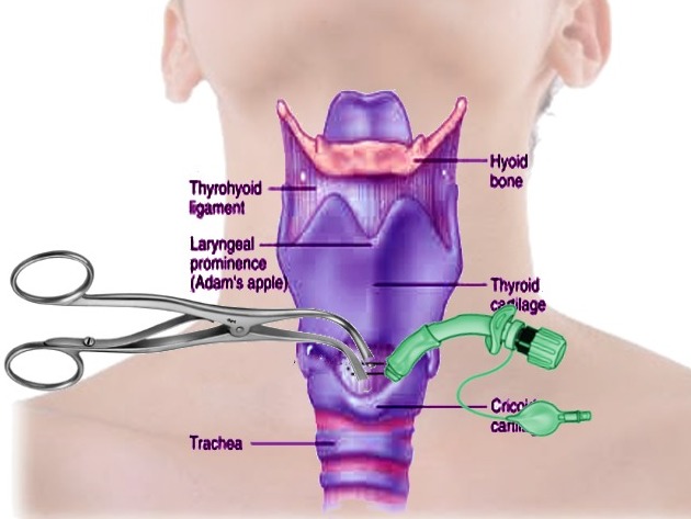
- Front-of-neck airway (Paramedic/RN/CCT, per protocol): if the tube cannot be placed, cricothyrotomy is the preferred invasive airway, and it is faster with lower morbidity than tracheostomy. Use a vertical incision when landmarks cannot be felt, as with a neck hematoma. If the larynx or cricoid is disrupted, cricothyrotomy is relatively contraindicated and a tracheal approach is preferred; emergency tracheostomy is indicated when the cricoid cartilage or superior trachea is extensively damaged. Cricothyrotomy is generally avoided in young children; use the pediatric protocol. See Cricothyrotomy.
- Tube into a laceration: a tube placed into an existing tracheal laceration can be a temporizing airway until definitive repair. Use extreme caution to avoid creating a false passage.
- Hemorrhage (Paramedic/RN/CCT): tranexamic acid (TXA) 1 g IV/IO over 10 min, then 1 g over 8 hr, for significant trauma hemorrhage (clinical signs of hemorrhagic shock) given within 3 hours of injury; there is no evidence of benefit without significant bleeding. Do not give with hypersensitivity to TXA or active intravascular clotting. This is off-label for trauma in the US and per protocol. See Tranexamic Acid.
- Analgesia (Paramedic/RN/CCT): fentanyl 1 mcg/kg IV/IO slowly (max initial 100 mcg), or morphine 0.1 mg/kg IV/IO slowly (max 10 mg initial dose), titrated to effect. See Fentanyl and Morphine. Sub-dissociative ketamine 0.1–0.3 mg/kg IV over 10–15 min (max 0.35 mg/kg) is an alternative. Reassess breathing after each dose.
- Access and monitoring: two large-bore IV or IO lines when shock is present. Continuous SpO₂ and ETCO₂ monitoring. See IV Insertion and IO Access.
ED & ICU Management
- Imaging: CT angiography of the neck, with chest when needed. A 2021 systematic review of nine cohort studies found a "no zone" pathway (CT angiography, then selective exploration) safe for stable patients. It came with fewer negative neck explorations. A near-miss CTA result needs further evaluation per local practice, such as angiography and endoscopy.
- Definitive airway: the team picks oral or nasal intubation, submental intubation or a surgical airway. The fracture pattern and surgical plan decide. Submental intubation is used in panfacial fractures, but cervical hematoma, infection and severe anterior-neck swelling are relative contraindications.
- Tracheostomy: for extensive damage to the cricoid cartilage or superior trachea, emergency tracheostomy is the indicated route. In acute maxillofacial trauma it is slow and difficult.
- Laryngotracheal injury: endoscopic evaluation of the larynx, trachea and esophagus belongs in the workup. Repair needs early ENT input. Never attempt blind intubation in these patients. The surgical airway comes first.
- Vascular injury: most low-velocity penetrating vascular injuries can be repaired primarily. In an unstable patient, one of a pair of arteries may be ligated, provided back-bleeding is adequate. Zone III vertebral injuries are often managed with embolization.
- Expanding hematoma: a hard sign. Transfer to the operating room for exploration.
- Consults: vascular surgery, ENT, maxillofacial surgery, neurosurgery and cardiothoracic surgery, once the injury runs beyond the attending surgeon's comfort zone.
- Facial fractures: open reduction and internal fixation is the usual approach for mandible fractures. The surgical team's plan sets the timing and sequence.
Transport Considerations
- Destination: the nearest trauma center that has ENT, maxillofacial and vascular surgery, plus an operating room. A hard sign or airway threat needs the operating room. Not a delayed workup.
- Continue in transit: continuous capnography and SpO₂. Reassess the airway after every move and at protocol intervals. Recheck swelling, bleeding and tracheal position under the collar.
- Surgical airway kit: build it around a scalpel, a bougie, a 6.0 cuffed tube and suction, with a dedicated suction system.
- Air transport: confirm the airway plan and surgical airway equipment before flight, per protocol. Altitude expands trapped gas. Subcutaneous emphysema, pneumothorax and pneumocephalus from a skull base fracture can all grow in flight. Secure the airway before loading when airway risk is high. A helicopter cabin has little room for a surgical airway or suction once you're airborne.
- Handoff: mechanism and time of injury, zone and platysma status, hard and soft signs present, airway assessment, device and attempts, ETCO₂ trend, TXA dose and time, analgesic doses and times, and cervical precautions.
Special Populations
- Pediatrics: use pediatric airway protocols and length-based equipment sizing (see Pediatric Airway and Broselow). A child's occiput and tongue are proportionally larger, so head position and jaw thrust matter more than in adults. Dose every drug by weight, using Weight-Based Dosing. TXA for pediatric trauma is off-label, and no standardized pediatric dosing is established. See the Tranexamic Acid page. Fentanyl and morphine doses are weight-based. See the drug pages.
- Pregnancy: expect a harder airway and faster desaturation. Pregnancy cuts functional residual capacity and raises oxygen demand. It also adds airway edema and aspiration risk. Preoxygenate fully and have a rescue plan ready. Use left uterine displacement from about 20 weeks onward, and involve obstetrics early, per protocol. After trauma beyond 20 weeks, the Trauma in Pregnancy page covers fetal monitoring.
- Patients on Anticoagulants: bleeding risk runs higher. Reverse per protocol and current product labeling. See Hemorrhagic Shock.
- Renal Impairment: the kidneys clear tranexamic acid. Don't delay or reduce the first trauma bolus for renal function. Renal adjustment applies to repeat and labeled doses. Check creatinine and adjust per protocol and label.
Pitfalls & Safety Checks
- Error: trusting a patient who talks at first contact. Swelling, hematoma and tongue displacement keep progressing, so reassess the airway over time.
- Error: passing a blind nasal tube with a Le Fort II or III, skull base or ethmoid fracture, or unknown cribriform plate status. The same goes for placing a nasopharyngeal airway or nasogastric tube with suspected midface or skull base fracture. Use an oropharyngeal airway. Use an orogastric tube after intubation.
- Error: attempting blind intubation in penetrating neck injury. Blind intubation is never the answer here. Go to the surgical airway early.
- Error: treating the collar as protection. Meanwhile it can hide a growing hematoma, subcutaneous emphysema or tracheal deviation.
- Error: missing a tongue obstruction after a bilateral mandible fracture. Use a jaw thrust. Lateral positioning only if spinal precautions allow.
- Error: clearing a neck wound with no platysma check and no CT angiography, especially when the patient cannot be examined.
- Error: paralyzing a patient before the rescue plan is ready. In a distorted or bleeding airway, set up the front-of-neck kit first. Then consider keeping spontaneous breathing.
- Error: giving TXA late. The window closes 3 hours after injury.
Clinical Pearls
References
- Ahmad I, El-Boghdadly K, Iliff H, Dua G, Higgs A, Huntington M, et al. (2026; online 2025-11-07). Difficult Airway Society 2025 guidelines for management of unanticipated difficult tracheal intubation in adults. Br J Anaesth, 136(1), 283–307. https://doi.org/10.1016/j.bja.2025.10.006
- Loss L, Henry R, White A, Matsushima K, Barrett C, Lammers D, Schreiber M, Inaba K. (2025). Penetrating neck trauma: a comprehensive review. Trauma Surg Acute Care Open, e001619. https://doi.org/10.1136/tsaco-2024-001619
- Chandrananth ML, Zhang A, Voutier CR, Skandarajah A, Thomson BNJ, Shakerian R, Read DJ. (2021). 'No zone' approach to the management of stable penetrating neck injuries: a systematic review. ANZ J Surg, 1083–1090. https://doi.org/10.1111/ans.16600
- Marí-Roig A, McLeod NMH, De Lange J, Dubois L, García Reija MF, Van Minnen B, Essig H. (2024). Controversies in the management of the airway in panfacial fractures: a literature review and algorithm proposal. J Clin Med. https://doi.org/10.3390/jcm13237294
- Saini S, Singhal S, Prakash S. (2021). Airway management in maxillofacial trauma. J Anaesthesiol Clin Pharmacol, 319–327. https://doi.org/10.4103/joacp.joacp_315_19
- Velopulos CG, Shihab HM, Lottenberg L, Feinman M, Raja A, Salomone J, Haut ER. (2018). Prehospital spine immobilization/spinal motion restriction in penetrating trauma: a practice management guideline from the Eastern Association for the Surgery of Trauma (EAST). J Trauma Acute Care Surg, 84(5), 736–744. https://doi.org/10.1097/TA.0000000000001764
- Fischer PE, Perina DG, Delbridge TR, Fallat ME, Salomone JP, Dodd J, Bulger EM, Gestring ML. (2018). Spinal Motion Restriction in the Trauma Patient – A Joint Position Statement. Prehosp Emerg Care, 22(6), 659–661. https://doi.org/10.1080/10903127.2018.1481476
- All India Difficult Airway Association. (2025). All India Difficult Airway Association 2025 guidelines for the management of unanticipated difficult airway in obstetrics under general anaesthesia. Indian J Anaesth, 69, 1142–1166. https://doi.org/10.4103/ija.ija_1082_25
- Talmy T, Almog O, Nadler R, Bendor CD, Akler D, Radomislensky I, Beer Z, Yosef Y, Haddad N, Kiso S, Epstein D, Brzezinski Sinai Y. (2026). Prehospital management of impending airway obstruction in combat trauma: a registry study. BMJ Mil Health. https://doi.org/10.1136/military-2025-003231
- CRASH-2 collaborators, Roberts I, Shakur H, Afolabi A, Brohi K, Coats T, et al. (2011). The importance of early treatment with tranexamic acid in bleeding trauma patients: an exploratory analysis of the CRASH-2 randomised controlled trial (landmark trial). The Lancet, 377(9771), 1096–1101. https://doi.org/10.1016/S0140-6736(11)60278-X
Medical Disclaimer
- For Educational Purposes Only: This content is intended for educational reference and should not be used for clinical decision-making.
- Not a Substitute for Professional Judgment: Always consult your local protocols, institutional guidelines, and supervising physicians.
- Verify Before Acting: Users are responsible for verifying information through authoritative sources before any clinical application.