

At a Glance
- Pattern
- A waveform that is not cardiac activity (motion, muscle, electrodes or interference) or a wrong electrode position, recorded as if it came from the heart.
- Normal (Adult)
- A stable baseline, labelled electrodes and a consistent QRS and P-wave polarity across the limb leads.
- Confirm It
- The pulse, the trace with the patient still, lead I and the P-wave polarity in II and III, V1 and V2 placement, and a repeat 12-lead.
- Abnormal → Think
- Chaotic trace in a well patient with a pulse, an inverted lead I, II or III, or an infarct pattern that changes with lead position: check artifact or lead reversal first. Unstable regular wide-complex tachycardia is VT.
- Red Flags
- Shocking, cardioverting or giving antiarrhythmics for artifact in a patient with a pulse who looks well; treating unstable wide-complex tachycardia as artifact; treating an inverted lead as ischemia without a repeat 12-lead.
- Bedside Action
- Check the patient first. Re-record with the patient still, check electrode labels and V1 and V2 placement, and get a repeat 12-lead.
Physiology & What It Represents
The ECG never sees the heart directly. It measures voltage between pairs of electrodes on the skin, and each lead looks at the heart from one fixed direction. The limb leads (I, II, III, aVR, aVL and aVF) look at the frontal plane. V1 to V6 look at the horizontal plane. Depolarization is the signal. Anything else that moves the voltage between two electrodes is artifact.
Artifact comes from three places. The patient's muscles are one: shivering, tremor and movement. Electrode-to-skin contact is another: poor adhesion, sweat and a moving cable. The third is external interference, such as 60-cycle mains noise, which an implanted device can read as a fast rhythm. Lead reversal is a different problem. The heart and the rhythm haven't changed. Swap two limb electrodes, though, and every lead records from a new direction. QRS and T waves can look inverted, shifted or like an infarct.
Normal Values & How to Measure
A usable recording needs four things. Clean skin contact. Electrodes on the labelled limbs and chest positions. A stable baseline. QRS and P waves with consistent polarity across the limb leads.
| Check | Correct Finding | Pediatric Note | Where / How to Check |
|---|---|---|---|
| Limb electrodes | One on each arm and each leg (RA, LA, LL, RL) | See Pediatric & Special Populations | Match each label at the electrode and at the cable |
| V1 and V2 | 4th intercostal space at the sternal edges | Same landmarks; the 4th space is harder to find in infants, so confirm on the trace | Find the 2nd rib and count down to the 4th space |
| Baseline | Stable and flat between beats | Repeat when the child is still | Look at the trace before reading any wave |
| Lead I and aVR | Lead I upright; aVR mostly negative | See Pediatric & Special Populations | Compare the two leads on the same beat |
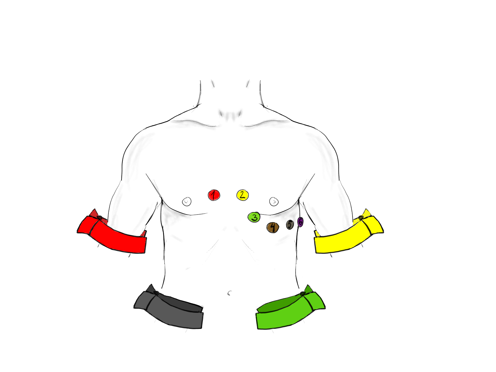
- Confirm contact: clean, dry skin and firm electrodes before you read the trace.
- Read the labels: match each limb electrode to its label at the electrode and at the cable.
- Check lead I: lead I inverted, but R waves build normally from V1 to V6? Think LA/RA reversal. Poor progression points to dextrocardia instead.
- Check II and III: lead II inverted with the other leads normal points to RA/LL reversal. Lead III inverted points to LA/LL reversal.
- Check V1 and V2: an odd P wave in V1 and V2 is your clue that the electrodes sit too high on the chest.
- Repeat: record again with the patient still and compare with a second lead before you act.
Artifact That Mimics VF or VT
Non-cardiac waveforms can pass for VF, VT or asystole on the monitor. The dangerous ones show up during resuscitation, or in a patient who still has a pulse.
ECG Criteria
- Chaotic baseline without an organized QRS-T: looks like VF or fine VF.
- Regular, high-amplitude deflections during compressions: stop when the compressions stop.
- Irregular, random high-frequency activity: tremor or shivering.
- Regular 50 or 60 Hz mains interference: a uniform, fine, rapid oscillation of the baseline, from a nearby power source or device.
- Flat line: a disconnected or loose lead, or low gain, can look like asystole. Check the pulse and a second lead before you call asystole.
Causes
| Category | Examples |
|---|---|
| CPR | Chest compressions and ventilation; oscillations of great amplitude |
| Movement | Patient movement, limb motion and baseline irregularity |
| Tremor | Parkinson disease, cerebellar or intention tremor, anxiety, hyperthyroidism, amphetamines, xanthines (including caffeine), beta-agonists, lithium, and benzodiazepine or alcohol withdrawal |
| Shivering | Hypothermia, fever and rigors |
| Electrodes and cables | Poor skin contact, a loose electrode or a moving cable |
| Lead off or low gain | A disconnected lead or a gain set too low gives a flat line that can look like asystole |
| External interference | 50 or 60 Hz mains signal and equipment such as a hot-tub engine |
| Implanted device | A deep brain stimulator has produced a VF-like artifact in a prehospital case report |
Clinical Significance
- A false VF or VT call can mean unnecessary shocks, antiarrhythmic drugs and interrupted CPR.
- One prehospital series of software STEMI calls found ECG artifact was the top cause of false-positive reads: 20% of 711.
- One reported case: an implanted cardioverter-defibrillator read 60-cycle interference as fast VF and fired an inappropriate shock.
Look-Alikes
- True VF, compression artifact and tremor are compared in the table under Differential Diagnosis & Mimics.
Limb Lead Reversal
Limb lead reversal means two limb electrodes sit on the wrong limbs. It's common enough that any unexpected axis, inverted lead or new infarct pattern should make you check. One tertiary-care audit of adult ECGs (patients under 18 and dextrocardia were excluded) found lead misplacement on 15 of 1,000 ECGs (1.5%). Those were 11 limb lead reversals (8 LA/LL and 3 RA/LA) and 4 chest lead reversals.
ECG Criteria
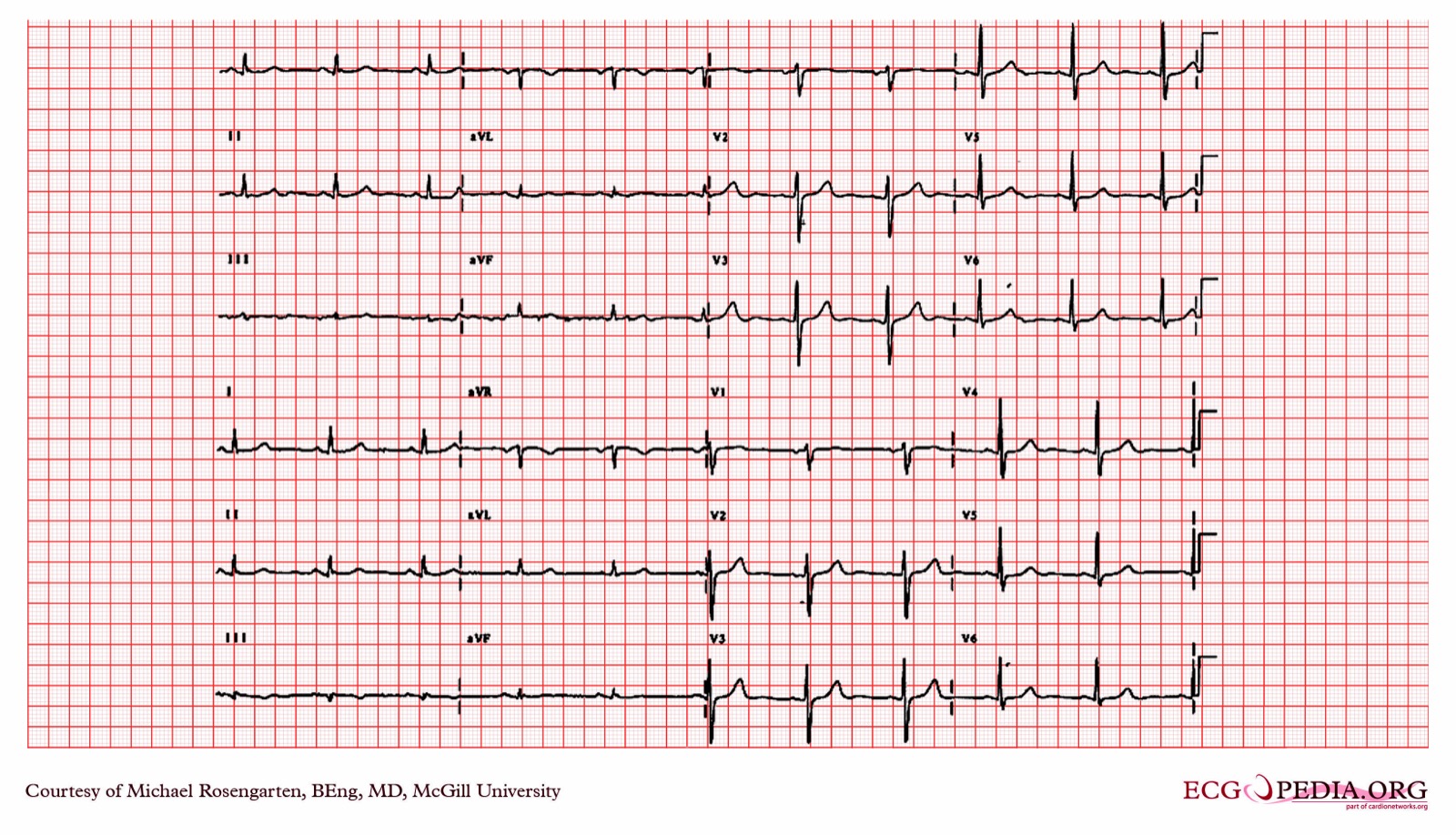
- LA/RA reversal: lead I inverted; aVR and aVL swapped; II and III swapped; aVF and V1 to V6 unchanged.
- RA/LL reversal: lead II inverted; lead I shows the inverted III and lead III shows the inverted I; aVR and aVF swapped; aVL unchanged.
- LA/LL reversal: lead III inverted; I and II swapped; aVL and aVF swapped; aVR and V1 to V6 unchanged.


Causes
- An electrode placed on the wrong limb.
- A cable connected to the wrong electrode.
Clinical Significance
- Reversed limb leads can mimic myocardial infarction, ischemia and axis deviation. They're hard to spot without a systematic check.
- A reversed 12-lead can send a patient down the STEMI pathway or into a false axis diagnosis. Unless ongoing symptoms say otherwise, repeat the tracing with correct placement before you act on it.
Look-Alikes
- Dextrocardia, true lateral or inferior infarction and axis deviation: see Differential Diagnosis & Mimics and the Q Wave page.
Precordial Lead Misplacement
Put chest electrodes too high and R-wave progression shifts, along with the ST and T waves in the anterior leads.
ECG Criteria
- High V1 and V2: an incomplete right bundle branch block pattern, anterior T-wave inversion, septal Q waves or ST elevation.
- Clue: an unusual P-wave morphology in V1 and V2.
Causes
- Electrodes placed too high on the chest, usually V1 and V2.
Clinical Significance
- High V1 and V2 can pass for ischemia, pulmonary embolism or a Brugada pattern.
- Rarely, high placement of V1 and V2 uncovers a true type 1 Brugada pattern. Don't assume every abnormal V1 is artifact.
- Compare with a prior tracing and re-record with correct landmarks before you activate a pathway. Unless the patient is unstable, that's the order.
Differential Diagnosis & Mimics
Look at the patient before you believe the monitor. Use the table to tell a true arrhythmia from the mimics you will meet most often.
| Feature | True VF | Compression artifact | Tremor or shivering |
|---|---|---|---|
| Waveform | Chaotic, no organized QRS-T | Regular, high-frequency, high-amplitude deflections | Irregular, random high-frequency activity |
| With movement stopped | Unchanged | Stops when compressions stop | Improves when the patient is still |
| Pulse | Absent | Absent during compressions; check in pauses | Usually present |
Artifact & Lead Misplacement
- Limb lead reversal: think of it when lead I, II or III flips on a tracing that otherwise looks normal (see Limb Lead Reversal).
- Precordial misplacement: chest leads placed too high can fake ST elevation or T-wave inversion (see Precordial Lead Misplacement).
- Tremor and movement: Parkinson disease, shivering and drugs such as amphetamines, caffeine, beta-agonists or benzodiazepine withdrawal can all fake a rhythm.
- Interference: one reported hot-tub engine set off an inappropriate ICD shock.
Bedside Management & Escalation
Treat the patient, not the waveform. Pulse, breathing and responsiveness come first, then the monitor.
- Pulseless with VF or pulseless VT on a rhythm check during a pause: CPR, defibrillation and drugs per the AHA 2025 ALS algorithm (Paramedic, RN, CCT). Pediatric: follow PALS.
- Pulse check: no more than 10 seconds, and only during a pause. A VF-like trace on the monitor needs a second source where you have one: end-tidal CO₂, an arterial-line waveform, a pulse oximetry waveform or point-of-care ultrasound.
- Palpable pulse with a chaotic or VF-like trace in a patient who looks well: treat it as artifact until shown otherwise. Re-record with the patient still, check a second lead and get a 12-lead ECG.
- Palpable pulse with a regular wide-complex tachycardia and any instability (hypotension, altered mental status, ischemic chest pain or acute heart failure): treat it as ventricular tachycardia. Use synchronized cardioversion per the AHA 2025 adult tachycardia algorithm. Don't delay it to hunt for artifact.
- Do not defibrillate, cardiovert or give antiarrhythmic drugs for artifact in a patient with a pulse who looks well. Unstable wide-complex tachycardia is the exception.
- Flat line: before you call asystole, check the pulse, the leads, the cable and the gain, and view a second lead.
- AED or monitor-defibrillator analysis: stop the vehicle if it is safe to do so, keep hands off the patient during analysis, and re-analyze if motion artifact is suspected.
- Suspected lead reversal: correct the electrodes and repeat the 12-lead. Ongoing ischemic symptoms don't wait for the repeat. Follow the local STEMI pathway while the tracing is repeated.
Follow local protocols and medical direction.
Pediatric & Special Populations
Pediatrics
- Limb electrode checks apply to children too. Chest positions use the same landmarks, but the 4th interspace is harder to find in infants and young children. Confirm placement on the trace. Pediatric ECGs may add right-sided leads (V3R, V4R) when right-heart disease is suspected.
- Movement and shivering produce artifact at any age. Re-record once the child is still. Don't treat the waveform first.
- A chaotic trace in a well-looking child with a pulse is artifact until checked. A wide-complex tachycardia with instability is treated as a real arrhythmia. Follow PALS.
Other Populations
- Implanted ICD: in one reported case, 60-cycle interference was read as fast VF and delivered an inappropriate shock. Ask about implanted devices. Where possible, move the patient away from the interference.
- Deep brain stimulator: a prehospital case report describes one that produced VF-simulated artifact.
- Parkinson disease and tremor: tremor is a recognized source of motion artifact. Repeat the tracing when the patient is still.
- Female patients, obesity and pregnancy: place chest electrodes beneath the breast tissue, not on it. Obesity and pregnancy shift the landmarks, which makes landmark errors more likely.
- Hypothermia and rigors: shivering produces artifact. Check the trace once it stops, if that can be done safely.
Monitoring & Transport Considerations
| Issue | What to Do |
|---|---|
| Patient moving, shivering or tremor | Re-record when still; compare a second lead before acting. |
| Chest compressions | Read the rhythm during a pause, not during compressions, per the AHA 2025 ALS algorithm. |
| Patient moved or cable changed | Check lead I and the P waves in I and II again. An inverted lead I with normal R-wave progression suggests LA/RA reversal; poor progression suggests dextrocardia. |
| Implanted device or nearby equipment | Ask about implanted devices and move the patient away from the interference source where possible. |
| Flat line | Check the pulse, leads, cable and gain, and view a second lead before calling asystole. |
| Automated rhythm or STEMI call | Confirm on the raw trace before you act on the call. |
| Vehicle in motion | If the rhythm is unclear and it is safe, re-record with the vehicle stopped. Stop before AED or monitor-defibrillator analysis, keep hands off, and re-analyze if motion artifact is suspected. |
Step-by-Step Approach & Pitfalls
- Check the patient: pulse (10 seconds or less), breathing and responsiveness come before the monitor.
- Look at the trace: is the baseline organized, does the pattern change when movement or compressions stop, and is a flat line real asystole or a disconnected lead?
- Check the limb electrodes: match each label to its limb and look for an inverted lead I, II or III.
- Check the chest electrodes: find the 4th interspace beside the sternum for V1 and V2, and look for an unusual P wave in V1 and V2.
- Re-record: with the patient still, compare a second lead with the first.
- Get a 12-lead ECG: with correct placement, and compare with a prior tracing if one exists.
- Act on the corrected trace: treat a confirmed rhythm per protocol and do not treat artifact.
Common Pitfalls
- Calling a chaotic, VF-like trace VF in a patient with a pulse who looks well. The reverse: calling an unstable regular wide-complex tachycardia artifact.
- Missing a limb reversal because the axis or infarct pattern looks real. That is the trap.
- Reading the rhythm during compressions or while the patient moves.
- Trusting an automated STEMI or rhythm call without a look at the raw trace.
- Calling asystole on a flat line that is really a disconnected lead or low gain.
- Assuming someone rechecked the electrodes after the patient was moved.
ECG Examples
Clinical Pearls
References
- Wigginton JG, et al. Part 9: Adult Advanced Life Support: 2025 American Heart Association Guidelines for Cardiopulmonary Resuscitation and Emergency Cardiovascular Care. Circulation. 2025;152(16 suppl 2):S538–S577. https://doi.org/10.1161/CIR.0000000000001376
- Kligfield P, et al. Recommendations for the standardization and interpretation of the electrocardiogram: part I: the electrocardiogram and its technology (landmark standards statement). Circulation. 2007;115(10):1306–1324. https://doi.org/10.1161/CIRCULATIONAHA.106.180200
- Bravo F, Moukabary T. Blips and flips for quick identification of limb-lead reversal. Am J Med. 2026 Aug 26; online ahead of print. https://doi.org/10.1016/j.amjmed.2026.08.007
- Pérez-Riera AR, Barbosa-Barros R, Daminello-Raimundo R, de Abreu LC. Main artifacts in electrocardiography. Ann Noninvasive Electrocardiol. 2018;23(2):e12494. https://doi.org/10.1111/anec.12494
- Bosson N, Sanko S, Stickney RE, et al. Causes of prehospital misinterpretations of ST elevation myocardial infarction. Prehosp Emerg Care. 2017;21(3):283–290. https://doi.org/10.1080/10903127.2016.1247200
- Strum RP, Drennan IR, Hillier M, Cheskes S. Ventricular fibrillation simulated electrocardiogram artifact by a deep brain stimulator. Prehosp Emerg Care. 2023;27(8):1115–1117. https://doi.org/10.1080/10903127.2023.2194407
- Walsh B. Misplacing V1 and V2 can have clinical consequences. Am J Emerg Med. 2018;36(5):865–870. https://doi.org/10.1016/j.ajem.2018.02.006
- Shenasa M, Heidary S, Shenasa H. Inappropriate ICD shock due to hot tub-induced external electrical interference. J Electrocardiol. 2018;51(5):852–855. https://doi.org/10.1016/j.jelectrocard.2018.06.016
- Ramadurai S, Varadarajan V, Lasrado AA, et al. A study of the frequency of lead reversal at a tertiary care institution. Cureus. 2025;17(9):e92345. https://doi.org/10.7759/cureus.92345
Medical Disclaimer
- For Educational Purposes Only: This content is intended for educational reference and should not be used for clinical decision-making.
- Not a Substitute for Professional Judgment: Always consult your local protocols, institutional guidelines, and supervising physicians.
- Verify Before Acting: Users are responsible for verifying information through authoritative sources before any clinical application.