

At a Glance
- Pattern
- Acute coronary occlusion hidden by a wide QRS from left bundle branch block (LBBB) or ventricular pacing, where the usual STEMI thresholds do not apply.
- Normal (Adult)
- Discordant ST-T is expected: ST and T run opposite the QRS. No Sgarbossa criterion met.
- Confirm It
- Measure ST at the J point in every lead against one baseline on every tracing (PR, as in the Smith 2012 derivation, or TP). Compare with a prior ECG; check V1–V3 and upright-QRS leads.
- Abnormal → Think
- Concordant STE ≥1 mm: possible occlusion
- Concordant STD ≥1 mm in V1–V3: possible occlusion
- Excessive discordant STE: ≥5 mm, or ST/S ratio −0.25 or less
- Red Flags
- Any one criterion with symptoms: possible occlusion, escalate per protocol. A negative result does not rule it out.
- Bedside Action
- Activate the STEMI pathway per local protocol when suspicion is high; send the prior ECG and repeat the 12-lead.
Physiology & What It Represents
The impulse normally runs from the SA node through the atria and AV node, then down the His–Purkinje system. That system splits into right and left bundles. In left bundle branch block (LBBB), the left bundle is blocked. The septum and right ventricle activate first. The left ventricle is then activated cell to cell. Ventricular pacing starts activation from one site, and the QRS comes out wide the same way. Repolarization follows that abnormal sequence, so the ST segment and T wave normally point opposite the QRS. That discordance is expected. It is not ischemia.
Acute coronary occlusion adds a separate injury current. Look for two patterns. The ST segment moves in the same direction as the QRS (concordant). Or it rises far more than the depth of the S wave would predict. In either case, the conduction defect alone does not explain the change. The Sgarbossa criteria are built to pick out those two patterns.
Normal Values & How to Measure
Measure every threshold at the J point. That is where the QRS ends and the ST segment begins. Use a flat baseline. Smith 2012 measured ST against the PR segment, the flat stretch between the end of the P wave and the QRS. The Smith thresholds were derived on that baseline. The TP segment is also used locally, as the flat baseline between a T wave and the next P wave. Either is acceptable, as long as the same baseline is used on every tracing. Amplitudes are in mm (1 mm = 0.1 mV).
| Parameter | Adult Threshold | Pediatric Note | Where / How to Measure |
|---|---|---|---|
| Baseline | PR segment (the Smith 2012 derivation baseline) or TP segment; the same on every tracing | Not established in children | Every lead, before the QRS |
| Concordant ST change | ST moves in the same direction as the main QRS deflection | Not established in children | Positive QRS (upright) or negative QRS (deep S) |
| Concordant STE | ≥1 mm in a lead with a positive QRS | Not established in children | J point, every lead with an upright QRS |
| Concordant STD | ≥1 mm in V1, V2 or V3 | Not established in children | J point in V1–V3 |
| Discordant STE | ≥5 mm (original rule), or ST/S ratio of −0.25 or less (Smith-modified rule) | Not established in children | Leads with an S wave (or R wave) of opposite polarity; measure the S-wave depth in the same lead |
- Confirm the QRS: LBBB or ventricular pacing, not just any wide complex. Check that every pacing spike is followed by a paced QRS.
- Set the baseline (PR or TP, the same on every tracing) and measure the J point in each lead, in mm.
- Mark each lead as concordant or discordant with the main QRS deflection.
- Apply both rule sets: the original score (concordant STE, concordant STD in V1–V3, excessive discordant STE) and the Smith-modified rule (any one criterion). For discordant STE, measure the S-wave depth in the same lead.
- Compare with a prior ECG whenever one exists. Chronic LBBB shows the same ST-T pattern on every tracing. New concordant change does not.
Original Sgarbossa Criteria
Sgarbossa's original criteria came from patients with left bundle branch block (LBBB) and acute myocardial infarction (MI) in the GUSTO-1 trial. That trial enrolled 26,003 North American patients. Three criteria carried independent value. Each one scores points in a weighted total.
ECG Criteria
| Criterion | Threshold | Points |
|---|---|---|
| Concordant STE | ≥1 mm in a lead with a positive QRS | 5 |
| Concordant STD | ≥1 mm in V1, V2 or V3 | 3 |
| Excessive discordant STE | ≥5 mm in a lead with a negative QRS | 2 |
| Positive score | Total of 3 or more points | — |
Causes
- Acute coronary occlusion: this is the finding the criteria are built to detect.
- Expected LBBB repolarization: discordant ST-T that earns no points. It is the baseline the criteria are measured against.
Clinical Significance
- Derivation: 131 of 26,003 GUSTO-1 patients had acute MI with LBBB. The derivation group was small.
- Specificity over sensitivity: the rule is specific. A negative score still does not exclude occlusion. A 2026 scoping review (reference 5) cites about 40% sensitivity and 97% specificity for the original rule in LBBB. Treat those figures as secondary. They are not in the abstract of the 2025 meta-analysis (reference 6).
- Practical use: a positive score is a reason to escalate per local protocol. It is not a diagnosis by itself.
Smith-Modified Sgarbossa Criteria
Smith and colleagues dropped the fixed 5 mm cutoff. Instead, they measured the discordant ST elevation against the depth of the S wave in the same lead. The modified rule is unweighted. Any one criterion is positive.
ECG Criteria
- Concordant STE: ≥1 mm in a lead with a positive QRS.
- Concordant STD: ≥1 mm in V1, V2 or V3.
- Discordant STE: ST/S ratio of −0.25 or less, meaning the ST elevation is at least 25% of the S-wave depth in that lead. Use only leads with discordant ST elevation of at least 1 mm and an opposite-polarity QRS.
Causes
- Acute coronary occlusion: the same target finding as above.
- Deep S waves with modest discordant elevation: the proportional rule can flag ST elevation that is too small for the original 5 mm threshold. Against a deep S wave, though, that elevation is large.
Clinical Significance
- Derivation: 33 ECGs with angiographic coronary occlusion were compared with 129 control ECGs. The controls had LBBB and came from emergency department patients without occlusion. Sensitivity was 91% (95% CI 76–98%). The weighted original rule reached 52%, and the unweighted original reached 67%.
- Trade-off: specificity was 90% (95% CI 83–95%). That is lower than the 98% (95% CI 93–100%) of the weighted original rule, and equal to the 90% of the unweighted original. The derivation set was small, with 33 occlusion ECGs. Don't read the figures as a precise estimate.
- Pooled data: the 2025 meta-analysis of LBBB studies (reference 6) reported 83.6% sensitivity (95% CI 55.4–95.5%) and 92.6% specificity (95% CI 78.9–97.7%) for the modified rule. The reference standard was angiographically confirmed occlusion. The confidence intervals are wide.
Ventricular Paced Rhythm
A paced QRS looks like LBBB, so the same concordant-change logic applies. Expect the ST-T to run opposite the paced QRS. In a 2021 multicenter case-control study of symptomatic patients with ventricular paced rhythm, 59 of them had occlusion MI.
ECG Criteria
- Sensitivity: the modified criteria found 81% of occlusion MI (95% CI 69–90%). The original criteria found 56% (95% CI 42–69%).
- Lead range: Dodd 2021 used concordant STD in V1–V3 with the modified criteria. Adding concordant STD in V4–V6 raised sensitivity to 86% (95% CI 75–94%) in that study. The abstract does not report specificity. Follow local protocol for the lead range.
- Pacing spike: confirm each spike is followed by a paced QRS before you read the ST segment.
Causes
- Acute coronary occlusion: the finding the criteria are looking for.
- Paced repolarization: expected discordant ST-T. It carries no points.
Clinical Significance
- Missed occlusion: the original rule missed about 44% of paced occlusion MI in that cohort. A negative result does not clear the patient.
- Conduction system pacing: the criteria were developed for LBBB and ventricular pacing. They were not developed for His bundle or left bundle branch area pacing. When the pacing type is unknown, ask cardiology for a read.
- Prior ECG: comparing with the pre-pacing or earlier paced tracing is the most useful step.
Differential Diagnosis & Mimics
The main mimic is the expected LBBB or paced pattern. New LBBB on its own is not diagnostic. The Fifth Universal Definition of MI counts new or presumed-new bundle branch block as an equivalent. It does so only with other signs of myocardial ischemia.
| Feature | Expected LBBB or Paced ST-T | Sgarbossa-Positive Pattern |
|---|---|---|
| ST-T direction | Opposite to the main QRS | Same direction as the QRS (concordant) |
| Discordant STE size | Proportional to the S wave; ratio above −0.25 | 5 mm or more, or ratio −0.25 or less |
| Compared with prior ECG | Unchanged | New or changed concordant ST change |
| New LBBB alone | Not diagnostic without ischemic ST-T change | New LBBB with a Sgarbossa-positive ST-T pattern |
Artifact & Lead Misplacement
- Limb lead reversal: it flips QRS and ST-T in the limb leads, and it can make an LBBB pattern look like an infarct. See the Cardiac Axis page.
- Precordial electrodes placed high: they change the V1–V3 morphology that the concordant STD criterion depends on.
- Tremor, motion and transport vibration: these can mimic ST change. Re-acquire the tracing in a still moment.
- Pacing spike misread: a spike on the ST segment can pass for a QRS or an ST change.
Bedside Management & Escalation
A positive Sgarbossa result changes the call. It points to acute coronary occlusion. Per the Fifth Universal Definition of MI, other ECG patterns may count as STEMI equivalents in the right clinical setting. Sgarbossa's criteria appear on that list of patterns suggestive of acute coronary occlusion. Doses are not on this page. Follow local protocols and medical direction.
- Positive criteria with symptoms: activate the STEMI pathway per local protocol and send the 12-lead with the notification.
- Negative criteria with high suspicion: a negative result does not rule occlusion out. Repeat the 12-lead, add serial tracings and escalate per protocol.
- Posterior and right-sided leads: add V7–V9 when V1–V3 show depression, and V4R when right ventricular infarction is suspected.
- Prior ECG: bring it or transmit it. The comparison often decides the read.
Pediatric & Special Populations
Pediatrics
- Derivation population: the criteria were derived in adults. They are not established in children. Use pediatric cardiology input and the pediatric ECG reference used locally.
- Congenital heart disease: LBBB or a paced QRS in a child with a surgical repair starts from a different baseline. Compare with the child's prior ECG and with pediatric vital signs for context.
Other Populations
- Older adults: chronic LBBB and prior infarction can show up on earlier tracings. Compare before you call a change new.
- Pacemaker or ICD patients: a paced QRS is the usual setting for these criteria. Check the pacing mode and capture.
- Conduction system pacing: the criteria were not developed for this setting. Ask cardiology for an ECG read.
Monitoring & Transport Considerations
| Issue | What to Do |
|---|---|
| Prior ECG | Send or transmit it with the first 12-lead; the comparison decides many reads |
| Serial 12-leads | Repeat while symptoms continue, at the interval set by local protocol |
| Lead set | Standard 12-lead; add V7–V9 for posterior change and V4R when right ventricular infarction is suspected |
| Pacing | Check that each spike is followed by a paced QRS before reading ST-T; recheck after any rhythm change |
| Artifact | Tremor and vibration in transport can mimic ST change; re-acquire when the patient is still |
| Transmission | Send the tracing when the criteria are positive or suspicion is high, per local protocol |
Step-by-Step Approach & Pitfalls
- Confirm the QRS is LBBB or ventricular pacing, not just any wide complex.
- Find the prior ECG and compare the ST-T pattern before calling anything new.
- Set the baseline and measure the J point in each lead, in mm.
- Mark each lead concordant or discordant with the main QRS deflection.
- Apply the original score (STE, STD in V1–V3, and discordant STE of 5 mm or more) and the Smith-modified rule (ST/S ratio of −0.25 or less).
- Escalate per local protocol when any criterion is positive, or when suspicion stays high after a negative result.
Common Pitfalls
- Applying the usual 1 mm STEMI threshold to expected discordant ST-T in LBBB.
- Treating a negative Sgarbossa result as ruling out occlusion.
- Measuring ST from the QRS peak or from the wrong baseline.
- Skipping the prior ECG, so chronic LBBB looks new or new change is missed.
- Forgetting that the Smith rule uses the ratio to S-wave depth, not an absolute millimeter value.
ECG Examples
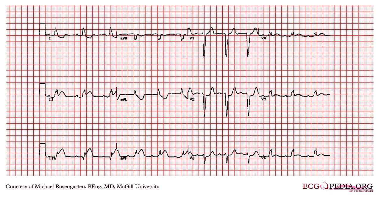
Clinical Pearls
References
- Mills, Newby, Zaman, Almahmeed, et al. Fifth Universal Definition of Myocardial Infarction (2026). Global Heart. 2026;21:64. https://doi.org/10.5334/gh.1578
- Sgarbossa EB, Pinski SL, Barbagelata A, Underwood DA, Gates KB, Topol EJ, et al. Electrocardiographic diagnosis of evolving acute myocardial infarction in the presence of left bundle-branch block. N Engl J Med. 1996;334(8):481–487 (landmark derivation study). https://doi.org/10.1056/NEJM199602223340801
- Smith SW, Dodd KW, Henry TD, Dvorak DM, Pearce LA. Diagnosis of ST-elevation myocardial infarction in the presence of left bundle branch block with the ST-elevation to S-wave ratio in a modified Sgarbossa rule. Ann Emerg Med. 2012;60(6):766–776 (derivation study). https://doi.org/10.1016/j.annemergmed.2012.07.119
- Dodd KW, Zvosec DL, Hart MA, Glass G, Bannister LE, Body RM, et al. Electrocardiographic diagnosis of acute coronary occlusion myocardial infarction in ventricular paced rhythm using the modified Sgarbossa criteria. Ann Emerg Med. 2021;78(4):517–529. https://doi.org/10.1016/j.annemergmed.2021.03.036
- Grimes AP, Maria SJ. Evidence for electrocardiographic patterns identifying acute coronary occlusion in non-ST-elevation acute coronary syndromes: a scoping review. Acad Emerg Med. 2026:e70389. https://doi.org/10.1111/acem.70389
- Alencar JN, Lima GWF, Geraldo HADS, Fernandes RC, Scheffer MK, Felicioni SP, Marchi MFN. Accuracy of Left Bundle Branch Block Chronology and Electrocardiography Criteria for Acute Myocardial Infarction Diagnosis: A Systematic Review and Meta-analysis. Arq Bras Cardiol. 2025;e20250109. https://doi.org/10.36660/abc.20250109
Medical Disclaimer
- For Educational Purposes Only: This content is intended for educational reference and should not be used for clinical decision-making.
- Not a Substitute for Professional Judgment: Always consult your local protocols, institutional guidelines, and supervising physicians.
- Verify Before Acting: Users are responsible for verifying information through authoritative sources before any clinical application.