Sodium (Na⁺)
Normal: 135–145 mEq/L
Main extracellular cation that sets serum osmolality. Low values come from water excess, SIADH or diuretics; high values from water loss or hypertonic intake.
Learn More

Eight results that catch dangerous potassium, sodium and glucose, and show how the kidneys are doing at the bedside.
The Basic Metabolic Panel (BMP) measures eight analytes: glucose, calcium, sodium, potassium, chloride, total CO₂, BUN and creatinine. Anion gap, eGFR and BUN:Cr are calculated from these results and are not counted as components. The BMP is the most common chemistry panel at ED triage, on ICU morning labs and before transfer. Chem-7 is an older name for a seven-analyte version of the same core chemistry. Check whether your laboratory's panel includes calcium.
The Renal Panel adds phosphorus and albumin to the BMP, and the Comprehensive Metabolic Panel (CMP) adds albumin, total protein and the liver tests to it. Because the BMP has no albumin, it cannot correct calcium.
Main extracellular cation that sets serum osmolality. Low values come from water excess, SIADH or diuretics; high values from water loss or hypertonic intake.
Learn MoreSets resting membrane potential. Hemolysis or delayed processing can falsely raise it; ECG changes and rhythm matter more than the number alone.
Learn MoreMain extracellular anion. Interpret with sodium and CO₂: a high chloride with low CO₂ suggests a non-gap acidosis.
Learn MoreSerum bicarbonate plus dissolved CO₂. Low values indicate metabolic acidosis or compensated respiratory alkalosis; high values suggest contraction or vomiting.
Learn MoreUrea from protein catabolism. Rises with volume depletion, GI bleeding, high-protein intake and steroids as well as kidney injury; interpret with creatinine.
Learn MoreMuscle-derived marker of GFR. Rises late after kidney injury, and a normal value can mask low GFR in older adults or low muscle mass.
Learn MoreMain energy substrate and the first value to check in any altered patient. Low values need treatment before other causes are considered; high values need ketones and osmolality.
Learn MoreTotal calcium includes protein-bound fractions. The BMP has no albumin, so correct with albumin from another panel or measure ionized calcium.
Learn MoreNa⁺ − (Cl⁻ + total CO₂). Raised in high-anion-gap acidosis (lactate, ketones, uremia, toxic alcohols, salicylate); normal in non-gap acidosis.
Learn MoreEstimated GFR from creatinine, age and sex using the race-free CKD-EPI 2021 equation. Less accurate in acute kidney injury, where the value is not at steady state.
Learn MoreHelps separate prerenal azotemia (ratio above 20:1) from intrinsic kidney injury. Unreliable with GI bleeding, steroids or high-protein intake.
Learn More| Test | Adult Normal (conventional) | SI Units | Critical Low | Critical High | Notes |
|---|---|---|---|---|---|
| Sodium | 135–145 mEq/L | 135–145 mmol/L | <120 | >160 | Serum osmolality-linked |
| Potassium | 3.5–5.0 mEq/L | 3.5–5.0 mmol/L | <2.5 | ≥6.5 | Hemolysis-prone |
| Chloride | 98–106 mEq/L | 98–106 mmol/L | — | — | Interpret with Na⁺ and CO₂ |
| Total CO₂ | 22–29 mEq/L | 22–29 mmol/L | <10 | >40 | Arterial HCO₃⁻ is 22–26 |
| BUN | 7–20 mg/dL | 2.5–7.1 mmol/L (urea) | — | — | Age- and protein-dependent |
| Creatinine | M 0.7–1.3 | F 0.6–1.1 mg/dL | M 62–115 | F 53–97 µmol/L | — | — | Sex-specific; lab-dependent |
| Glucose | 70–99 mg/dL (fasting) | 3.9–5.5 mmol/L | <40 | >450 | Fasting interval |
| Total Calcium | 8.5–10.5 mg/dL | 2.12–2.62 mmol/L | <6.0 | >13.0 | Albumin-dependent; no albumin on BMP |
Every pattern here is a combination of BMP results, not a single value. Check the clinical picture, then run the next test on the list.
| Pattern | Key Findings | Common Causes | Next Step |
|---|---|---|---|
| Prerenal AKI | Creatinine up, BUN:Cr above 20:1, low urine sodium | Volume loss, low perfusion, NSAIDs, ACE inhibitor or ARB | Volume status, urine studies, fluid challenge |
| Intrinsic AKI | Creatinine up, BUN:Cr near 10–15:1 | Ischemic or toxic tubular injury, sepsis, contrast | Urinalysis, urine sodium, nephrology if persistent |
| High-anion-gap acidosis | Anion gap above 12 with low CO₂ | Lactate, ketones (DKA, starvation), uremia, toxic alcohols, salicylate | Lactate, beta-hydroxybutyrate, serum osmolality, toxicology |
| Normal-anion-gap acidosis | Normal gap, high chloride, low CO₂ | Diarrhea, renal tubular acidosis, large volumes of saline | Urine anion gap, stool or urine electrolytes |
| Hypovolemic hyponatremia | Low Na⁺, low serum osmolality, low urine sodium | Diuretics, GI losses, third-spacing | Volume exam, urine sodium and osmolality |
| SIADH | Low Na⁺ and osmolality, urine osmolality inappropriately high, normal volume | Medications, CNS disease, pulmonary disease, malignancy | Exclude hypothyroidism and adrenal insufficiency first |
| Hypercalcemia | Total Ca above 10.5 mg/dL, with albumin needed to correct it | Hyperparathyroidism, malignancy, thiazides, vitamin D excess | Albumin, ionized calcium, PTH, medication review |
| Pseudohyperkalemia | K⁺ above 5.0 mEq/L with no ECG change, hemolyzed or fist-clenched sample | Hemolysis, prolonged tourniquet, delayed processing, high platelet or WBC counts | Repeat on a clean draw with a short tourniquet time |
| Analyte | Changes After | Peak or Nadir | Repeat Interval | Monitoring Target |
|---|---|---|---|---|
| Potassium | Insulin-glucose, beta-agonist or potassium-binder treatment | Early effect, then rebound | At least 1, 2, 4, 6 and 24 hr after identifying and treating moderate or severe hyperkalemia (UK guideline 2026) | Falling K⁺ with no ECG change |
| Glucose | Insulin-glucose infusion | Falls within the first hour; late hypoglycemia possible | 0, 30, 60, 90, 120, 180, 240, 300 and 360 min after insulin-glucose (UK guideline 2026) | Glucose kept above 70 mg/dL |
| Sodium | Hypertonic saline bolus | Rises after each bolus | After each bolus, then per the hypertonic saline page schedule | Rise kept within the 24-hour limits (see Critical Findings) |
| Creatinine | Kidney injury | Rises over 24–48 hr | Repeat daily until the trend is clear | Stable or falling creatinine |
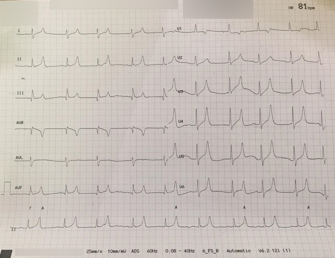
Recognize: peaked T waves, PR prolongation, lost P waves, QRS widening, bradycardia or sine wave. The ECG changes get treated now. Nobody waits for a second level. Follow local protocols and medical direction.
IV sodium bicarbonate has no routine role for acute hyperkalemia outside cardiac arrest (UK guideline 2026). Calcium protects the myocardium. It won't lower K⁺. With ECG changes, moderate hyperkalemia (K⁺ 6.0–6.4 mEq/L) gets the same calcium. If ECG changes are absent, the UK guideline reaches for insulin-glucose and nebulized albuterol. US practice varies on several doses, so go by local protocol and medical direction.
If the arrest is suspected to be hyperkalemic, the ACLS arrest algorithm still runs. Calcium chloride 10 mL (1 g) of 10% goes in IV or IO as a push. Sodium bicarbonate is 50 mEq IV or IO, repeated every 5–10 min as the blood gas guides (UK guideline 2026, 25.1–25.3; US label). Regular insulin is 10 units IV over 15–30 min with dextrose 50 g (AHA 2025 Table 3). IV calcium is COR 2b, LOE C-LD. IV sodium bicarbonate is COR 2b, LOE C-EO. IV insulin with glucose is COR 2b, LOE C-EO. AHA's 2025 CPR and ECC guidance, Part 10, calls all three effectiveness not well established in this setting. The UK guideline 2026 and ERC pair insulin with dextrose 25 g. Inhaled beta-2 agonists earn a no-benefit grade in arrest (COR 3: No Benefit, LOE C-EO; recommendation 4).
On digoxin? Give IV calcium slowly, with close ECG monitoring (calcium chloride and calcium gluconate labels).
Recognize: hypotonic hyponatremia with neurologic symptoms. Treat it now, acute or chronic. A pending sodium result is never a reason to delay.
Recognize: sweating, confusion, seizure or unresponsiveness. Every altered patient gets a glucose check before other causes come into play.
Recognize: carpopedal spasm, laryngospasm, seizure, QT prolongation or hypotension. Get an ionized calcium. Total calcium on a BMP can't be corrected without albumin. The goal is ionized calcium back in the reference range (4.5–5.3 mg/dL) and symptoms gone.
Recognize: total Ca above 13.0 mg/dL with confusion, vomiting or polyuria. Or low CO₂ with a high anion gap and a low pH on blood gas.
Recognize: weakness, paralysis, arrhythmia, or ECG changes such as flattened T waves, U waves or ST depression. Manage it under local protocol and medical direction.
Recognize: DKA means ketosis with a pH below 7.30 or bicarbonate below 18 mmol/L, with glucose at 200 mg/dL or higher, or a known diabetes history (2024 ADA consensus). Glucose can sit near normal in euglycemic DKA, most often with SGLT2 inhibitors. HHS brings markedly high glucose, dehydration and altered mental status, with little or no ketosis. The BMP gives you the glucose, CO₂, anion gap and potassium. Ketones, blood gas and serum osmolality confirm it (2024 ADA consensus).
Recognize: thirst, confusion, weakness, seizure or coma, usually from water loss. Check volume status and glucose. The sodium result alone won't tell you the cause.
| Device / Method | Analytes | Specimen | Limitations |
|---|---|---|---|
| i-STAT CHEM8+ | Na⁺, K⁺, Cl⁻, ionized Ca, glucose, BUN, creatinine, TCO₂ | Whole blood, heparinized | No total calcium and no liver tests; eGFR reporting depends on analyzer configuration; cartridge temperature and storage limits |
| Glucometer | Glucose only | Capillary blood | Unreliable in shock or hypoperfusion; interference from maltose, icodextrin and high-dose vitamin C |
| Laboratory BMP | All 8 analytes | Serum or lithium heparin plasma | Turnaround of hours; results are often not back before transfer, so send the draw and the time |
Pediatric reference intervals shift with age for several BMP analytes. This page has no age-band tables, so use your lab's pediatric table. Weight-based doses are below (see weight-based dosing and pediatric vital signs).
| Indication | Pediatric Dose | Max / Repeat | Notes |
|---|---|---|---|
| Symptomatic hypocalcemia | Calcium chloride 10%: 2.7–5 mg/kg IV slowly (0.027–0.05 mL/kg of 10%) | Repeat as ionized calcium guides; label cites no trial data on repeat dosing | DailyMed label; no faster than 1 mL/min |
| Hyperkalemia with ECG changes | Calcium chloride 10%: 20 mg/kg (0.2 mL/kg) IV/IO slowly; IV/IO push in arrest | Max 1 g per dose; repeat if ECG changes persist | NASEMSO v3.0, Cardiac Arrest item 8b (p. 119). Differs from FDA label: the label gives no hyperkalemia dose; the pediatric label dose is for hypocalcemia only. |
| Hyperkalemic arrest (insulin) | Regular insulin 0.1 units/kg IV over 15–30 min with dextrose 0.5 g/kg (D10 5 mL/kg IV) | Max 10 units (severe hyperkalemia) | AHA 2025 Part 10, Table 3 (children, arrest). No pediatric insulin dose is established here for non-arrest hyperkalemia; use the pediatric protocol and medical direction. |
| Hypoglycemia | Dextrose 0.5–1 g/kg IV/IO (D10 5–10 mL/kg preferred) | Max 25 g per dose; reassess after each dose | D10 preferred at every age (dextrose page) |
| Severe symptomatic hyponatremia | 3% NaCl 2 mL/kg IV over 10 min | Max 100 mL per dose; repeat per protocol and recheck Na⁺ after each dose | High-alert never free-flow (hypertonic saline page) |
The hypocalcemia row is the labeled pediatric range. That label covers only hypocalcemic disorders. So the hyperkalemia calcium row (20 mg/kg) is off-label, and its source is NASEMSO v3.0 (Cardiac Arrest, item 8b).
Pregnancy changes how you read the panel. Respiratory alkalosis is normal here, with PaCO₂ around 28–32 mmHg and HCO₃⁻ around 18–21 mEq/L. A low total CO₂ alone does not mean metabolic acidosis. Dilution lowers sodium. A creatinine above 0.8 mg/dL is abnormal in pregnancy. Treat hyperkalemia and hypoglycemia the way you would in a non-pregnant adult.
A normal creatinine can mask a markedly reduced GFR when muscle mass is low. Use eGFR instead. In this group, the usual culprits behind hyperkalemia, hyponatremia and AKI are volume depletion and polypharmacy (diuretics, ACE inhibitors, ARBs, potassium-sparing drugs).
Read K⁺ and HCO₃⁻ against the dialysis schedule. The pre-dialysis sample is the reference. Severe hyperkalemia in patients on dialysis calls for urgent dialysis (UK guideline 2026). With ECG changes present, give IV calcium even when dialysis is immediately available (UK guideline 2026).
| Error or Interferent | Effect on Results | Prevention |
|---|---|---|
| Hemolysis | Falsely high K⁺ (pseudohyperkalemia) | Clean venipuncture; repeat without hemolysis, and treat now if the ECG shows toxic changes |
| Drawing above an IV line | Dilution with saline (low K⁺, calcium, glucose, BUN, creatinine; high Na⁺ and Cl⁻); dextrose-containing fluid falsely raises glucose | Draw from the opposite arm; if not possible, stop the infusion and follow lab flushing policy |
| Prolonged tourniquet or fist clenching | Falsely high K⁺ | Limit tourniquet time; no fist pumping |
| Delayed processing | Glucose falls; K⁺ rises | Process promptly or use the lab's glucose-preserving tube |
| EDTA (lavender) carryover | Falsely high K⁺ and very low calcium | Follow the order of draw; avoid tube carryover |
| Lipemia or icterus | Method-dependent interference | Ask the lab for its interference index |
| Point-of-care meter interference | Falsely high glucose (maltose, icodextrin, vitamin C) | Use a laboratory glucose when the meter result does not fit the patient |