

Bedside Snapshot
- Indications
- Every advanced airway, from placement to extubation or handoff
- CPR: EtCO₂ trend for compression quality and ROSC
- Sedation, opioids, and severe TBI ventilation
- Contraindications / Abort
- No waveform after placement in a perfusing patient: remove the tube and re-confirm
- In cardiac arrest or very low flow, a flat trace does not prove misplacement: confirm first
- Flat line during ventilation: check circuit and tube first
- A number with no waveform is not confirmation
- Critical Steps
- Confirm a bag waveform before the tube goes in
- Read the shape on every breath, not only the number
- Recheck after every move, suction pass or sedation change
- Confirm & Monitor
- Sustained waveform on every breath
- Trend EtCO₂ with SpO₂, heart rate and BP
- Abrupt EtCO₂ rise during CPR can signal ROSC: check a pulse
Key Numbers
Capnography confirms the tube, shows ventilation and tracks CPR; read the waveform shape before the number.
| Parameter | Value |
|---|---|
| Tube confirmation | Adults in cardiac arrest: continuous waveform capnography with clinical assessment is the most reliable confirmation (2025 AHA Part 9, COR 1, LOE C-LD). Infants and children with a perfusing rhythm: exhaled CO₂ detection, colorimetric or capnography (2025 AHA Part 8, COR 1, LOE C-LD). A sustained waveform on every breath is the teaching standard; about 6 breaths is common teaching, not an AHA number |
| Typical EtCO₂ | 35–45 mmHg in a perfusing patient with normal ventilation |
| TBI eucapnia target | EtCO₂ 35–40 mmHg; avoid below 35 mmHg |
| TBI herniation (temporary) | EtCO₂ 30–35 mmHg, only for signs of active herniation |
| Prolonged prophylactic hyperventilation | Not recommended (PaCO₂ 25 mmHg or less), BTF 4th edition |
| Low EtCO₂ during CPR (intubated adults) | Failure to exceed 10 mmHg by waveform capnography after 20 minutes of ALS resuscitation may be one component of a multimodal decision to end resuscitation (2025 AHA Part 9, COR 2b, LOE C-LD). Never the sole criterion. In nonintubated adults, no specific EtCO₂ cutoff should be used to end resuscitation (COR 3: Harm) |
| ROSC during CPR | An abrupt increase in EtCO₂ may detect ROSC (2025 AHA Part 9, COR 2b, LOE B-NR). A rise greater than 10 mmHg may indicate ROSC, but ROSC can occur with smaller rises. Epinephrine and sodium bicarbonate can change EtCO₂, so confirm with a pulse check |
| DKA screen | Reported cut-offs range 21–36 mmHg; use as a screen, not a diagnosis |
Overview & Principles
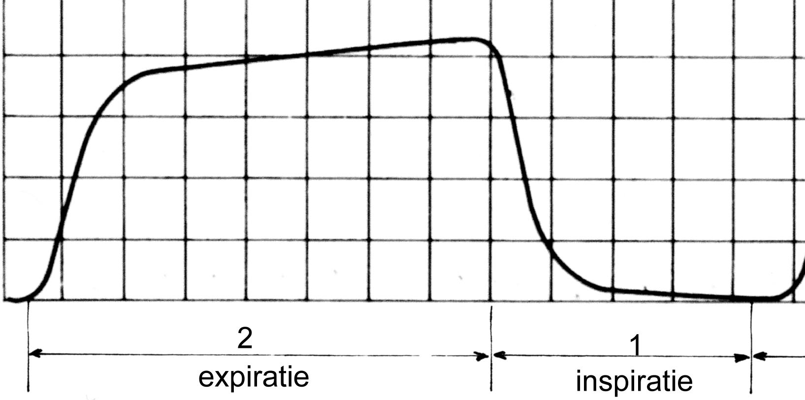
Capnography reads the CO₂ in every exhaled breath and draws it as a waveform over time. The end-tidal CO₂ (EtCO₂) is the peak at the end of exhalation. That CO₂ reaches the lungs by way of the blood, so the number reflects ventilation and pulmonary blood flow. Think of it as a ventilation and perfusion signal. It is not a measure of oxygenation.
How It Works
- Baseline: sits near zero while the patient breathes in. If it never drops back to zero, the patient is re-breathing CO₂.
- Expiratory upstroke: dead-space gas from the airways leaves first. Alveolar gas follows it.
- Alveolar plateau: this is gas from the lungs itself. Read the EtCO₂ at the far end of it.
- Plateau height: follows pulmonary blood flow. That means during CPR it also tells you about cardiac output.
- Sampling: a mainstream sensor sits in the airway circuit. A sidestream sampler pulls gas down a sample line. Follow the device instructions for use.
Where It Fits
You use capnography to confirm and watch advanced airways, guide ventilation, track CPR quality and signal return of spontaneous circulation (ROSC). It also catches hypoventilation during sedation. The American Society of Anesthesiologists (ASA) standard asks for continual quantitative monitoring of expired CO₂, from tube placement until extubation or transfer. Pulse oximetry (SpO₂) still needs watching, because it cannot show ventilation.
Indications & Contraindications
Indications
- Every advanced airway (endotracheal tube or supraglottic airway) from placement until extubation or handoff
- Any ventilated patient during transport, including after every move
- CPR: EtCO₂ trend for compression quality and ROSC
- Procedural sedation, opioids and sedatives, where hypoventilation is a risk
- Severe traumatic brain injury (TBI), to avoid hypocapnia
- Asthma or COPD with bronchospasm, to follow the waveform slope
Absolute Contraindications
None. Nothing about capnography breaks the skin. Pick a sensor and sample line that fit both the patient and the device.
Relative Contraindications & Cautions
- Dead-space states: pulmonary embolism, low cardiac output, COPD and ARDS all widen the gap to PaCO₂. A normal EtCO₂ does not rule out hypercapnia.
- Leaks or blockage: a leaking circuit, a kinked sample line or a full water trap gives false or dampened readings.
- Numbers without a waveform: a value with no shape behind it is not confirmation.
- Cardiac arrest or very low flow: EtCO₂ can be absent or very low even with a correctly placed tube. Little blood reaches the lungs, so there is little CO₂ to exhale. See Confirmation.
Scope & Team Roles
| Provider Level | Typical Role | Notes |
|---|---|---|
| EMT | Assists | Connects the sensor and keeps tubing patent; reports the monitor |
| AEMT | Monitors & maintains | Reads the waveform and EtCO₂ per protocol |
| Paramedic | Performs | Confirms tube placement, monitors CPR and reads the waveform |
| RN (ED/ICU) | Monitors & maintains | Alarms, trends, troubleshoots and escalates changes |
| Critical Care Transport (CFRN / CTRN / FP-C / CCP-C) | Performs | Interprets the waveform during transport and reassesses after every move |
| Physician / APP | Performs | Interprets waveforms and sets targets with the team |
Team Roles During the Procedure
- Operator: places the tube and calls for confirmation.
- Ventilator or bag operator: keeps the mask seal and the rate steady.
- Monitor reader: watches the waveform and calls the shape out loud.
- Recorder: notes the time of the first sustained waveform and each value.
Equipment & Preparation
Equipment
- Capnography monitor with a waveform display and a numeric EtCO₂ readout
- Sensor: mainstream (in the circuit) or sidestream (sample line)
- Low-dead-space airway adapter that matches the tube or airway device (a large adapter adds dead space to small tubes)
- Water trap and filter, as the device instructions specify
- Colorimetric detector as a backup
- Bag-valve mask, suction and a spare sample line
Setup
Power up the monitor and set the alarm limits before you connect the patient. Then get a waveform on the bag, before the tube goes in. Now you know what normal looks like for this patient.
Non-Intubated Patients
Sidestream sampling through a nasal or nasal-oral cannula suits patients on oxygen, sedation or opioids, and patients in respiratory distress. Mouth breathing and supplemental oxygen flow can dilute or lower the reading. The trend and the waveform carry more weight than the exact number. For the cannula and sampling flow, go by the device instructions.
Pre-Procedure Checklist
- Monitor on; sampler ready as the device instructions describe
- Sample line, filter and water trap connected with no kinks
- Waveform visible on bag-mask ventilation before placement
- Adult or pediatric adapter matches the tube
- Alarm limits set and audible to the team
- Colorimetric backup at hand
- Spare sample line and water trap available
Step-by-Step Procedure
Setup takes seconds. The tube is confirmed at the first sustained waveform, not at the first number. Work the phases in order, and stop at the warning box whenever the waveform is missing.
Phase 1: Prepare
- Set up the monitor: connect the sensor or sample line and the water trap, and confirm the device is reading.
- Check the bag waveform: ventilate by bag-mask and confirm a normal waveform before the tube is placed.
Phase 2: Perform
- Place the airway adapter: between the tube and the bag or ventilator, after the tube is in place.
- Ventilate and watch the shape: look for a waveform on each breath, not only a number.
- Look for a sustained waveform: count at least 5–6 breaths with a waveform on each one.
Phase 3: Confirm & Secure
- Record the first sustained waveform: note the time, the EtCO₂ value and the shape.
- Secure the sample line: route it so it cannot kink or pull the tube.
- Keep the monitor on: the waveform stays on screen from placement until extubation or handoff.
Record the baseline shape before any transfer. Any change after the move will then stand out.
Alternative Technique: Colorimetric Detector
A colorimetric detector tells you CO₂ is there. It gives no waveform and no trend. Keep it as a backup for when quantitative capnography is not available. The ASA anesthesia standard requires a quantitative method for continual end-tidal CO₂ monitoring: capnography, capnometry or mass spectrometry. That runs from tube placement until extubation or transfer. A colorimetric detector does not replace it.
Confirmation & Monitoring
Primary Confirmation
A sustained waveform on every breath confirms the tube is in the trachea. Look for about 6 breaths in common teaching, each with an EtCO₂ value and a shape. One breath is not enough. Breath sounds and chest rise support the result, but they don't replace it. In adults in cardiac arrest, continuous waveform capnography with clinical assessment is the most reliable way to confirm and monitor tube placement (2025 AHA Part 9, COR 1, LOE C-LD).
Secondary Confirmation
- Chest rise with each breath and bilateral breath sounds
- No gurgling over the stomach
- SpO₂ and heart rate improving or stable
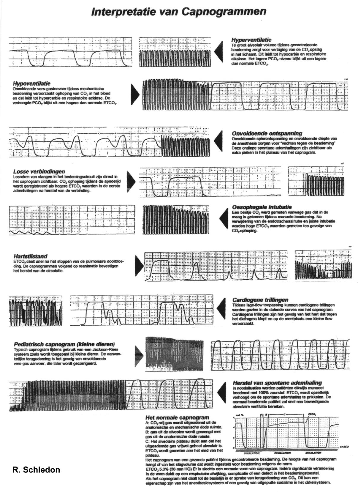
| Finding | Meaning | Action |
|---|---|---|
| Sustained waveform on each breath | Tube in the trachea and ventilating | Continue; secure and document |
| Brief CO₂ that fades after bag breaths | Gas from the stomach, possible esophageal tube | Remove and reposition; re-confirm |
| No waveform | Esophageal tube, dislodged tube, disconnected circuit or no pulmonary blood flow | Check connections; if perfusing, reposition or remove the tube. In arrest, confirm by direct or video laryngoscopy or ultrasound first |
| Rising slope in the plateau (shark fin) | Bronchospasm or obstruction | Treat per protocol; follow the slope |
| Notch in the plateau (curare cleft) | Breathing against the ventilator under neuromuscular blockade | Deepen sedation or paralysis per protocol |
| Baseline that does not return to zero | Re-breathing | Check valves, circuit and absorber |
| Rising EtCO₂ with a falling rate | Hypoventilation | Ventilate; check sedation and opioid effect |
| Abrupt rise in EtCO₂ during CPR | Possible ROSC | Check pulse and BP now; if ROSC, start post-arrest care. Confirm with a pulse check, because bicarbonate and epinephrine can change EtCO₂ |
Ongoing Monitoring
- Continuous waveform from placement until extubation or handoff
- Recheck the shape after every move, suction pass or sedation change
- Trend EtCO₂ with SpO₂, heart rate and BP
- Blood gas when the number and the clinical picture disagree
- Ventilation rate with an advanced airway: during CPR, adults, it may be reasonable to give 1 breath every 6 seconds (10 per minute) with continuous compressions (2025 AHA Part 9, COR 2b, LOE C-LD); infants and children, it may be reasonable to target 20–30 per minute (Part 8, COR 2b, LOE C-LD). For TBI, follow the protocol target.
Troubleshooting
| Problem | Likely Cause | Fix |
|---|---|---|
| No waveform after placement | Esophageal or dislodged tube | Check connections; if perfusing, remove the tube, bag-mask ventilate and re-attempt or place a supraglottic airway. In arrest, confirm placement first (see Confirmation) |
| Waveform lost during ventilation | Disconnected circuit or leak at the adapter | Check every connection; check tube depth at the teeth |
| Dampened or low waveform | Water or secretions in the sample line or trap | Clear or replace the trap and line per the device instructions |
| Waveform with no number | Sampler not reading or device setting wrong | Check the device settings and instructions for use |
| Notch in the plateau | Breathing against the ventilator under paralysis | Deepen sedation or paralysis per protocol |
| Baseline that does not return to zero | Re-breathing from a valve or absorber fault | Check valves and absorber; test the circuit |
| Rising slope (shark fin) | Bronchospasm or airway obstruction | Treat per protocol; check for kinks and secretions |
| Low EtCO₂ with a high rate | Over-ventilation, or low cardiac output or pulmonary embolism | Reduce the rate to target; look for the cause |
Complications
| Complication | Recognition | Prevention | Management |
|---|---|---|---|
| Immediate: unrecognized esophageal intubation | No sustained waveform; gurgling | Waveform confirmation every time | If perfusing, remove the tube; ventilate; re-attempt or place a supraglottic airway. In arrest, confirm placement before removing |
| Immediate: unrecognized hypoventilation after sedation | Rising EtCO₂ with a falling rate; falling SpO₂ | Continuous EtCO₂ during sedation | Support ventilation; reversal per protocol |
| Delayed: hypocapnia after transport or TBI ventilation | EtCO₂ below 35 mmHg | Target 35–40 mmHg; trend with the monitor | Reduce the rate to target; reassess |
| Delayed: missed deterioration in CPR | Low EtCO₂ that does not rise | Trend EtCO₂ and check compression quality | Review CPR quality and the reversible causes per protocol |
| Rare: false reassurance from a number alone | Value on screen, no waveform | Read the shape on each breath | Treat as unconfirmed until the waveform appears |
| Rare: blocked sample line | Dampened or absent waveform | Check the trap and line at setup and after moves | Replace per the device instructions |
Special Populations
This page does not cover pregnancy, obesity or hypothermia. For those patients, follow local protocol.
Pediatrics
- Dead space: a large adapter adds dead space to a small tube, and that can dampen the waveform. Size a low-dead-space adapter to the tube.
- Airway confirmation: in infants and children with a perfusing rhythm, you should use exhaled CO₂ detection (colorimetric or capnography) to confirm ETT placement (2025 AHA Part 8, section 17.4, COR 1, LOE C-LD). With low pulmonary blood flow or in cardiac arrest, exhaled CO₂ may not be reliable.
- Adapters and sampling: use the pediatric airway adapter and sample line for the tube size. See equipment sizing.
- Interpretation: the same waveform rules apply to children. This page gives no separate pediatric EtCO₂ target. Follow the pediatric protocol.
- CPR with an advanced airway: a target of 20–30 breaths per minute (one breath every 2–3 seconds) may be reasonable. Hyperventilation may compromise hemodynamics (2025 AHA Part 8, section 6, COR 2b, LOE C-LD). For children with an invasive airway, ETCO₂ monitoring may be considered to judge CPR quality (Part 8, section 9, COR 2b, LOE B-NR). A specific ETCO₂ cutoff alone should not be used to end resuscitation (COR 3: Harm, LOE C-LD).
- After ROSC: the 2025 AHA pediatric guideline (Part 8) sets no PaCO₂ target. For comatose adults after ROSC, the target is an arterial PaCO₂ of 35–45 mmHg (2025 AHA Part 11, COR 1, LOE B-R). That is an arterial value, not an EtCO₂ target. For children, follow the pediatric post-arrest protocol.
- Sedation: routine capnography in procedural sedation was associated with fewer hypoxic episodes and adverse events, according to a 2026 pediatric review.
- Airway: see the pediatric airway page for tube and device choices.
Metabolic Acidosis (DKA and Sepsis)
- DKA: a 2025 systematic review and meta-analysis (8 studies) puts the pooled specificity of EtCO₂ for diagnosing DKA at 0.88 (95% CI 0.79–0.93). The pooled sensitivity is printed with a CI that does not contain the point estimate. Don't quote it as fact. Study-level sensitivities ran from 0.71 to 0.99. Cut-offs ran from 21 to 36 mmHg, so use EtCO₂ as a screen only.
- Confirm: the diagnosis rests on blood gas and glucose.
- Sepsis (limited evidence): a 2026 narrative review reports that EtCO₂ falls as lactate rises. It is less reliable in multisystem shock.
Traumatic Brain Injury
| Situation | EtCO₂ Target | Notes |
|---|---|---|
| Routine ventilation | 35–40 mmHg | Avoid hypocapnia; monitor with capnography |
| Active herniation (temporary) | 30–35 mmHg | Only for signs of active herniation; reverse as soon as the cause is treated |
| Prophylactic hyperventilation | Avoid | Avoid without herniation signs (BTF prehospital 2023); prolonged PaCO₂ 25 mmHg or less not recommended (BTF 4th edition) |
Signs of active herniation are Cushing's triad, a dilated or asymmetric unreactive pupil, extensor posturing, or an acute, progressive fall in GCS. A GCS of 8 or less defines severe TBI. By itself, though, it is not a herniation sign.
Obstructive Lung Disease
- Shark fin: a rising phase III slope points to bronchospasm. In one intubated asthma patient, the slope dropped before EtCO₂ did as treatment took hold (single case, limited evidence).
- Tiring: in a patient who is working hard, a normal or rising EtCO₂ is a warning sign.
Post-Procedure Care & Transport
Immediate Aftercare
- Confirm the waveform and EtCO₂ after the first transfer, and again after every move.
- Check that the sample line, trap and tube are secure.
Ongoing Maintenance
- Empty or replace the water trap per the device instructions.
- Keep the sample line free of kinks, and replace it when it is damaged.
Transport Considerations
- Noise and vibration can mask alarms. Look at the waveform, not only the sound.
- Recheck the shape after every move, sedation change or suction pass.
- Watch for a falling rate with a rising EtCO₂, which is hypoventilation.
Document & Hand Off
- Time of the first sustained waveform, and the EtCO₂ value
- Device, sampling type and airway adapter size
- Any alarms, troubleshooting steps and the result
- Waveform shape at handoff, and the trend since placement
Clinical Pearls & Pitfalls
Pearls
- Shape first: read the waveform on every breath before you read the number.
- Sustained, not single: one breath is not enough. Confirmation needs a waveform on every breath across a sustained run (about 6 breaths in common teaching).
- Trend, do not snapshot: one value tells you less than the direction it takes over minutes.
- Plateau tells the story: the slope, the notch and the baseline each point to a cause.
Common Pitfalls
- Number without a shape: the value looks normal, and the tube is still not confirmed. Fix: wait for the waveform.
- Using EtCO₂ alone for PaCO₂: dead space widens the gap between the two. Fix: check a blood gas when the picture does not fit.
- Ignoring the baseline: a baseline that never returns to zero means re-breathing. Fix: check the circuit.
- Flat trace in arrest: low pulmonary blood flow can produce a low or absent EtCO₂ even with a correct tube. Fix: confirm placement by direct or video laryngoscopy or ultrasound before you remove it.
- Bicarbonate bump: sodium bicarbonate and epinephrine can shift EtCO₂ and make it look like ROSC (2025 AHA Part 9 lists both as influences on EtCO₂). Fix: check a pulse before you call ROSC.
Education & Skill Maintenance
Skill Maintenance
- Practice naming each waveform shape with a partner, using the figures on this page.
- Run a simulation that begins with an esophageal tube and asks the team to find it from the waveform.
- Check the device and the trap at each shift handoff.
- Debrief any alarm or waveform loss, and record what changed the waveform.
References
- Wigginton JG, Agarwal S, Bartos JA, et al. Part 9: Adult Advanced Life Support: 2025 American Heart Association Guidelines for Cardiopulmonary Resuscitation and Emergency Cardiovascular Care. Circulation. 2025;152(16 suppl 2):S538–S577. https://doi.org/10.1161/cir.0000000000001376
- American Society of Anesthesiologists. Standards for Basic Anesthetic Monitoring. Last amended October 15, 2025. https://www.asahq.org/standards-and-guidelines/standards-for-basic-anesthetic-monitoring
- Lulla A, Lumba-Brown A, Totten AM, et al. Prehospital Guidelines for the Management of Traumatic Brain Injury, 3rd Edition. Prehosp Emerg Care. 2023;27(5):507–538. https://doi.org/10.1080/10903127.2023.2187905
- Carney N, Totten AM, O'Reilly C, et al. Guidelines for the Management of Severe Traumatic Brain Injury, Fourth Edition. Neurosurgery. 2017. https://doi.org/10.1227/NEU.0000000000001432
- Hajipoor Kashgsaray N, Jamei K, Kabiri N. Diagnostic Accuracy of End-Tidal Carbon Dioxide for Assessing Diabetic Ketoacidosis: A Systematic Review and Meta-Analysis. Arch Acad Emerg Med. 2025;13(1):e71. PMC12702517. https://pmc.ncbi.nlm.nih.gov/articles/PMC12702517/
- Rath S, Alsabri M, Tawfik AM, et al. Seeing beyond the numbers: capnography as a vital tool in pediatric emergency care. Int J Emerg Med. 2026;19:9. https://doi.org/10.1186/s12245-025-01110-z
- Shikama M, Yamamoto M, Osawa I, et al. Monitoring the Resolution of Acute Exacerbation of Airway Bronchoconstriction in an Asthma Attack Using Capnogram Waveforms. Crit Care Explor. 2023;5(4):e0899. https://doi.org/10.1097/CCE.0000000000000899
- 2025 American Heart Association Guidelines for CPR and ECC, Part 8: Pediatric Advanced Life Support. American Heart Association; 2025. https://cpr.heart.org/en/resuscitation-science/cpr-and-ecc-guidelines/pediatric-advanced-life-support
- 2025 American Heart Association Guidelines for CPR and ECC, Part 11: Post-Cardiac Arrest Care. American Heart Association; 2025. https://cpr.heart.org/en/resuscitation-science/cpr-and-ecc-guidelines/post-cardiac-arrest-care
Medical Disclaimer
- For Educational Purposes Only: This content is intended for educational reference and should not be used for clinical decision-making.
- Not a Substitute for Professional Judgment: Always consult your local protocols, institutional guidelines, and supervising physicians.
- Verify Before Acting: Users are responsible for verifying information through authoritative sources before any clinical application.