

Bedside Snapshot
- Recognize
- Ischemic chest pain or equivalent with ST elevation ≥1 mm in 2 contiguous leads (STEMI; higher V2–V3 thresholds by sex and age), or ischemic symptoms with a rise and/or fall of troponin (NSTEMI, UA)
- Red Flags
- SBP <90 mmHg, cool or clammy skin, pulmonary edema, sustained VT, heart block or cardiac arrest
- First Actions
- 12-lead ECG within 10 min; aspirin 162–325 mg chewed; oxygen if SpO₂ <90%; IV access; notify the STEMI center
- Key Doses
- Aspirin 162–325 mg PO chewed; clopidogrel 300–600 mg PO load; heparin 60 units/kg IV (max 4,000 units); nitroglycerin 0.4 mg SL every 5 min
- Don't Miss
- A normal first ECG does not exclude STEMI; nitrates in hypotension or RV infarct; a wrong lytic dose
- Escalate & Transport
- PCI-capable STEMI center when FMC-to-device can be ≤90 min (≤120 min if transferred); fibrinolysis when PCI cannot start within 120 min; cardiogenic shock calls for emergent revascularization and shock-team involvement
Definition & Pathophysiology
Acute coronary syndrome (ACS) is not one diagnosis. It is a range of acute myocardial ischemia. Most often a plaque ruptures or erodes, and a thrombus forms on top of it. The range includes ST-elevation myocardial infarction (STEMI), non-ST-elevation myocardial infarction (NSTEMI) and unstable angina (UA). Under the Fourth Universal Definition, an MI needs a rise and/or fall of cardiac troponin with at least one value above the 99th percentile upper reference limit. It also needs evidence of ischemia.
- STEMI: a persistent ST-elevation pattern on the ECG. Treat it as an occlusion until proven otherwise. Reperfusion is time-critical.
- NSTEMI and UA: no persistent ST elevation. Patients are risk-stratified. Treatment is antithrombotic therapy plus an invasive or selective strategy.
- Mechanism: the plaque breaks and exposes material that clots. A complete occlusion (most STEMI) or a partial obstruction (NSTEMI, UA) cuts the supply. The muscle that loses supply is injured.
Recognition & Red Flags
Typical and Atypical Presentation
- Typical: pressure, squeezing or heaviness in the chest. It often radiates to the jaw, neck, arm or back. It lasts more than a few minutes, and rest does not relieve it.
- Atypical: epigastric pain, dyspnea, nausea, diaphoresis, fatigue or syncope. Women, older adults, people with diabetes and people with kidney disease present this way more often.
Red Flags
- ST elevation at the J point in 2 or more contiguous leads (thresholds in Assessment and Diagnostics)
- Ischemic chest pain with SBP <90 mmHg, cool or clammy skin, or altered mentation (possible cardiogenic shock)
- Pulmonary edema, new heart murmur or new heart failure signs (possible mechanical complication)
- Sustained ventricular arrhythmia, complete heart block or cardiac arrest
Assessment & Diagnostics
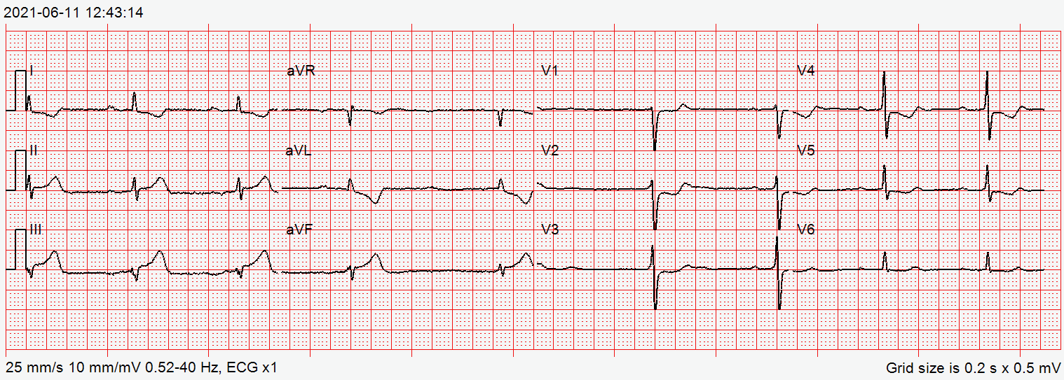
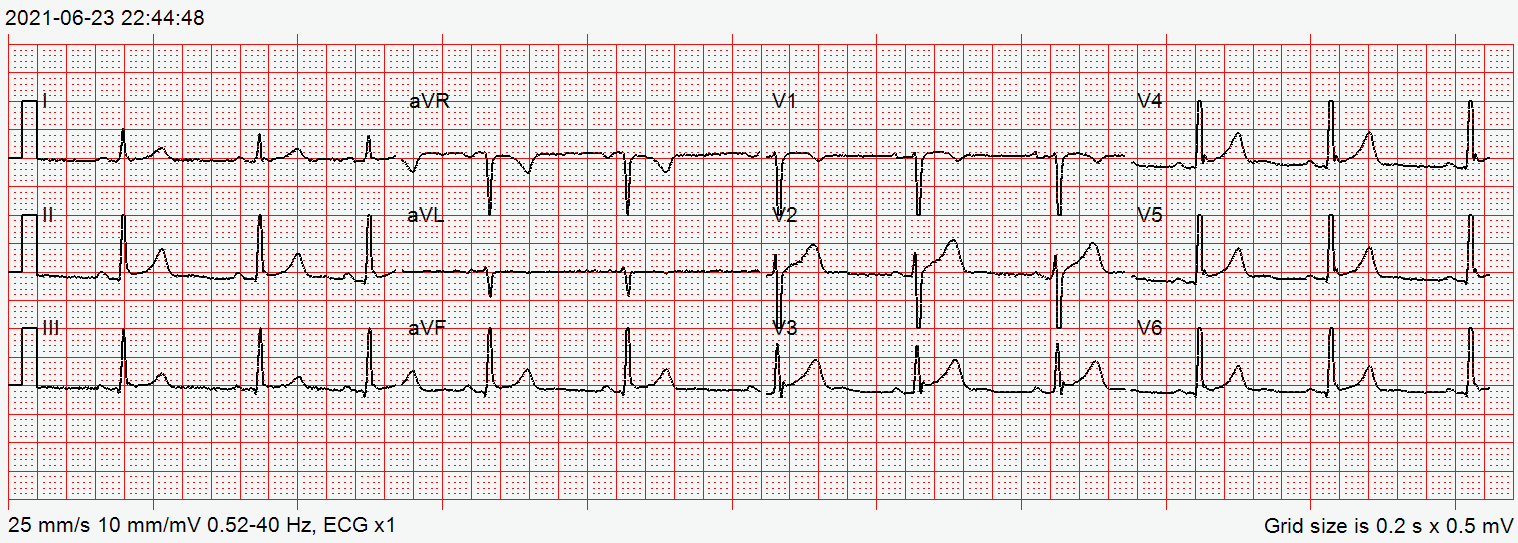
STEMI is diagnosed on the 12-lead ECG, not on the lab. Troponin confirms myocardial injury, and it means the most when you read it over time.
| Check | Why | Act If |
|---|---|---|
| 12-lead ECG | Identifies STEMI and STEMI equivalents; the time-critical test | Within 10 min of first medical contact. ST elevation ≥1 mm in 2 contiguous leads: activate the STEMI pathway (V2–V3 thresholds in the list below) |
| Serial ECG | Ischemia evolves; a first ECG can be non-diagnostic | Repeat with any change in pain, rhythm or hemodynamics, and per protocol |
| BP, HR and SpO₂ | Shock, bradycardia, heart block and hypoxemia | SBP <90 mmHg: shock pathway. SpO₂ <90%: oxygen (see Oxygen) |
| High-sensitivity troponin | Confirms myocardial injury (Fourth Universal Definition) | Rise or fall per the lab protocol (0/1 h or 0/2 h rapid pathway where used); a single early value does not rule ACS out |
| Blood glucose | Hypoglycemia mimics ACS symptoms and can coexist | Low: treat per protocol; recheck after treatment |
12-Lead STEMI Criteria (Fourth Universal Definition)
- Most leads: J-point ST elevation ≥1 mm in 2 contiguous leads
- V2–V3: ≥2 mm in men 40 years or older, ≥2.5 mm in men under 40, ≥1.5 mm in women
- Posterior and right-sided leads: ST depression in V1–V3 or ST elevation in V4R suggests a posterior or RV infarct; obtain these leads per protocol
Differential & Mimics
- Pericarditis: diffuse ST elevation with PR depression and pleuritic, positional pain. The ST elevation does not sit in one coronary territory.
- Left bundle branch block: discordant ST changes do not establish STEMI. Apply the Smith-modified Sgarbossa criteria and compare with prior ECGs. See Conduction Blocks
- LV hypertrophy or early repolarization: broad, widespread ST elevation with high voltage. There is no reciprocal pattern. Compare with prior tracings.
- Pulmonary embolism: tachycardia, hypoxemia, right heart strain and dyspnea. The ECG shows no coronary pattern.
- Aortic dissection: tearing pain, a pulse or BP difference between the arms, or a new aortic regurgitation murmur. Avoid anticoagulation and fibrinolysis until dissection is excluded.
- Takotsubo syndrome: ST changes after an emotional or physical trigger, with apical ballooning. The coronary pattern is often non-occlusive.
BLS Management
- Scene safety and recognition: identify chest pain or ACS symptoms early and request ALS support.
- Position: semi-recumbent and comfortable; limit exertion; do not let the patient walk.
- Oxygen: apply oxygen when SpO₂ is below the protocol threshold or breathing is labored; do not give routine oxygen to a normoxic patient.
- Aspirin: assist with the patient's own chewed aspirin when no allergy or bleeding is present and the dose has not already been taken; follow the protocol for a home dose already taken.
- Nitroglycerin: assist with the patient's own prescription only where state and agency protocol allow, after a BP check and a PDE-5 inhibitor screen (no sildenafil or vardenafil within 24 hours, no tadalafil within 48 hours, no riociguat).
- Monitor and transport: keep the patient calm and monitored; keep the time of symptom onset and every medication time; report early.
Key dose: aspirin 162–325 mg PO, chewed, once. See Aspirin.
ALS Management
Doses vary by protocol; follow local protocols and medical direction.
- 12-lead ECG within 10 min (Paramedic/RN/CCT): acquire, interpret and transmit; activate the STEMI pathway at the first ST-elevation finding. See 12-Lead ECG Acquisition
- Access and monitoring (Paramedic/RN/CCT): IV or IO access; continuous rhythm monitoring; defibrillator pads on when a STEMI is likely to be complicated by arrhythmia
- Aspirin: 162–325 mg PO, chewed, once, unless contraindicated or already taken; then 81 mg PO daily per the receiving team (keep the maintenance dose at or below 100 mg with ticagrelor). See Aspirin
- P2Y12 inhibitor (Paramedic/RN/CCT): clopidogrel 300–600 mg PO load, then 75 mg PO daily; STEMI with primary PCI 600 mg; STEMI with fibrinolysis, age 75 or under 300 mg; over 75 no load. Ticagrelor or prasugrel is preferred where the cardiology team chooses PCI; choice and timing belong to cardiology. Prasugrel is contraindicated with active pathological bleeding or prior TIA or stroke (boxed warning), is generally not recommended at 75 or older, and the label suggests 5 mg maintenance under 60 kg; in NSTE-ACS its loading dose waits until coronary anatomy is established. Ticagrelor carries bleeding and dyspnea cautions. See Clopidogrel
- Anticoagulant (Paramedic/RN/CCT): unfractionated heparin 60 units/kg IV bolus (max 4,000 units), then 12 units/kg/hr IV (max 1,000 units/hr) for NSTE-ACS or with a fibrinolytic; or enoxaparin 1 mg/kg subcut every 12 hr for NSTE-ACS. For STEMI heading to primary PCI, heparin is given per the PCI center protocol (70–100 units/kg IV bolus). With fibrinolysis, enoxaparin is 30 mg IV bolus plus 1 mg/kg subcut under 75 years (no IV bolus and 0.75 mg/kg at 75 or older), started 15 min before to 30 min after the lytic. Anticoagulant choice and timing follow the receiving cardiology team. Titrate heparin by nomogram. See Heparin and Enoxaparin
- Nitroglycerin for ongoing pain (Paramedic/RN/CCT): 0.4 mg SL every 5 min, up to 3 doses in 15 min, reassessing after each, while SBP is at least 90 mmHg (many protocols require above 100 mmHg) and not 30 mmHg or more below baseline. No sildenafil or vardenafil within 24 hours, no tadalafil within 48 hours, and no riociguat. Use with caution in aortic stenosis. Stop for hypotension and do not give in suspected RV infarct. See Nitroglycerin
- Opioid analgesia (second line): for pain not relieved by nitrates, with caution for blood pressure and respiratory effects. Opioids delay and reduce absorption of oral P2Y12 inhibitors, so use the lowest effective titrated dose. Some protocols prefer fentanyl. See Fentanyl and Morphine
- Beta-blocker: no routine prehospital IV beta-blocker; hold for hypotension, shock, bradycardia, heart block or decompensated heart failure. Oral dosing is set by the receiving team. See Metoprolol
- Fibrinolysis (STEMI with persistent ST elevation and symptom onset 12 hours or less, when PCI cannot start within 120 min of first medical contact): single weight-band IV bolus of tenecteplase over 5 seconds (30 mg under 60 kg up to 50 mg at 90 kg or more; maximum 50 mg), with aspirin and an anticoagulant, under the regional program. Aim for needle within 30 minutes of arrival. Screen first for absolute contraindications: prior intracranial hemorrhage; ischemic stroke within 3 months; intracranial neoplasm, arteriovenous malformation or aneurysm; active internal bleeding or bleeding diathesis; intracranial or intraspinal surgery within 2 months; significant closed-head or facial trauma within 3 months; suspected aortic dissection; severe uncontrolled hypertension (label bleeding-risk warning: SBP above 175 or DBP above 110 mmHg). The US label has no age-based dose reduction; a half dose at 75 or older applies only where the regional STREAM-based protocol says so. See Tenecteplase and Fibrinolytics
- Oxygen: start when SpO₂ is below 90%; titrate to the protocol target of at least 90% and avoid hyperoxia (the general target on the Oxygen page is 94–98%; follow the local ACS protocol where they differ). See Oxygen
- Ventricular fibrillation or pulseless VT: defibrillate and continue ACLS per the 2025 AHA adult advanced life support guideline. See Defibrillation
ED & ICU Management
- Reperfusion decision: primary PCI is the preferred route for STEMI when it can start within 120 min of first medical contact (goal 90 min for direct transport). If it cannot, fibrinolysis goes in without delay. After successful fibrinolysis, transfer for angiography. Typically that is 3 to 24 hours after lysis, and not within the first 2–3 hours. ESC guidance uses 2 to 24 hours. Failed fibrinolysis, shock or heart failure means immediate transfer for rescue PCI.
- Cardiac arrest with ROSC: get a 12-lead ECG once circulation returns. ST elevation needs emergent angiography, per the 2025 AHA adult advanced life support guideline.
- NSTE-ACS: serial troponin and ECG do the risk stratifying. High-risk features get an early invasive strategy, decided with cardiology.
- Cardiogenic shock: for hypotension, norepinephrine is the first-line vasopressor. Once MAP is supported, add dobutamine for low output (see Norepinephrine and Dobutamine). Give fluid in small boluses, and reassess after each one. Consider RV infarct or a mechanical complication. Emergent revascularization, with the shock team involved early, is the core treatment. A microaxial flow pump (see Impella) may be considered in selected patients (DanGer Shock). A routine intra-aortic balloon pump is not recommended (IABP-SHOCK II; see Intra-aortic balloon pump).
- Heparin infusion: follow the institutional nomogram, with units/kg/hr from the drug library. See Heparin.
- Airway: intubate early for failure or shock. Optimize hemodynamics before the tube goes in. See RSI.
- Observation: keep telemetry running, keep serial ECGs and troponins going, and reassess pain, BP and perfusion until disposition.
Transport Considerations
- Destination: a PCI-capable STEMI center, if FMC-to-device time can be 90 min or less on direct transport (120 min or less if transferred). If not, fibrinolysis under the regional system, then transfer for angiography. Pre-notify the receiving PCI center and request cath-lab activation. The 120-minute FMC-to-device figure is the cutoff for choosing PCI over lysis. It is not the performance goal. When lysis is chosen, the goal is needle within 30 minutes of arrival.
- Bypass: skip the non-PCI facility for STEMI when a PCI center sits within the regional transport plan.
- In transit: continuous rhythm monitoring with 12-lead ECGs per protocol; defibrillator pads on; reassess BP, SpO₂ and pain.
- Infusions: nitroglycerin and heparin on pumps with the drug library; watch for hypotension and bleeding.
- Handoff: symptom onset, first ECG time and findings, each medication with dose and time, lytic bolus time and dose, dosing weight, allergies and bleeding history.
Special Populations
- Pediatrics: ACS is uncommon in children. Coronary anomaly and Kawasaki disease are the key mimics. Dosing for aspirin, heparin, enoxaparin and tenecteplase is not established in children. Bring in pediatric cardiology and pediatric emergency care guidance. See weight-based dosing.
- Pregnancy: the same ECG-based pathway applies. Obstetrics and cardiology individualize the choice of antithrombotic therapy and fibrinolysis. In later pregnancy, position the patient left-lateral. Spontaneous coronary artery dissection (SCAD) is a major cause of ACS in pregnancy and the postpartum period. Fibrinolysis is controversial in SCAD. Avoid it unless PCI is unavailable, and make that call with cardiology (2018 AHA SCAD statement).
- Older Adults: symptoms are often atypical. At 75 and older, bleeding risk is higher, and that changes the clopidogrel and enoxaparin doses on their medication pages. Prasugrel is generally not recommended at 75 or older. Fibrinolysis carries more bleeding and intracranial hemorrhage risk (see Tenecteplase).
- Stimulant Use: cocaine or methamphetamine can cause ACS, with sympathomimetic chest pain. Benzodiazepines are first-line for agitation and chest pain. In the acute phase, beta-blockers should be avoided unless medical direction says otherwise.
- Renal Impairment: enoxaparin builds up in the body. Reduce the dose per the enoxaparin page, and check renal function.
- Obesity: heparin and enoxaparin are dosed on actual body weight. Put the dosing weight on the handoff.
- Patients on Anticoagulants: therapeutic anticoagulation raises bleeding risk. It can rule out fibrinolysis (see the contraindications under ALS Management). Check the regional exclusion criteria.
- Patients on Beta-Blockers: a beta-blocker can blunt the compensatory tachycardia in shock. Don't add a second negative chronotrope without medical direction.
Pitfalls & Safety Checks
- Error: reading a non-diagnostic first ECG as ruling out STEMI. It doesn't. Repeat the ECG and act on any new ST elevation.
- Error: nitroglycerin given in hypotension or RV infarct, inside the PDE-5 windows (sildenafil or vardenafil 24 hours, tadalafil 48 hours), or with riociguat. Check BP and the medication history before every dose.
- Error: mixing up tenecteplase stroke and STEMI doses. Name the drug, the indication and the mg dose. Have a second clinician check the weight band.
- Error: misreading the heparin concentration (1,000, 5,000 or 10,000 units/mL) or the enoxaparin strength (100 or 150 mg/mL). Read the units or mg on every container.
- Error: locking onto anxiety, reflux or musculoskeletal pain, especially in women and older adults. Go by the ECG and the risk, not the label.
- Error: giving morphine or another opioid before an oral P2Y12 inhibitor and forgetting that absorption slows. Use the lowest effective titrated dose.
- Error: holding aspirin for a transport or a second assessment. Give it early unless contraindicated.
Clinical Pearls
References
- Rao, S. V., O'Donoghue, M. L., Ruel, M., et al. (2025). 2025 ACC/AHA/ACEP/NAEMSP/SCAI Guideline for the Management of Patients With Acute Coronary Syndromes. Circulation, 151. https://doi.org/10.1161/CIR.0000000000001309
- Wigginton, J. G., Agarwal, S., Bartos, J. A., et al. (2025). Part 9: Adult Advanced Life Support: 2025 American Heart Association Guidelines for Cardiopulmonary Resuscitation and Emergency Cardiovascular Care. Circulation, 152 (suppl 2). https://doi.org/10.1161/cir.0000000000001376
- Thygesen, K., Alpert, J. S., Jaffe, A. S., et al. (2018). Fourth Universal Definition of Myocardial Infarction (2018). Circulation, 138, e618–e651. https://doi.org/10.1161/CIR.0000000000000617
- O'Gara, P. T., Kushner, F. G., Ascheim, D. D., et al. (2013). 2013 ACCF/AHA Guideline for the Management of ST-Elevation Myocardial Infarction. Journal of the American College of Cardiology, 61, e78–e140. https://doi.org/10.1016/j.jacc.2012.11.019
- Hayes, S. N., Kim, E. S. H., Saw, J., et al. (2018). Spontaneous Coronary Artery Dissection: Current State of the Science: A Scientific Statement From the American Heart Association. Circulation, 137, e523–e557. https://doi.org/10.1161/CIR.0000000000000564
- Møller, J. E., Engstrøm, T., Jensen, L. O., et al. (2024). Microaxial flow pump or standard care in infarct-related cardiogenic shock (DanGer Shock). New England Journal of Medicine, 390, 1382–1393. https://doi.org/10.1056/NEJMoa2312572
- Thiele, H., Zeymer, U., Neumann, F.-J., et al. (2012). Intraaortic balloon support for myocardial infarction with cardiogenic shock (IABP-SHOCK II). New England Journal of Medicine, 367, 1287–1296. https://doi.org/10.1056/NEJMoa1208410
- TNKase (tenecteplase) for injection: US prescribing information (current label). DailyMed. https://dailymed.nlm.nih.gov/dailymed/drugInfo.cfm?setid=e647640d-c395-4b4b-a0be-1162f9c21d84
- Nitrostat (nitroglycerin sublingual tablets): US prescribing information (current label). DailyMed. https://dailymed.nlm.nih.gov/dailymed/drugInfo.cfm?setid=9a52da75-d0af-4237-96c0-0b12adfa8636
- Effient (prasugrel) tablets: US prescribing information with boxed warning (revised 2022). DailyMed. https://dailymed.nlm.nih.gov/dailymed/drugInfo.cfm?setid=69ea2d58-5353-4222-b61f-cab976fa7e5e
Medical Disclaimer
- For Educational Purposes Only: This content is intended for educational reference and should not be used for clinical decision-making.
- Not a Substitute for Professional Judgment: Always consult your local protocols, institutional guidelines, and supervising physicians.
- Verify Before Acting: Users are responsible for verifying information through authoritative sources before any clinical application.