

Bedside Snapshot
- Recognize
- One-sided absent or reduced breath sounds with dyspnea. Tension adds hypotension, hypoxemia or rising ventilator pressures. The diagnosis is clinical; do not wait for imaging.
- Red Flags
- Hypotension, falling SpO₂, rising peak airway pressure with a one-sided exam, or arrest after chest trauma
- First Actions
- Open chest wound: vented seal per protocol; oxygen to target; decompress the affected side per protocol; reassess after each attempt; IV or IO access; analgesia once decompressed; call for ALS and transport
- Key Doses
- Oxygen to SpO₂ 94–98% per protocol (higher flow if hypoxic or in shock); fentanyl 25–100 mcg IV, titrate to effect, after decompression if hypotensive (see Fentanyl). Catheter size per protocol
- Don't Miss
- A stable-looking BP does not exclude it; a hiss is not proof of re-expansion; blood on needle withdrawal means a chest tube
- Escalate & Transport
- Trauma center for hemothorax or chest trauma; chest tube at the receiving center; thoracic surgery for massive hemothorax or persistent air leak; reassess after each move and at each altitude change
Definition & Pathophysiology
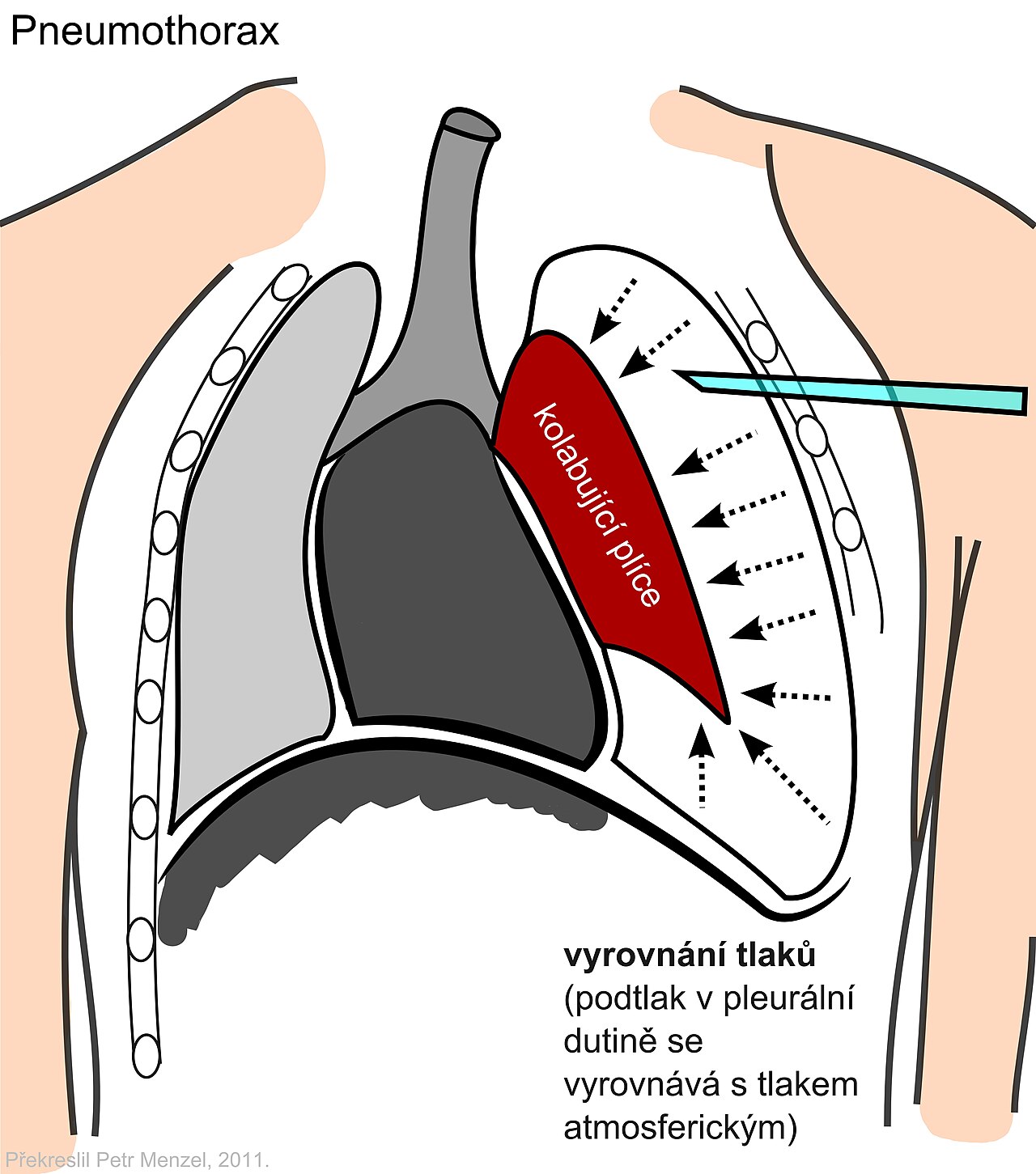
Air in the pleural space is a pneumothorax. That air sits between the lung and the chest wall, and the lung falls away from the wall. Spontaneous pneumothorax is primary (no known lung disease) or secondary (underlying lung disease). A blunt or penetrating chest injury causes a traumatic one. Procedures and positive-pressure ventilation cause the iatrogenic and ventilator-associated kinds. Blood in that same space is a hemothorax. Air and blood together make a hemopneumothorax.
- Tension physiology: Every breath adds air to the pleural space, and none of it can leave. A lung tear, a chest wound or a valve-like flap traps it. Pleural pressure climbs, the mediastinum shifts, venous return drops and the opposite lung gets compressed.
- Open pneumothorax (sucking chest wound): The wound is a direct door. Air passes straight between the atmosphere and the pleural space. Seal it with a non-vented dressing and that air can become trapped and create tension.
- Common causes: rib fracture or penetrating wound, central-line or thoracic procedures, positive-pressure ventilation with high airway pressures, and an open chest wound
- Course: A simple pneumothorax can become tension quickly. Especially under positive-pressure ventilation or during transport to altitude.
Recognition & Red Flags
Pneumothorax
- Spontaneous: sudden pleuritic chest pain and dyspnea, often at rest, with reduced or absent breath sounds on one side
- Traumatic or iatrogenic: chest injury, rib fractures or a procedure followed by new dyspnea, pain or falling SpO₂
Tension Pneumothorax (Clinical Diagnosis)
- One-sided absent or reduced breath sounds plus hypotension, hypoxemia or obstructed ventilation
- Ventilated patient: rising peak airway pressures with falling SpO₂ or BP and a one-sided exam
Stable vitals do not exclude it: A normal SpO₂ or a stable-looking BP does not rule out a pneumothorax. Go by the exam and the trend. When tension signs are present, do not wait for imaging.
Red Flags
- Hypotension, falling SpO₂ or new cyanosis
- Rising ventilator pressures with a one-sided exam, or hemodynamic collapse after a chest injury
- Cardiac arrest in a patient with chest trauma (decompress both sides per protocol)
Hemothorax
- Signs: dull percussion and reduced breath sounds on one side, with shock (falling BP, tachycardia, pallor) after chest trauma or a procedure. Blood loss hides easily. It can sit in the chest and in other body cavities.
- Hemorrhage versus tension: Flat neck veins favor blood loss. Distended neck veins with tracheal shift favor tension. Both can cause one-sided silence and falling BP, so reassess after every intervention.
Assessment & Diagnostics
Tension is diagnosed at the bedside. Decompress and drain first. The chest X-ray or ultrasound comes afterward to confirm size and tube position.
| Check | Why | Act If |
|---|---|---|
| Breath sounds and chest movement | One-sided loss of air entry | Absent or reduced on one side with hypotension: treat the side as tension per protocol |
| BP, HR and perfusion | Obstructed venous return causes shock | Hypotension with one-sided signs: decompress without waiting for imaging |
| SpO₂ and work of breathing | Shunt and failing ventilation | Falling SpO₂: oxygen to target (see ALS Management) |
| Waveform EtCO₂ (intubated) | Confirms tube position and ventilation | Falling EtCO₂ or waveform change: check tube and the chest, see Capnography |
| Ventilator pressures (intubated) | Peak pressure rises with tension | Rising peak pressure with a one-sided exam: decompress; check auto-PEEP or circuit problems only as a quick differential |
| Chest X-ray or ultrasound | Size, side and tube position | Use after decompression or when stable; never a reason to delay decompression in tension |
Differential & Mimics
- Auto-PEEP or bronchospasm (ventilated): Rising pressures, equal breath sounds and a patent airway. Check the ventilator circuit and expiratory time.
- Endotracheal tube in one main bronchus: Breath sounds on one side only, and the tube depth is wrong. Check the depth and the waveform with capnography.
- Anaphylaxis: Bronchospasm and hypotension, with skin or mucosal signs after an exposure. See Anaphylaxis.
- Cardiac tamponade: hypotension with muffled heart sounds and normal breath sounds on both sides
BLS Management
- Position for breathing: let the patient sit or lie in the position that eases breathing, unless hypotension or unconsciousness requires otherwise.
- Open chest wound: expose the wound and apply a vented chest seal per protocol. If a vented seal is not available, a non-vented seal is used and monitored closely. Reassess breath sounds, SpO₂ and BP after sealing.
- Oxygen: apply oxygen per protocol to reach SpO₂ 94–98%. Use 88–92% only where the patient is at risk of hypercapnic failure (see the Oxygen page).
- Monitor and ventilate gently: watch SpO₂, respiratory rate and effort, and BP where the unit has it. If you are assisting with a bag-valve-mask, avoid over-ventilation. It can convert a simple pneumothorax to tension. Record the time of onset of symptoms.
- Do not decompress at BLS level: Needle decompression sits above BLS scope in most systems, per local protocol. If one-sided silence with falling BP or rising distress develops under a seal, lift or vent the seal per protocol and call ALS.
- Call ALS early and report one-sided findings, the mechanism of injury, any sealed wound, time of onset and the response to oxygen and position.
ALS Management
Doses vary by protocol; follow local protocols and medical direction.
- Oxygen (Paramedic/RN/CCT): titrate to SpO₂ 94–98%, which is the BTS 2017 target for most acutely ill adults. Trauma or shock protocols may call for higher-flow oxygen when the patient is hypoxic; follow protocol. See Oxygen
- Open chest wound (Paramedic/RN/CCT): confirm the vented seal is in place per protocol. If tension signs appear under a seal, lift or vent it and decompress per protocol.
- Tension: needle decompression (Paramedic/CCT per protocol): decompress the affected side at the 2nd intercostal space at the midclavicular line or the 4th or 5th intercostal space at the anterior axillary line. Catheter gauge and length are set by protocol. Reassess BP, SpO₂ and breath sounds after each attempt. See Needle Decompression & Finger Thoracostomy
- Analgesia (Paramedic/RN/CCT): give opioids after tension is relieved and only when the patient is hemodynamically stable. Fentanyl 25–100 mcg IV, titrated to effect and to respiratory drive, with reduced doses in hypotension (see Fentanyl). The fentanyl label warns of hypotension, especially with other CNS depressants, and advises considering hypovolemia. Ketamine is an alternative; consider it per protocol for hypotensive or hemorrhaging patients (see Ketamine).
- Access (Paramedic/RN/CCT): IV or IO access per protocol. See IO Access
- Ventilated patient (RN/CCT): decompress without delay when tension signs are present. Disconnect and hand-bag only as a quick check for auto-PEEP or circuit problems, never as a reason to delay decompression.
- Hemorrhage (Paramedic/RN/CCT): bleeding control and fluids or blood per the Hemorrhage Control page and local protocol
- Hemothorax (Paramedic/RN/CCT): needle decompression does not treat blood. Resuscitate with blood products or fluids per protocol, control bleeding and transport to a trauma center. Permissive hypotension is used in trauma only per protocol.
- Traumatic arrest (Paramedic/RN/CCT): decompress both sides per protocol, with hemorrhage control and airway management under the traumatic arrest protocol.
ED & ICU Management
- Definitive drainage: After tension decompression, a chest tube is the definitive drain. It is also the drain for a large or symptomatic pneumothorax or a hemothorax. See Chest Tubes.
- Drain size: Small-bore drains (about 12–14 Fr) go in for an uncomplicated spontaneous pneumothorax (secondary review). For a large air leak, surgical emphysema or mechanical ventilation, go larger. Trauma, hemothorax and ventilated patients need a larger bore per protocol. See Chest Tubes.
- Spontaneous pneumothorax, minimal symptoms: BTS 2023 supports conservative management for a minimally symptomatic primary pneumothorax. The guideline weighs symptoms and high-risk features (hemodynamic compromise, significant hypoxia, underlying lung disease) above size alone. Needle aspiration is an option in selected patients. Its immediate success is lower than a drain's (secondary review).
- Hemothorax: Drain it and measure the output. Surgical review is considered for an initial output over 1,500 mL, or for ongoing output of about 150–200 mL/h for 2–4 h. Treat that as a guide, not a rule (see Chest Tubes). Call surgery early if the patient is unstable or still needs transfusion. Transfuse per protocol (see Blood Transfusion).
- Ventilated patients: Reassess ventilator pressures after every drain or change in settings. Follow the ventilator protocol (see Mechanical Ventilation).
- Persistent air leak: Refer to the thoracic team. If the leak keeps going despite drainage for about 5–7 days, surgery is considered (secondary review).
Transport Considerations
- Before moving: Decompress and reassess first. Then secure the catheter or drain, and keep oxygen and ventilator settings in place.
- Altitude: Air expands on ascent (Boyle's law). An untreated pneumothorax can enlarge in flight. Reassess before flight and at each altitude change. Place a drain before flight when feasible. Cabin-altitude figures are on the Flight Transport Physiology page (see Flight Transport Physiology).
- Chest drains: Keep the unit below chest level, and the tubing free of kinks. A bubbling drain is never clamped unless a specialist directs it (see Chest Tubes).
- One-way valve or drain unit in flight: A one-way (Heimlich) valve suits a pneumothorax. A blood-bearing drain uses a three-chamber unit, set per protocol. Check for air leak and blood in the chamber at each reassessment.
- Noise and vibration: Trust the monitors and the breath sounds, not the sound of air. Reassess after each move and each ventilator change.
- Hand-off: time of recognition, side, site, catheter or drain used, response to each attempt, oxygen and ventilator settings, drain output, and any blood or fluid given.
Special Populations
- Pediatrics: The pediatric protocol and the child's chest-wall size set the needle site, catheter size and depth. Do not reach for an adult default. This page gives no pediatric catheter length. Early signs are tachycardia and tachypnea. Hypotension is a late sign, so act on the trend. Use Equipment Sizing and weight-based analgesia per Weight-Based Dosing. Monitor with Vital Signs.
- Pregnancy: The gravid uterus pushes the diaphragm up, so the drain site may need adjusting per protocol and with the trauma team. In a gravid patient beyond about 20 weeks, left lateral uterine displacement relieves aortocaval compression during resuscitation. Positioning for comfort alone is not enough in a hypotensive patient. Involve obstetric and trauma-center teams early.
- Obesity: Catheter length and site follow protocol. A thicker chest wall means higher failure rates, so consider a longer catheter or the alternative site per protocol (see Needle Decompression).
- Patients on Anticoagulants: Drain placement raises the bleeding risk. Correction must not delay decompression of tension or drainage of a massive hemothorax (see Chest Tubes).
- Ventilated Patients: Positive pressure can turn a small pneumothorax into tension. Check pressures with every change and decompress early (see Recognition).
Pitfalls & Safety Checks
- Error: waiting for imaging. A tension patient with hypotension or rising pressures gets decompressed first. Confirm afterward with BP, SpO₂ and pressures. A chest X-ray alone does not cut it.
- Error: judging by a hiss. A hiss is air moving through a catheter, and that does not prove the lung has re-expanded. Confirm with the clinical response.
- Error: assuming the needle worked. In a 2026 retrospective Omaha review of 214 prehospital needle decompressions, 30% of patients responded (48% after excluding the 63% who received CPR en route). Most of the indications were subjective, such as absent breath sounds. Complications occurred in 14%. The authors recommend objective criteria such as confirmed hypoxia. Needle decompression is still indicated for true tension. Check the response after each attempt.
- Error: deep or angled insertion. A 2026 case report describes a catheter that entered the left ventricle after prehospital needle thoracostomy. Use the protocol site and depth. Never angle the needle toward the heart.
- Error: clamping a bubbling drain. Clamping can turn a pneumothorax into tension. Keep the drain open unless a specialist directs otherwise.
- Error: missing re-tension. A decompressed patient can re-tension in transit or after ventilator changes. Reassess after every move. If the signs come back, re-decompress at once.
- Error: missing tension under a seal. Tension can develop under an occlusive chest seal. Lift or vent the seal and reassess. If the signs persist, decompress per protocol.
Hemothorax and tension look alike: One-sided silence and falling BP show up in both. Blood on needle withdrawal means plan a chest tube. The needle does not treat that bleeding.
Clinical Pearls
Decide early: You decide this one at the bedside. Silent on one side with hypotension? That justifies decompression before any image.
Ventilator discriminator (CCRN/CEN): Peak pressure climbing, BP falling, one side silent. Call it tension until you prove otherwise. Auto-PEEP gets a quick look, never a wait.
The needle is a bridge: A needle buys time, and only time. Start planning the chest tube the moment the needle goes in.
References
- British Thoracic Society (2023). British Thoracic Society Guideline for pleural disease. Thorax, 78 (Suppl 3), s1–s42. https://doi.org/10.1136/thorax-2022-219784
- O'Driscoll BR, Howard LS, Earis J, Mak V (2017). BTS guideline for oxygen use in adults in healthcare and emergency settings. Thorax, 72 (Suppl 1), ii1–ii90. https://doi.org/10.1136/thoraxjnl-2016-209729
- Cheng HS, Wong C, Chiu PH, Tong CW (2024). Management of spontaneous pneumothorax: a mini-review on its latest evidence. J Thorac Dis, 16, 4756–4763. https://doi.org/10.21037/jtd-24-415
- Boever JC, Ott MJ, Muldiiarov V, et al. (2026). Prehospital needle thoracostomy and the need to implement objective criteria for intervention: a retrospective study. Injury, 57(5), 112973. https://doi.org/10.1016/j.injury.2025.112973
- Ahmed K, Abdelrahman R, Alahmad M, et al. (2026). Iatrogenic cardiac injury following prehospital needle decompression: a case report. J Trauma Inj, online ahead of print. https://doi.org/10.20408/jti.2025.0136
- Hospira, Inc. (revised 2026). Fentanyl Citrate Injection, USP prescribing information. DailyMed, set ID bfa8018a-4cbc-434b-e09e-782872b0340a. https://dailymed.nlm.nih.gov/dailymed/drugInfo.cfm?setid=bfa8018a-4cbc-434b-e09e-782872b0340a
- Joint Trauma System (2024). Tactical Combat Casualty Care Guidelines, dated 25 Jan 2024, as listed in the Joint Trauma System Clinical Practice Guidelines Index. https://jts.health.mil/assets/docs/cpgs/CPG_Index.pdf
Medical Disclaimer
- For Educational Purposes Only: This content is intended for educational reference and should not be used for clinical decision-making.
- Not a Substitute for Professional Judgment: Always consult your local protocols, institutional guidelines, and supervising physicians.
- Verify Before Acting: Users are responsible for verifying information through authoritative sources before any clinical application.
AI Assistance Notice
AI was used to assist in organizing, paraphrasing, and formatting this information.